Posterior Triangle
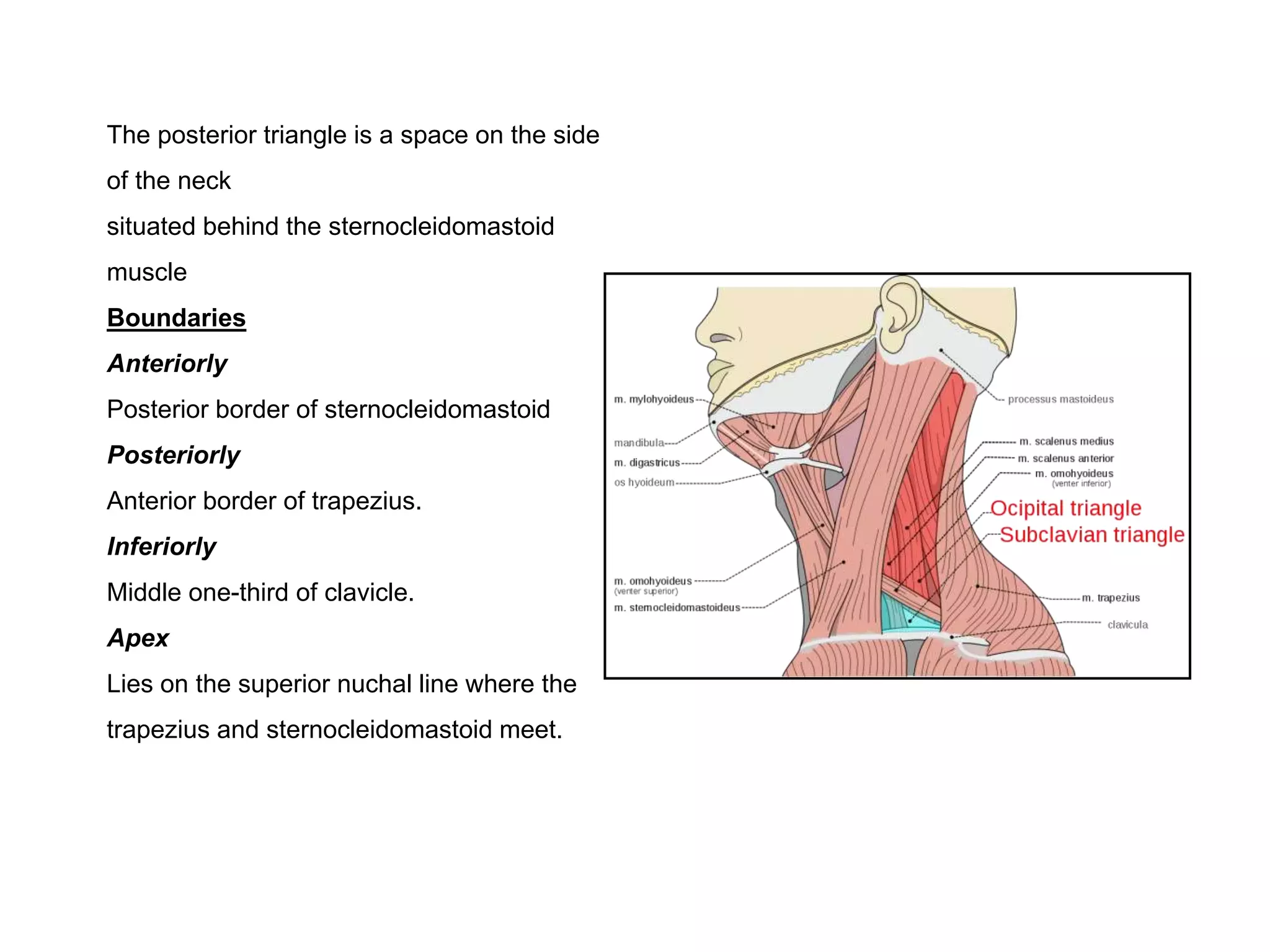
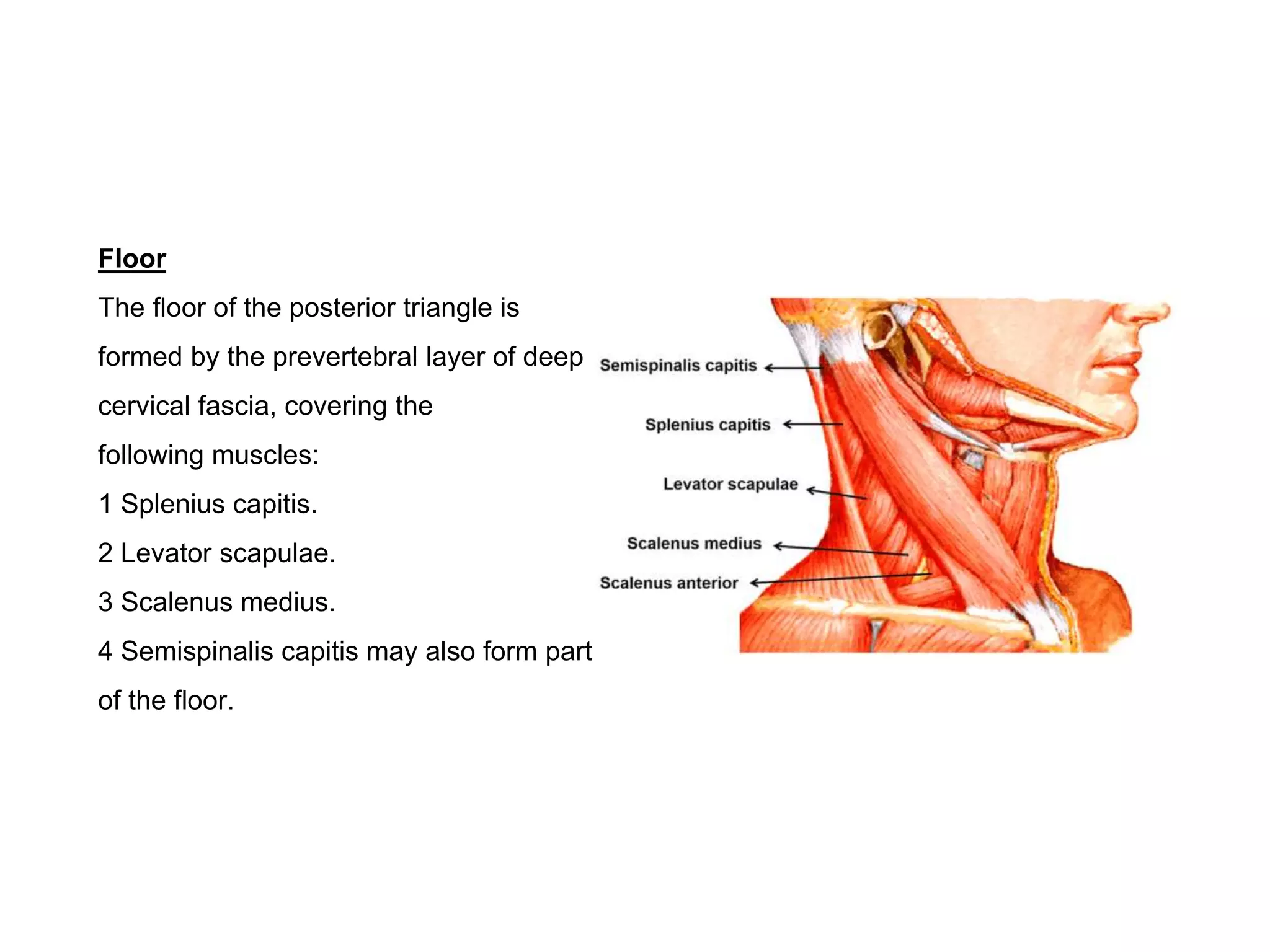
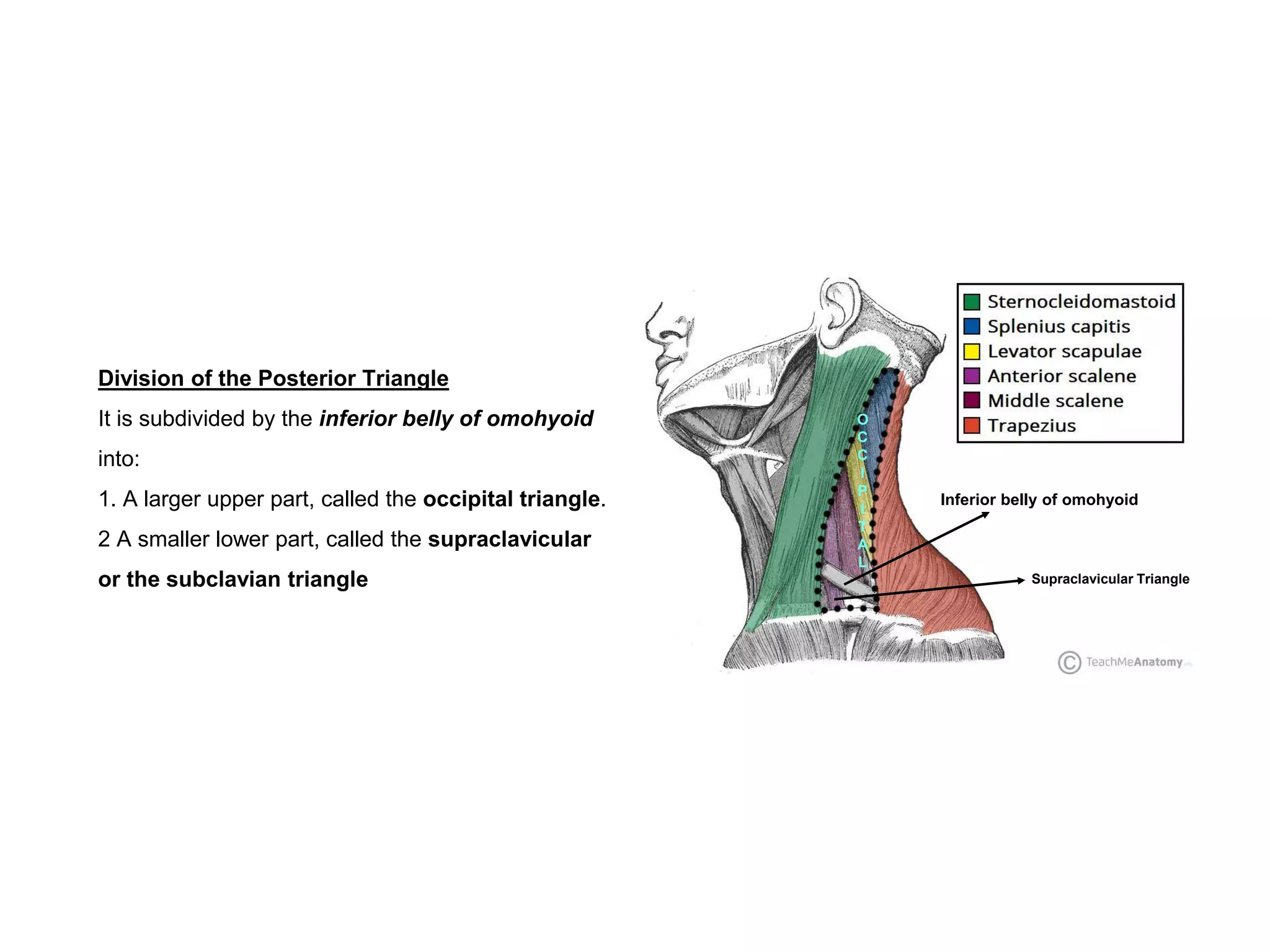
The posterior triangle is a space on the side
of the neck
situated behind the sternocleidomastoid
muscle
Boundaries
Anteriorly
Posterior border of sternocleidomastoid
Posteriorly
Anterior border of trapezius.
Inferiorly
Middle one-third of clavicle.
Apex
Lies on the superior nuchal line where the
trapezius and sternocleidomastoid meet.
Roof
The roof is formed by the investing layer of deep
cervical fascia,
The superficial fascia over the posterior triangle
contains:
1. The platysma.
2. The external jugular and posterior external
jugular veins.
3. Parts of the supraclavicular, great auricular,
transverse cutaneous and lesser occipital nerves
4. Unnamed arteries derived from the occipital,
transverse cervical and suprascapular arteries.
5. Lymph vessels which pierce the deep fascia to
end in the supraclavicular nodes.
Floor
The floor of the posterior triangle is
formed by the prevertebral layer of deep
cervical fascia, covering the
following muscles:
1 Splenius capitis.
2 Levator scapulae.
3 Scalenus medius.
4 Semispinalis capitis may also form part
of the floor.
Division of the Posterior Triangle
It is subdivided by the inferior belly of omohyoid
into:
1. A larger upper part, called the occipital triangle.
2 A smaller lower part, called the supraclavicular
or the subclavian triangle
Inferior belly of omohyoid
O
C
C
I
P
I
T
A
L
Supraclavicular Triangle
Contents of Posterior Triangle
Contents Occipital Triangle Supraclavicular Triangle
Nerves
1. Spinal accessory nerve
2. Four cutaneous branches of cervical plexus:
a. Lesser occipital (C2)
b. Great auricular (C2, C3)
c. Anterior cutaneous nerve of neck (C2, C3)
d. Supraclavicular nerves (C3, C4)
3. Muscular branches:
a. Two small branches to the levator scapulae
(c3, c4)
b. Two small branches to the trapezius (C3, C4)
c. Nerve to rhomboids (proprioceptive) (C5)
4. C5, C6 roots of the brachial plexus
1. Three trunks of brachial plexus
2. Nerve to serratus anterior (long thoracic, c5,
c6, c7)
3. Nerve to subclavius (C5, C6)
4. Suprascapular nerve (C5, C6)
Vessels
1. Transverse cervical artery and vein
2. Occipital artery
1. Third part of subclavian artery and subclavian
vein
2. Suprascapular artery and vein
3. Commencement of transverse cervical artery
and
termination of the corresponding vein
4. Lower part of external jugular vein
Lymph nodes
Along the posterior border of the
sternocleidomastoid, more in the lower part- the
supraclavicular nodes
and a few at the upper angle-the occipital nodes
A few members of the supraclavicular chaln
Clinical Anatomy
• The most common swelling in the posterior triangle is due to enlargement of the supraclayicular lymph nodes, While
doing biopsy of the lymph node, one must be careful in preserving the accessory nerve which,may. get entangled
amongst enlarged lymph nodes
• Supraclavicular lymph nodes are commonly enlarged in tuberculosis, Hodgkin's disease, and in malignant growths of
the breast arm or chest.
• Block dissection of the neck for malignant diseases is the removal of cervical lymph nodes along with other structures
involved in the growth. This procedure does not endanger those nerves of the posterior triangle which lie deep to the
prevertebral fascia, i.e- the brachial and cervical plexuses and their muscular branches.
• A cervical rib may compress the second part of subclavian artery. In these case blood supply to upper limb reaches via
anastomosis around the scapula.
• Dysphagia caused by compression of the oesophagus by an abnormal subclavian artery is called dysphagia lusoria
• Elective arterial surgery of the conrmon carotid artery is done for aneurysms, AV fistulae or arteriosclerotic occlusions. It
is better to expose the common carotid artery in its upper part where it is superficial. While ligating the artery. care
should be taken not to include the vagus nerve or the sympathetic chain.
• Second part of the subclavian artery may get pressed by the scalenus anterior muscle, resulting in decreased blood
supply to the upper limb. If the muscle is divided the effects are abolished.
Posterior triangle of the neck

Posterior triangle of the neck

  • 1.
  • 2.
    The posterior triangleis a space on the side of the neck situated behind the sternocleidomastoid muscle Boundaries Anteriorly Posterior border of sternocleidomastoid Posteriorly Anterior border of trapezius. Inferiorly Middle one-third of clavicle. Apex Lies on the superior nuchal line where the trapezius and sternocleidomastoid meet.
  • 3.
    Roof The roof isformed by the investing layer of deep cervical fascia, The superficial fascia over the posterior triangle contains: 1. The platysma. 2. The external jugular and posterior external jugular veins. 3. Parts of the supraclavicular, great auricular, transverse cutaneous and lesser occipital nerves 4. Unnamed arteries derived from the occipital, transverse cervical and suprascapular arteries. 5. Lymph vessels which pierce the deep fascia to end in the supraclavicular nodes.
  • 4.
    Floor The floor ofthe posterior triangle is formed by the prevertebral layer of deep cervical fascia, covering the following muscles: 1 Splenius capitis. 2 Levator scapulae. 3 Scalenus medius. 4 Semispinalis capitis may also form part of the floor.
  • 5.
    Division of thePosterior Triangle It is subdivided by the inferior belly of omohyoid into: 1. A larger upper part, called the occipital triangle. 2 A smaller lower part, called the supraclavicular or the subclavian triangle Inferior belly of omohyoid O C C I P I T A L Supraclavicular Triangle
  • 6.
    Contents of PosteriorTriangle Contents Occipital Triangle Supraclavicular Triangle Nerves 1. Spinal accessory nerve 2. Four cutaneous branches of cervical plexus: a. Lesser occipital (C2) b. Great auricular (C2, C3) c. Anterior cutaneous nerve of neck (C2, C3) d. Supraclavicular nerves (C3, C4) 3. Muscular branches: a. Two small branches to the levator scapulae (c3, c4) b. Two small branches to the trapezius (C3, C4) c. Nerve to rhomboids (proprioceptive) (C5) 4. C5, C6 roots of the brachial plexus 1. Three trunks of brachial plexus 2. Nerve to serratus anterior (long thoracic, c5, c6, c7) 3. Nerve to subclavius (C5, C6) 4. Suprascapular nerve (C5, C6) Vessels 1. Transverse cervical artery and vein 2. Occipital artery 1. Third part of subclavian artery and subclavian vein 2. Suprascapular artery and vein 3. Commencement of transverse cervical artery and termination of the corresponding vein 4. Lower part of external jugular vein Lymph nodes Along the posterior border of the sternocleidomastoid, more in the lower part- the supraclavicular nodes and a few at the upper angle-the occipital nodes A few members of the supraclavicular chaln
  • 8.
    Clinical Anatomy • Themost common swelling in the posterior triangle is due to enlargement of the supraclayicular lymph nodes, While doing biopsy of the lymph node, one must be careful in preserving the accessory nerve which,may. get entangled amongst enlarged lymph nodes • Supraclavicular lymph nodes are commonly enlarged in tuberculosis, Hodgkin's disease, and in malignant growths of the breast arm or chest. • Block dissection of the neck for malignant diseases is the removal of cervical lymph nodes along with other structures involved in the growth. This procedure does not endanger those nerves of the posterior triangle which lie deep to the prevertebral fascia, i.e- the brachial and cervical plexuses and their muscular branches. • A cervical rib may compress the second part of subclavian artery. In these case blood supply to upper limb reaches via anastomosis around the scapula. • Dysphagia caused by compression of the oesophagus by an abnormal subclavian artery is called dysphagia lusoria • Elective arterial surgery of the conrmon carotid artery is done for aneurysms, AV fistulae or arteriosclerotic occlusions. It is better to expose the common carotid artery in its upper part where it is superficial. While ligating the artery. care should be taken not to include the vagus nerve or the sympathetic chain. • Second part of the subclavian artery may get pressed by the scalenus anterior muscle, resulting in decreased blood supply to the upper limb. If the muscle is divided the effects are abolished.