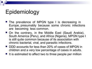
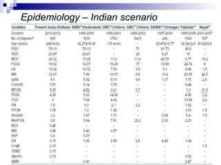
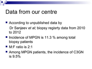
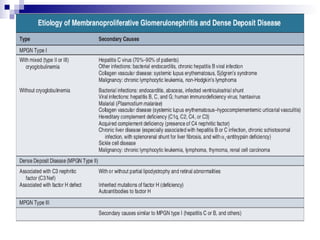
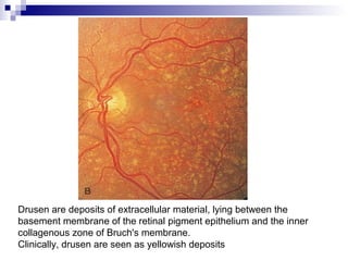
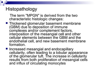
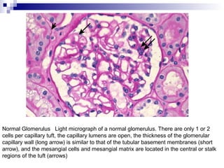
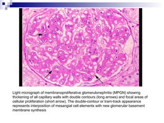
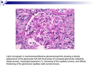
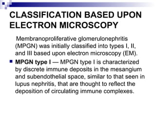
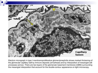
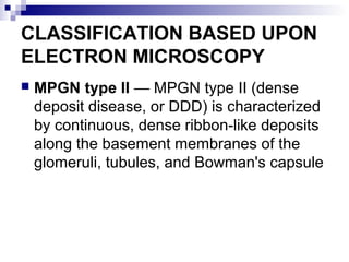
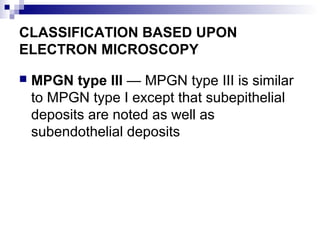
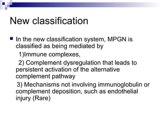
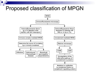
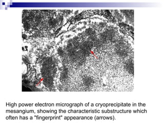
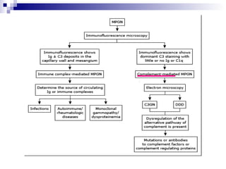
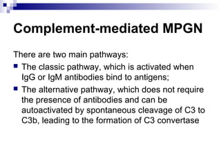
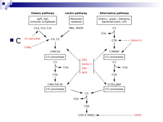
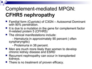
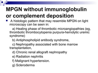
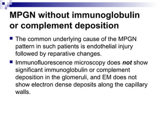
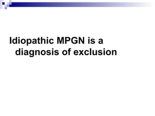
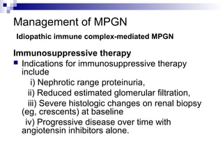
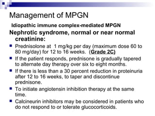
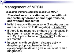
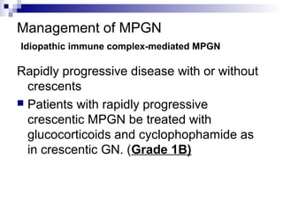
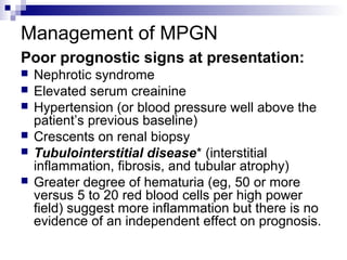
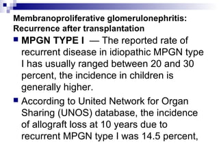
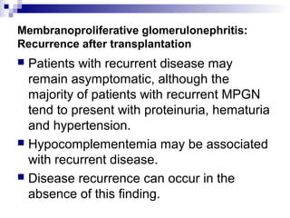
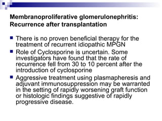
Membranoproliferative
Glomerulonephritis
Dr. Mohit Mathur
29/10/2013
Introduction
 Membranoproliferative glomerulonephritis
(MPGN), also known as mesangiocapillary
glomerulonephritis, is a pattern of glomerular
injury viewed by light microscopy.
 Its name is derived from the characteristic
histologic changes including hypercellularity and
thickening of the glomerular basement
membrane, often leading to a lobular
appearance of the glomerular tuft.
Epidemiology
 In North America and Europe, MPGN (types
I and III) and DDD constitute less than 5% of
all primary glomerulonephritis.
 MPGN accounts for 5% to 10% of primary renal
causes of nephrotic syndrome in children and
adults.
 It occurs equally in males and females and in
the United States is relatively more common
in Caucasians than in African Americans.
Epidemiology
 MPGN presenting in childhood (primarily
between the ages of 8 and 14 years)
includes types I and III MPGN and
DDD and is frequently idiopathic or
associated with nephritic factors.
 MPGN presenting in adults (typically older
than 18 years) is usually type I or type
III and is commonly associated with
cryoglobulinemia and HCV infection
Epidemiology
 The prevalence of MPGN type I is decreasing in
Europe, presumably because some chronic infections
are becoming less common.
 On the contrary, in the Middle East (Saudi Arabia),
South America (Peru), and Africa (Nigeria), MPGN type I
is still quite common because of its association with
chronic bacterial, viral, and parasitic infections.
 DDD accounts for less than 20% of cases of MPGN in
children and a very low percentage of cases in adults.
 It is estimated to affect two to three people per million
Epidemiology – Indian scenario
Data from our centre
 According to unpublished data by
Dr Sanjeev et al; biopsy regisrty data from 2010
to 2012
 Incidence of MPGN is 11.3 % among total
biopsy patients
 M:F ratio is 2:1
 Among MPGN patients, the incidence of C3GN
is 9.5%
Clinical features
 The clinical presentation is similar to that in other
types of glomerulonephritis.
 In patients with active disease, the urine
sediment reveals hematuria, typically with
dysmorphic red cells and occasionally with red
cell casts
 There is a variable degree of proteinuria;
 The serum creatinine may be normal or
elevated.
Clinical features…
 Occasional patients with indolent disease
present late in the course at a time when
active inflammation has subsided.
 Such patients may have a bland urine
sediment with a variable degree of
proteinuria and elevation in serum
creatinine.
 The diagnosis is made by renal biopsy
Clinical features
 Among patients who have the different forms of
MPGN there are generally no differences in
clinical presentation with the possible exception
of patients with DDD, which is a form of
complement-mediated MPGN that is associated
with drusen formation that may be seen on
fundoscopic examination and with partial
lipodystrophy
Lipodystrophy
Drusen are deposits of extracellular material, lying between the
basement membrane of the retinal pigment epithelium and the inner
collagenous zone of Bruch's membrane.
Clinically, drusen are seen as yellowish deposits
Histopathology
The term "MPGN" is derived from the two
characteristic histologic changes:
 Thickened glomerular basement membrane
(GBM) due to deposition of immune
complexes and/or complement factors,
interposition of the mesangial cell and other
cellular elements between the GBM and the
endothelial cell, and new basement membrane
formation.
 Increased mesangial and endocapillary
cellularity, often leading to a lobular appearance
of the glomerular tuft. The increase in cellularity
results from both proliferation of mesangial cells
and influx of circulating monocytes
Normal Glomerulus Light micrograph of a normal glomerulus. There are only 1 or 2
cells per capillary tuft, the capillary lumens are open, the thickness of the glomerular
capillary wall (long arrow) is similar to that of the tubular basement membranes (short
arrow), and the mesangial cells and mesangial matrix are located in the central or stalk
regions of the tuft (arrows)
Light micrograph of membranoproliferative glomerulonephritis (MPGN) showing
thickening of all capillary walls with double contours (long arrows) and focal areas of
cellular proliferation (short arrow). The double-contour or tram-track appearance
represents interposition of mesangial cell elements with new glomerular basement
membrane synthesis
Light micrograph in membranoproliferative glomerulonephritis showing a lobular
appearance of the glomerular tuft with focal areas of increased glomerular cellularity
(large arrows), mesangial expansion (*), narrowing of the capillary lumens, and diffuse
thickening of the glomerular capillary walls (small arrows).
CLASSIFICATION BASED UPON
ELECTRON MICROSCOPY
Membranoproliferative glomerulonephritis
(MPGN) was initially classified into types I, II,
and III based upon electron microscopy (EM).
 MPGN type I — MPGN type I is characterized
by discrete immune deposits in the mesangium
and subendothelial space, similar to that seen in
lupus nephritis, that are thought to reflect the
deposition of circulating immune complexes.
Normal glomerulus
Electron micrograph in type I membranoproliferative glomerulonephritis shows marked thickening of
the glomerular capillary wall by immune deposits (arrowhead) and by interposition of mesangial cell
processes (arrow). There are two layers of the glomerular basement membrane (GBM) surrounding
the mesangial interposition that account for the double-contour appearance on light microscopy.
CLASSIFICATION BASED UPON
ELECTRON MICROSCOPY
 MPGN type II — MPGN type II (dense
deposit disease, or DDD) is characterized
by continuous, dense ribbon-like deposits
along the basement membranes of the
glomeruli, tubules, and Bowman's capsule
Electron micrograph in dense deposit disease (DDD) showing dense, ribbon-
like appearance of subendothelial and intramembranous material (arrow) and
narrowing of the capillary lumen due to proliferation of cells (double arrow).
CLASSIFICATION BASED UPON
ELECTRON MICROSCOPY
 MPGN type III — MPGN type III is similar
to MPGN type I except that subepithelial
deposits are noted as well as
subendothelial deposits
Membranoproliferative glomerulonephritis type III. This electron
micrograph shows the irregular appearance of electron-dense,
presumed immune aggregates within, outside, and under the
glomerular capillary basement membrane.
CLASSIFICATION BASED UPON ELECTRON
MICROSCOPY
Limitations of this classification
 The EM-based classification can result in
overlap between types I and III.
 Not based on pathogenesis – Multiple
mechanisms of injury may result in a common
pattern of injury.
 In contrast, a classification that is based upon
the pathogenetic process helps to direct the
clinical evaluation and to provide disease-
specific treatments.
New classification
 Sethi S, Fervenza FC. Membranoproliferative
glomerulonephritis: pathogenetic heterogeneity
and proposal for a new classification. Semin
Nephrol 2011; 31:341.
 Sethi S, Fervenza FC. Membranoproliferative
glomerulonephritis--a new look at an old entity.
N Engl J Med 2012; 366:1119.
New classification
 In the new classification system, MPGN is
classified as being mediated by
1)Immune complexes,
2) Complement dysregulation that leads to
persistent activation of the alternative
complement pathway
3) Mechanisms not involving immunoglobulin or
complement deposition, such as endothelial
injury (Rare)
Proposed classification of MPGN
Immune complex-mediated
MPGN
 Immune complex-mediated MPGN is
characterized by the presence of immune
complexes and complement components.
 Immune complex-mediated MPGN results from
chronic antigenemia and/or circulating immune
complexes and can be seen in chronic
infections, autoimmune diseases, and
monoclonal gammopathies
Immune complex-mediated
MPGN
 Immunofluorescence microscopy may
suggest the underlying etiology of immune
complex-mediated MPGN
Immune complex-mediated MPGN
Infection
 Immune complex-mediated MPGN is most
commonly secondary to hepatitis C and B
viral infections* ??
 The incidence of HCV-associated MPGN
varies with location.
 HCV-induced MPGN is typically
associated with mixed cryoglobulinemia.
Immune complex-mediated
MPGN ; Infection
 MPGN resulting from hepatitis C virus
infection typically shows granular
deposition of IgM, C3, and both kappa
and lambda light chains.
 IgG may or may not be present, and C1q
is typically negative.
 This pattern may also be seen with MPGN
induced by other viral infections .
Immune complex-mediated MPGN;
Infection
 EM: Fingerprint pattern associated with
cryoprecipitates is seen in mixed
cryoglobulinemia*
*Rarely with Lupus
High power electron micrograph of a cryoprecipitate in the
mesangium, showing the characteristic substructure which
often has a "fingerprint" appearance (arrows).
Immune complex-mediated MPGN;
Infection
 Chronic bacterial (eg, endocarditis, shunt
nephritis, abscesses), fungal, and,
particularly in the developing world*,
parasitic infections (eg, schistosomiasis,
echinococcosis) can cause MPGN
Immune complex-mediated
MPGN
Autoimmune disorders
 Immune complex-mediated MPGN is
observed in patients with systemic lupus
erythematosus (particularly in the chronic
phase of lupus nephritis)
 Rarely in Sjögren's syndrome or
rheumatoid arthritis
Immune complex-mediated
MPGN: Autoimmune disorders
 MPGN resulting from autoimmune
diseases is typically characterized by the
"full house" pattern of immunoglobulin
deposition, including IgG, IgM, IgA, C1q,
C3, and kappa and lambda light chains
Immune complex-mediated
MPGN; Autoimmune disorders
 EM: Tubuloreticular structures in
endothelial cells in MPGN are suggestive
of lupus nephritis
Electron micrograph in diffuse proliferative lupus nephritis shows massive subendothelial
deposits (D) and characteristic tubuloreticular structures (arrow) in the endothelial cells
(En). The subendothelial deposits cause marked thickening of the glomerular capillary
wall, leading to a wire loop appearance on light microscopy
Immune complex-mediated
MPGN: Dysproteinemia
 Dysproteinemias are an important
cause of MPGN and can be
associated on immunofluorescence
microscopy with:
Monoclonal immunoglobulin deposition
Immune complex-mediated
MPGN: Dysproteinemia
 MPGN resulting from monoclonal
gammopathy is characterized by
deposition of monotypic kappa or lambda
light chains but not both.
 MPGN associated with heavy-chain
deposition may show immunoglobulin
deposition (heavy-chain isotypes) in the
absence of either light chain
Immune complex-mediated
MPGN: Dysproteinemia
The clinical features and prognosis of MPGN
associated with monoclonal gammopathy are:
 Nephrotic syndrome (49%)
 Renal insufficiency (68%)
 Hematuria (77%)
 End-stage renal disease (ESRD) (22%)
Immune complex-mediated
MPGN: Dysproteinemia
 Cases of MPGN in patients with chronic
lymphocytic leukemia have also been
described, usually in association with
mixed cryoglobulinemia or
noncryoprecipitating monoclonal IgG or
IgM
Immune complex-mediated
MPGN: Rare causes
Rare causes of MPGN include
 Non-Hodgkin lymphoma,
 Renal cell carcinoma,
 Snake venom
 Splenorenal shunt surgery for portal
hypertension
 Melanoma
 Alpha-1-antitrypsin deficiency.
Complement-mediated MPGN
There are two main pathways:
 The classic pathway, which is activated when
IgG or IgM antibodies bind to antigens;
 The alternative pathway, which does not require
the presence of antibodies and can be
autoactivated by spontaneous cleavage of C3 to
C3b, leading to the formation of C3 convertase
 Omplement path
Complement-mediated MPGN
 Complement-mediated MPGN is less
common than immune complex-mediated
MPGN and results from dysregulation and
persistent activation of the alternative
complement pathway.
Complement-mediated MPGN
 This form of MPGN is due to the
deposition of complement products along
the capillary walls and in the mesangium.
 Immunofluorescence microscopy of
kidney sections demonstrates bright C3
staining, but NO immunoglobulin staining,
in the mesangium and along the capillary
walls.
Complement-mediated MPGN
 Complement-mediated MPGN may be further
classified based upon ultrastructural features
observed on EM as DDD or C3GN.
 Some genetic mutations that have been
associated with these disorders are also
associated with atypical hemolytic uremic
syndrome.
 Hypocomplementemia is a characteristic finding
in most patients with MPGN.
Complement-mediated MPGN
 A normal C3 concentration does not
exclude complement-mediated MPGN,
and it is not unusual to find a normal C3
concentration in the chronic phases of the
disease, as in adults with DDD or patients
with C3GN.
Complement-mediated MPGN:
Dense deposit disease (DDD)
 Dense deposit disease (DDD, also called MPGN type II)
is a rare form of MPGN that affects both children and
young adults.
 It has also been associated with monoclonal
gammopathies in older adults.
 On light microscopy, patterns of injury other than MPGN
can also be seen:
a) Mesangial proliferative
b) Diffuse proliferative
c) Crescentic glomerulonephritis
d) Sclerosing glomerulopathy.
Complement-mediated MPGN:
Dense deposit disease (DDD)
 C3 convertase-stabilizing antibody (called
C3 nephritic factor or C3NeF) is found in
approximately 80 percent of patients with
DDD and results in activation of the
alternative complement pathway that
characterizes DDD
Complement-mediated MPGN:
Dense deposit disease (DDD)
 Immunofluorescence microscopy demonstrates
C3 deposits
 EM (required to establish the diagnosis) shows
the characteristic sausage-shaped, wavy,
densely osmophilic deposits along the
glomerular basement membranes (GBM) and
mesangium (from which the disease receives its
name)
Complement-mediated MPGN:
C3 Glomerulonephritis (C3GN)
 C3 glomerulonephritis (C3GN, also called
glomerulonephritis with isolated C3
deposits) is, like DDD, caused by
excessive activation of the alternative
complement cascade due to mutations in
or antibodies to complement regulating
proteins
 C3GN has also been reported in
association with monoclonal
gammopathies and anti-factor H activity.
Complement-mediated MPGN:
C3 Glomerulonephritis (C3GN)
 Light microscopy often shows an MPGN
pattern of injury
Other forms of glomerulonephritis may be seen (mesangial
proliferative, diffuse proliferative, crescentic
glomerulonephritis,sclerosing glomerulopathy)
 Immunofluorescence microscopy shows
extensive C3 deposition along the
capillary walls and mesangium
with NO significant immunoglobulin
deposition
Immunofluorescence microscopy showing bright granular C3 staining
on the mesangium and along capillary walls in a patient with C3
glomerulonephritis (40x).
 Fig
Complement-mediated MPGN:
C3 Glomerulonephritis (C3GN)
 EM demonstrates deposits that are similar
to those seen with immune complex-
mediated MPGN but does not show the
typical sausage-shaped intramembranous
and mesangial deposits observed in DDD
Complement-mediated MPGN:
C3 Glomerulonephritis (C3GN)
 Patients with C3GN typically present with proteinuria,
which can be associated with nephrotic syndrome,
hematuria (sometimes synpharyngitic), and variable
degrees of hypertension and azotemia.
 C3 levels are usually low, C4 levels are normal, and
some patients have a C3 convertase-stabilizing
autoantibody called C3 nephritic factor (or C3NeF),
which is also seen in DDD
 Normal C3 does not exclude C3GN
 There may be progression to ESRD, and C3GN can
recur after renal transplantation.
Complement-mediated MPGN:
CFHR5 nephropathy
 Familial form (Cypriots) of C3GN – Autosomal Dominant
with 90% penetration.
 It is due to a mutation in the gene for complement factor
H-related protein 5 (CFHR5)
 The clinical manifestations include:
- Hematuria in approximately 90 percent ( often
synpharyngitic)
- Proteinuria in 38 percent.
 Men are much more likely than women to develop
chronic kidney disease and ESRD
 Recurrent nephropathy can occur in transplanted
kidneys.
 There is no treatment of proven efficacy.
MPGN without immunoglobulin
or complement deposition
 A histologic pattern that may resemble MPGN on light
microscopy can be seen in:
a) Healing phase of thrombotic microangiopathies (eg,
thrombotic thrombocytopenia purpura-hemolytic uremic
syndrome)
b) Antiphospholipid antibody syndrome,
c) Nephropathy associated with bone marrow
transplantation,
d) Chronic renal allograft nephropathy
e) Radiation nephritis
f) Malignant hypertension.
g) Scleroderma
MPGN without immunoglobulin
or complement deposition
 The common underlying cause of the MPGN
pattern in such patients is endothelial injury
followed by reparative changes.
 Immunofluorescence microscopy does not show
significant immunoglobulin or complement
deposition in the glomeruli, and EM does not
show electron dense deposits along the capillary
walls.
Idiopathic MPGN is a
diagnosis of exclusion
Management of MPGN
 The management of MPGN depends upon
the underlying cause as most patients
have either a circulating immune complex
disease or dysregulation of the alternative
complement pathway.
Management of MPGN
 Immune complex-mediated MPGN —
Patients who have such findings should be
evaluated for the following
disorders before treatment for MPGN is
initiated.
Management of MPGN
Infections
 Hepatitis B and hepatitis C virus should be
excluded by serology.
 Chronic bacterial infections should be excluded
by culture, including blood cultures.
 Test for fungi in the presence of a suggestive
history (eg, fever of unknown origin, unexplained
pulmonary infiltrates)
 Test for Parasitic infections (eg, malaria,
schistosomiasis, leishmaniasis) as appropriate.
Management of MPGN
Autoimmune diseases
 Screening as appropriate according to age
and clinical scenario.
Management of MPGN
Monoclonal gammopathy
 Monoclonal gammopathies should be excluded
by serum protein electrophoresis or serum free
light chains and urine electrophoresis and
immunofixation.
 Most patients with MPGN and a monoclonal
gammopathy have no identifiable disease; this
disorder has been called MPGN associated with
monoclonal gammopathy of uncertain
significance (MGUS).
Management of MPGN
Monoclonal gammopathy
 Occasionally patients with a monoclonal gammopathy
and MPGN have a serious and potentially treatable
cause.
 These include multiple myeloma, low-grade B cell
lymphoma, and chronic lymphocytic leukemia.
 These disorders may be diagnosed at presentation or
later after an initial diagnosis of monoclonal gammopathy
of undetermined significance
 Treatment of these disorders can lead to improvement in
the MPGN.
Evaluation of Complement-
mediated MPGN
 Patients should undergo an evaluation for
activation of the alternative pathway of
complement.
 Including measuring C3, C4, CH50 (which
provides a measure of activation of the classic
complement pathway), and AH50 (which
provides a measure of activation of the
alternative complement pathway).
 Genetic analysis for mutations and allele
variants of complement factors are done if
indicated (Research laboratories)
 Normal C3 levels does not rule out a
complement-mediated MPGN
Management of MPGN
Treatment of the glomerulonephritis
 The treatment of secondary MPGN is directed at
treatment of the underlying condition since the renal
disease often resolves with treatment of causes such as
infection, autoimmune disorders, and monoclonal
gammopathy.
 When an inciting condition is present (eg, infection),
resolution of the MPGN usually occurs after successful
treatment of the primary disease, such as antiviral
therapy in MPGN due to hepatitis C or B virus.
 Immunosuppressive therapy is both unnecessary and
potentially deleterious in patients with hepatitis
 At least partial remission of MPGN can also be induced
with early antimicrobial therapy of bacterial endocarditis
or an infected ventriculoatrial shunt .
Management of MPGN
 Chemotherapy in chronic lymphocytic leukemia and
treatment of multiple myeloma leads to resolution of
MPGN.
 The optimal therapy of MPGN in patients with a
monoclonal gammopathy of undetermined significance
(MGUS) is uncertain.
 Guidelines suggest treating patients with a non-IgM
MGUS with a regimen used to treat multiple myeloma.
 In patients with an IgM MGUS, a regimen used to treat
Waldenstrom macroglobulinemia is advised.
Management of MPGN
Treatment of the glomerulonephritis
 Once treatable underlying causes of
MPGN have been excluded, three
conditions remain:
i) Idiopathic immune complex-mediated
MPGN
ii) C3 glomerulonephritis
iii) Dense deposit disease
Management of MPGN
Idiopathic immune complex-mediated MPGN
 The prevalence of idiopathic immune
complex-complex mediated MPGN has
not been well defined.
 There are no randomized trials upon
which to base treatment recommendations
for patients with idiopathic immune
complex-mediated MPGN
 Treatment is generally determined by the
severity of kidney dysfunction.
Management of MPGN
Idiopathic immune complex-mediated MPGN
 Patients who have a normal estimated
GFR and non-nephrotic range proteinuria
may be treated conservatively with
angiotensin inhibitors alone (Grade 1B) in
order to control blood pressure and
reduce proteinuria – To be followed up
regularly for progression.
Management of MPGN
Idiopathic immune complex-mediated MPGN
Immunosuppressive therapy
 Indications for immunosuppressive therapy
include
i) Nephrotic range proteinuria,
ii) Reduced estimated glomerular filtration,
iii) Severe histologic changes on renal biopsy
(eg, crescents) at baseline
iv) Progressive disease over time with
angiotensin inhibitors alone.
Management of MPGN
Idiopathic immune complex-mediated MPGN
Nephrotic syndrome, normal or near normal
creatinine:
 Prednisolone at 1 mg/kg per day (maximum dose 60 to
80 mg/day) for 12 to 16 weeks. (Grade 2C)
 If the patient responds, prednisone is gradually tapered
to alternate day therapy over six to eight months.
 If there is less than a 30 percent reduction in proteinuria
after 12 to 16 weeks, to taper and discontinue
prednisone.
 To initiate angiotensin inhibition therapy at the same
time.
 Calcineurin inhibitors may be considered in patients who
do not respond to or tolerate glucocorticoids.
Management of MPGN
Idiopathic immune complex-mediated MPGN
Elevated serum creatinine, with or without
nephrotic syndrome and/or hypertension,
and without crescents:
 Initial therapy with prednisone (1 mg/kg per day,
maximum dose 60 to 80 mg/day). (Grade 2C)
 If there is no response or there are increases in
the serum creatinine and/or proteinuria, to
add Cyclophosphamide (Grade 2C) for three to
six months.
 In patients with persistent disease activity
despite cyclophosphamide, to stop
cyclophosphamide and give a trial of rituximab
(ungraded)
Management of MPGN
Idiopathic immune complex-mediated MPGN
Rapidly progressive disease with or without
crescents
 Patients with rapidly progressive
crescentic MPGN be treated with
glucocorticoids and cyclophophamide as
in crescentic GN. (Grade 1B)
Management of MPGN
Idiopathic immune complex-mediated MPGN
No proven role in current scenario*
 Mycophenolate mofetil
 Antiplatelet agents
 Anticoagulants
*Previous studies showed uncertain benefit; studies
conducted before elucidation of underlying pathogenetic
mechanisms
Management of MPGN
C3 glomerulonephritis
There are no trials that have evaluated therapy in C3
glomerulonephritis. Patients who have nephrotic range
proteinuria or a decreased estimated GFR may be
treated according to the cause, if one is identified:
 Patients with C3 glomerulonephritis due to
autoantibodies to a complement protein may benefit from
immunosuppressive therapy
(eg: glucocorticoids, rituximab)
 Patients with a genetic mutation in the complement
cascade may benefit from treatment with drugs that
inhibit formation of the membrane attack complex (MAC)
such as eculizumab.
 Patients with MPGN secondary to factor H deficiency
may benefit from treatment with plasma infusion
Management of MPGN
Treatment of DDD/C3GN
 Plasma infusion or exchange is first line therapy
for patients with DDD or C3 glomerulopathy who
have factor H defects, elevated C3NeF, or
monoclonal gammopathy.
 There are no data, other than at the case report
level, that this intervention alters disease
prognosis.
 Immunosuppressive therapies not specifically
targeted to DDD, such as
corticosteroids, cyclophophamide, and
calcineurin inhibitors, have either been
unsuccessful or not studied adequately.
Management of MPGN
Treatment of DDD/C3GN
 Factor H defects — treated with periodic
infusions of fresh frozen plasma (FFP) to
replace the missing or mutant protein
 Elevated C3NeF and normal factor
H — Patients who have circulating C3NeF
levels but normal factor H levels and
activity should undergo plasma exchange
with albumin.
 Duration and frequency of the above
regimens unclear, to be continued
indefinitely or till there is remission.
Management of MPGN
Poor prognostic signs at presentation:
 Nephrotic syndrome
 Elevated serum creainine
 Hypertension (or blood pressure well above the
patient’s previous baseline)
 Crescents on renal biopsy
 Tubulointerstitial disease* (interstitial
inflammation, fibrosis, and tubular atrophy)
 Greater degree of hematuria (eg, 50 or more
versus 5 to 20 red blood cells per high power
field) suggest more inflammation but there is no
evidence of an independent effect on prognosis.
Membranoproliferative glomerulonephritis:
Recurrence after transplantation
 MPGN TYPE I — The reported rate of
recurrent disease in idiopathic MPGN type
I has usually ranged between 20 and 30
percent, the incidence in children is
generally higher.
 According to United Network for Organ
Sharing (UNOS) database, the incidence
of allograft loss at 10 years due to
recurrent MPGN type I was 14.5 percent,
Membranoproliferative glomerulonephritis:
Recurrence after transplantation
 Patients with recurrent disease may
remain asymptomatic, although the
majority of patients with recurrent MPGN
tend to present with proteinuria, hematuria
and hypertension.
 Hypocomplementemia may be associated
with recurrent disease.
 Disease recurrence can occur in the
absence of this finding.
Membranoproliferative glomerulonephritis:
Recurrence after transplantation
 There is no proven beneficial therapy for the
treatment of recurrent idiopathic MPGN
 Role of Cyclosporine is uncertain. Some
investigators have found that the rate of
recurrence fell from 30 to 10 percent after the
introduction of cyclosporine
 Aggressive treatment using plasmapheresis and
adjuvant immunosuppression may be warranted
in the setting of rapidly worsening graft function
or histologic findings suggestive of rapidly
progressive disease.
Membranoproliferative glomerulonephritis:
Recurrence after transplantation
 DENSE DEPOSIT DISEASE:
 It recurs more frequently than MPGN type I,
ranging from 50 to 100 percent in various series
 Affected patients typically present within one
year following transplantation with non-nephrotic
range proteinuria.
 In the UNOS analysis, graft loss due to
recurrent MPGN type II was 29.5 percent
 Pediatric patients have worse prognosis and
outcomes.
Membranoproliferative glomerulonephritis:
Recurrence after transplantation
TREATMENT OF Recurrent DDD
 There is no known effective therapy
 Suggested interventions included
a) Plasmapheresis,
b) Substitution of tacrolimus for cyclosporine
c) Reduction in the dose or discontinuation of the
calcineurin inhibitor,
d) Increase in the corticosteroid dose, or the
administration of pulse methylprednisolone
 Successful treatment of recurrent MPGN II has been
described with eculizumab.
 Other treatment measures as discussed before for
DDD/C3GN
THANK YOU

MPGN

  • 1.
  • 2.
    Introduction  Membranoproliferative glomerulonephritis (MPGN),also known as mesangiocapillary glomerulonephritis, is a pattern of glomerular injury viewed by light microscopy.  Its name is derived from the characteristic histologic changes including hypercellularity and thickening of the glomerular basement membrane, often leading to a lobular appearance of the glomerular tuft.
  • 3.
    Epidemiology  In NorthAmerica and Europe, MPGN (types I and III) and DDD constitute less than 5% of all primary glomerulonephritis.  MPGN accounts for 5% to 10% of primary renal causes of nephrotic syndrome in children and adults.  It occurs equally in males and females and in the United States is relatively more common in Caucasians than in African Americans.
  • 4.
    Epidemiology  MPGN presentingin childhood (primarily between the ages of 8 and 14 years) includes types I and III MPGN and DDD and is frequently idiopathic or associated with nephritic factors.  MPGN presenting in adults (typically older than 18 years) is usually type I or type III and is commonly associated with cryoglobulinemia and HCV infection
  • 5.
    Epidemiology  The prevalenceof MPGN type I is decreasing in Europe, presumably because some chronic infections are becoming less common.  On the contrary, in the Middle East (Saudi Arabia), South America (Peru), and Africa (Nigeria), MPGN type I is still quite common because of its association with chronic bacterial, viral, and parasitic infections.  DDD accounts for less than 20% of cases of MPGN in children and a very low percentage of cases in adults.  It is estimated to affect two to three people per million
  • 6.
  • 7.
    Data from ourcentre  According to unpublished data by Dr Sanjeev et al; biopsy regisrty data from 2010 to 2012  Incidence of MPGN is 11.3 % among total biopsy patients  M:F ratio is 2:1  Among MPGN patients, the incidence of C3GN is 9.5%
  • 9.
    Clinical features  Theclinical presentation is similar to that in other types of glomerulonephritis.  In patients with active disease, the urine sediment reveals hematuria, typically with dysmorphic red cells and occasionally with red cell casts  There is a variable degree of proteinuria;  The serum creatinine may be normal or elevated.
  • 10.
    Clinical features…  Occasionalpatients with indolent disease present late in the course at a time when active inflammation has subsided.  Such patients may have a bland urine sediment with a variable degree of proteinuria and elevation in serum creatinine.  The diagnosis is made by renal biopsy
  • 11.
    Clinical features  Amongpatients who have the different forms of MPGN there are generally no differences in clinical presentation with the possible exception of patients with DDD, which is a form of complement-mediated MPGN that is associated with drusen formation that may be seen on fundoscopic examination and with partial lipodystrophy
  • 12.
  • 13.
    Drusen are depositsof extracellular material, lying between the basement membrane of the retinal pigment epithelium and the inner collagenous zone of Bruch's membrane. Clinically, drusen are seen as yellowish deposits
  • 14.
    Histopathology The term "MPGN"is derived from the two characteristic histologic changes:  Thickened glomerular basement membrane (GBM) due to deposition of immune complexes and/or complement factors, interposition of the mesangial cell and other cellular elements between the GBM and the endothelial cell, and new basement membrane formation.  Increased mesangial and endocapillary cellularity, often leading to a lobular appearance of the glomerular tuft. The increase in cellularity results from both proliferation of mesangial cells and influx of circulating monocytes
  • 15.
    Normal Glomerulus Lightmicrograph of a normal glomerulus. There are only 1 or 2 cells per capillary tuft, the capillary lumens are open, the thickness of the glomerular capillary wall (long arrow) is similar to that of the tubular basement membranes (short arrow), and the mesangial cells and mesangial matrix are located in the central or stalk regions of the tuft (arrows)
  • 16.
    Light micrograph ofmembranoproliferative glomerulonephritis (MPGN) showing thickening of all capillary walls with double contours (long arrows) and focal areas of cellular proliferation (short arrow). The double-contour or tram-track appearance represents interposition of mesangial cell elements with new glomerular basement membrane synthesis
  • 17.
    Light micrograph inmembranoproliferative glomerulonephritis showing a lobular appearance of the glomerular tuft with focal areas of increased glomerular cellularity (large arrows), mesangial expansion (*), narrowing of the capillary lumens, and diffuse thickening of the glomerular capillary walls (small arrows).
  • 18.
    CLASSIFICATION BASED UPON ELECTRONMICROSCOPY Membranoproliferative glomerulonephritis (MPGN) was initially classified into types I, II, and III based upon electron microscopy (EM).  MPGN type I — MPGN type I is characterized by discrete immune deposits in the mesangium and subendothelial space, similar to that seen in lupus nephritis, that are thought to reflect the deposition of circulating immune complexes.
  • 19.
  • 20.
    Electron micrograph intype I membranoproliferative glomerulonephritis shows marked thickening of the glomerular capillary wall by immune deposits (arrowhead) and by interposition of mesangial cell processes (arrow). There are two layers of the glomerular basement membrane (GBM) surrounding the mesangial interposition that account for the double-contour appearance on light microscopy.
  • 21.
    CLASSIFICATION BASED UPON ELECTRONMICROSCOPY  MPGN type II — MPGN type II (dense deposit disease, or DDD) is characterized by continuous, dense ribbon-like deposits along the basement membranes of the glomeruli, tubules, and Bowman's capsule
  • 22.
    Electron micrograph indense deposit disease (DDD) showing dense, ribbon- like appearance of subendothelial and intramembranous material (arrow) and narrowing of the capillary lumen due to proliferation of cells (double arrow).
  • 23.
    CLASSIFICATION BASED UPON ELECTRONMICROSCOPY  MPGN type III — MPGN type III is similar to MPGN type I except that subepithelial deposits are noted as well as subendothelial deposits
  • 24.
    Membranoproliferative glomerulonephritis typeIII. This electron micrograph shows the irregular appearance of electron-dense, presumed immune aggregates within, outside, and under the glomerular capillary basement membrane.
  • 25.
    CLASSIFICATION BASED UPONELECTRON MICROSCOPY Limitations of this classification  The EM-based classification can result in overlap between types I and III.  Not based on pathogenesis – Multiple mechanisms of injury may result in a common pattern of injury.  In contrast, a classification that is based upon the pathogenetic process helps to direct the clinical evaluation and to provide disease- specific treatments.
  • 26.
    New classification  SethiS, Fervenza FC. Membranoproliferative glomerulonephritis: pathogenetic heterogeneity and proposal for a new classification. Semin Nephrol 2011; 31:341.  Sethi S, Fervenza FC. Membranoproliferative glomerulonephritis--a new look at an old entity. N Engl J Med 2012; 366:1119.
  • 27.
    New classification  Inthe new classification system, MPGN is classified as being mediated by 1)Immune complexes, 2) Complement dysregulation that leads to persistent activation of the alternative complement pathway 3) Mechanisms not involving immunoglobulin or complement deposition, such as endothelial injury (Rare)
  • 28.
  • 29.
    Immune complex-mediated MPGN  Immunecomplex-mediated MPGN is characterized by the presence of immune complexes and complement components.  Immune complex-mediated MPGN results from chronic antigenemia and/or circulating immune complexes and can be seen in chronic infections, autoimmune diseases, and monoclonal gammopathies
  • 30.
    Immune complex-mediated MPGN  Immunofluorescencemicroscopy may suggest the underlying etiology of immune complex-mediated MPGN
  • 31.
    Immune complex-mediated MPGN Infection Immune complex-mediated MPGN is most commonly secondary to hepatitis C and B viral infections* ??  The incidence of HCV-associated MPGN varies with location.  HCV-induced MPGN is typically associated with mixed cryoglobulinemia.
  • 32.
    Immune complex-mediated MPGN ;Infection  MPGN resulting from hepatitis C virus infection typically shows granular deposition of IgM, C3, and both kappa and lambda light chains.  IgG may or may not be present, and C1q is typically negative.  This pattern may also be seen with MPGN induced by other viral infections .
  • 33.
    Immune complex-mediated MPGN; Infection EM: Fingerprint pattern associated with cryoprecipitates is seen in mixed cryoglobulinemia* *Rarely with Lupus
  • 34.
    High power electronmicrograph of a cryoprecipitate in the mesangium, showing the characteristic substructure which often has a "fingerprint" appearance (arrows).
  • 35.
    Immune complex-mediated MPGN; Infection Chronic bacterial (eg, endocarditis, shunt nephritis, abscesses), fungal, and, particularly in the developing world*, parasitic infections (eg, schistosomiasis, echinococcosis) can cause MPGN
  • 36.
    Immune complex-mediated MPGN Autoimmune disorders Immune complex-mediated MPGN is observed in patients with systemic lupus erythematosus (particularly in the chronic phase of lupus nephritis)  Rarely in Sjögren's syndrome or rheumatoid arthritis
  • 37.
    Immune complex-mediated MPGN: Autoimmunedisorders  MPGN resulting from autoimmune diseases is typically characterized by the "full house" pattern of immunoglobulin deposition, including IgG, IgM, IgA, C1q, C3, and kappa and lambda light chains
  • 38.
    Immune complex-mediated MPGN; Autoimmunedisorders  EM: Tubuloreticular structures in endothelial cells in MPGN are suggestive of lupus nephritis
  • 39.
    Electron micrograph indiffuse proliferative lupus nephritis shows massive subendothelial deposits (D) and characteristic tubuloreticular structures (arrow) in the endothelial cells (En). The subendothelial deposits cause marked thickening of the glomerular capillary wall, leading to a wire loop appearance on light microscopy
  • 40.
    Immune complex-mediated MPGN: Dysproteinemia Dysproteinemias are an important cause of MPGN and can be associated on immunofluorescence microscopy with: Monoclonal immunoglobulin deposition
  • 41.
    Immune complex-mediated MPGN: Dysproteinemia MPGN resulting from monoclonal gammopathy is characterized by deposition of monotypic kappa or lambda light chains but not both.  MPGN associated with heavy-chain deposition may show immunoglobulin deposition (heavy-chain isotypes) in the absence of either light chain
  • 42.
    Immune complex-mediated MPGN: Dysproteinemia Theclinical features and prognosis of MPGN associated with monoclonal gammopathy are:  Nephrotic syndrome (49%)  Renal insufficiency (68%)  Hematuria (77%)  End-stage renal disease (ESRD) (22%)
  • 43.
    Immune complex-mediated MPGN: Dysproteinemia Cases of MPGN in patients with chronic lymphocytic leukemia have also been described, usually in association with mixed cryoglobulinemia or noncryoprecipitating monoclonal IgG or IgM
  • 44.
    Immune complex-mediated MPGN: Rarecauses Rare causes of MPGN include  Non-Hodgkin lymphoma,  Renal cell carcinoma,  Snake venom  Splenorenal shunt surgery for portal hypertension  Melanoma  Alpha-1-antitrypsin deficiency.
  • 45.
  • 47.
    Complement-mediated MPGN There aretwo main pathways:  The classic pathway, which is activated when IgG or IgM antibodies bind to antigens;  The alternative pathway, which does not require the presence of antibodies and can be autoactivated by spontaneous cleavage of C3 to C3b, leading to the formation of C3 convertase
  • 48.
  • 49.
    Complement-mediated MPGN  Complement-mediatedMPGN is less common than immune complex-mediated MPGN and results from dysregulation and persistent activation of the alternative complement pathway.
  • 50.
    Complement-mediated MPGN  Thisform of MPGN is due to the deposition of complement products along the capillary walls and in the mesangium.  Immunofluorescence microscopy of kidney sections demonstrates bright C3 staining, but NO immunoglobulin staining, in the mesangium and along the capillary walls.
  • 51.
    Complement-mediated MPGN  Complement-mediatedMPGN may be further classified based upon ultrastructural features observed on EM as DDD or C3GN.  Some genetic mutations that have been associated with these disorders are also associated with atypical hemolytic uremic syndrome.  Hypocomplementemia is a characteristic finding in most patients with MPGN.
  • 52.
    Complement-mediated MPGN  Anormal C3 concentration does not exclude complement-mediated MPGN, and it is not unusual to find a normal C3 concentration in the chronic phases of the disease, as in adults with DDD or patients with C3GN.
  • 53.
    Complement-mediated MPGN: Dense depositdisease (DDD)  Dense deposit disease (DDD, also called MPGN type II) is a rare form of MPGN that affects both children and young adults.  It has also been associated with monoclonal gammopathies in older adults.  On light microscopy, patterns of injury other than MPGN can also be seen: a) Mesangial proliferative b) Diffuse proliferative c) Crescentic glomerulonephritis d) Sclerosing glomerulopathy.
  • 54.
    Complement-mediated MPGN: Dense depositdisease (DDD)  C3 convertase-stabilizing antibody (called C3 nephritic factor or C3NeF) is found in approximately 80 percent of patients with DDD and results in activation of the alternative complement pathway that characterizes DDD
  • 55.
    Complement-mediated MPGN: Dense depositdisease (DDD)  Immunofluorescence microscopy demonstrates C3 deposits  EM (required to establish the diagnosis) shows the characteristic sausage-shaped, wavy, densely osmophilic deposits along the glomerular basement membranes (GBM) and mesangium (from which the disease receives its name)
  • 56.
    Complement-mediated MPGN: C3 Glomerulonephritis(C3GN)  C3 glomerulonephritis (C3GN, also called glomerulonephritis with isolated C3 deposits) is, like DDD, caused by excessive activation of the alternative complement cascade due to mutations in or antibodies to complement regulating proteins  C3GN has also been reported in association with monoclonal gammopathies and anti-factor H activity.
  • 57.
    Complement-mediated MPGN: C3 Glomerulonephritis(C3GN)  Light microscopy often shows an MPGN pattern of injury Other forms of glomerulonephritis may be seen (mesangial proliferative, diffuse proliferative, crescentic glomerulonephritis,sclerosing glomerulopathy)  Immunofluorescence microscopy shows extensive C3 deposition along the capillary walls and mesangium with NO significant immunoglobulin deposition
  • 58.
    Immunofluorescence microscopy showingbright granular C3 staining on the mesangium and along capillary walls in a patient with C3 glomerulonephritis (40x).  Fig
  • 59.
    Complement-mediated MPGN: C3 Glomerulonephritis(C3GN)  EM demonstrates deposits that are similar to those seen with immune complex- mediated MPGN but does not show the typical sausage-shaped intramembranous and mesangial deposits observed in DDD
  • 60.
    Complement-mediated MPGN: C3 Glomerulonephritis(C3GN)  Patients with C3GN typically present with proteinuria, which can be associated with nephrotic syndrome, hematuria (sometimes synpharyngitic), and variable degrees of hypertension and azotemia.  C3 levels are usually low, C4 levels are normal, and some patients have a C3 convertase-stabilizing autoantibody called C3 nephritic factor (or C3NeF), which is also seen in DDD  Normal C3 does not exclude C3GN  There may be progression to ESRD, and C3GN can recur after renal transplantation.
  • 61.
    Complement-mediated MPGN: CFHR5 nephropathy Familial form (Cypriots) of C3GN – Autosomal Dominant with 90% penetration.  It is due to a mutation in the gene for complement factor H-related protein 5 (CFHR5)  The clinical manifestations include: - Hematuria in approximately 90 percent ( often synpharyngitic) - Proteinuria in 38 percent.  Men are much more likely than women to develop chronic kidney disease and ESRD  Recurrent nephropathy can occur in transplanted kidneys.  There is no treatment of proven efficacy.
  • 62.
    MPGN without immunoglobulin orcomplement deposition  A histologic pattern that may resemble MPGN on light microscopy can be seen in: a) Healing phase of thrombotic microangiopathies (eg, thrombotic thrombocytopenia purpura-hemolytic uremic syndrome) b) Antiphospholipid antibody syndrome, c) Nephropathy associated with bone marrow transplantation, d) Chronic renal allograft nephropathy e) Radiation nephritis f) Malignant hypertension. g) Scleroderma
  • 63.
    MPGN without immunoglobulin orcomplement deposition  The common underlying cause of the MPGN pattern in such patients is endothelial injury followed by reparative changes.  Immunofluorescence microscopy does not show significant immunoglobulin or complement deposition in the glomeruli, and EM does not show electron dense deposits along the capillary walls.
  • 64.
    Idiopathic MPGN isa diagnosis of exclusion
  • 65.
    Management of MPGN The management of MPGN depends upon the underlying cause as most patients have either a circulating immune complex disease or dysregulation of the alternative complement pathway.
  • 66.
    Management of MPGN Immune complex-mediated MPGN — Patients who have such findings should be evaluated for the following disorders before treatment for MPGN is initiated.
  • 67.
    Management of MPGN Infections Hepatitis B and hepatitis C virus should be excluded by serology.  Chronic bacterial infections should be excluded by culture, including blood cultures.  Test for fungi in the presence of a suggestive history (eg, fever of unknown origin, unexplained pulmonary infiltrates)  Test for Parasitic infections (eg, malaria, schistosomiasis, leishmaniasis) as appropriate.
  • 68.
    Management of MPGN Autoimmunediseases  Screening as appropriate according to age and clinical scenario.
  • 69.
    Management of MPGN Monoclonalgammopathy  Monoclonal gammopathies should be excluded by serum protein electrophoresis or serum free light chains and urine electrophoresis and immunofixation.  Most patients with MPGN and a monoclonal gammopathy have no identifiable disease; this disorder has been called MPGN associated with monoclonal gammopathy of uncertain significance (MGUS).
  • 70.
    Management of MPGN Monoclonalgammopathy  Occasionally patients with a monoclonal gammopathy and MPGN have a serious and potentially treatable cause.  These include multiple myeloma, low-grade B cell lymphoma, and chronic lymphocytic leukemia.  These disorders may be diagnosed at presentation or later after an initial diagnosis of monoclonal gammopathy of undetermined significance  Treatment of these disorders can lead to improvement in the MPGN.
  • 71.
    Evaluation of Complement- mediatedMPGN  Patients should undergo an evaluation for activation of the alternative pathway of complement.  Including measuring C3, C4, CH50 (which provides a measure of activation of the classic complement pathway), and AH50 (which provides a measure of activation of the alternative complement pathway).  Genetic analysis for mutations and allele variants of complement factors are done if indicated (Research laboratories)  Normal C3 levels does not rule out a complement-mediated MPGN
  • 72.
    Management of MPGN Treatmentof the glomerulonephritis  The treatment of secondary MPGN is directed at treatment of the underlying condition since the renal disease often resolves with treatment of causes such as infection, autoimmune disorders, and monoclonal gammopathy.  When an inciting condition is present (eg, infection), resolution of the MPGN usually occurs after successful treatment of the primary disease, such as antiviral therapy in MPGN due to hepatitis C or B virus.  Immunosuppressive therapy is both unnecessary and potentially deleterious in patients with hepatitis  At least partial remission of MPGN can also be induced with early antimicrobial therapy of bacterial endocarditis or an infected ventriculoatrial shunt .
  • 73.
    Management of MPGN Chemotherapy in chronic lymphocytic leukemia and treatment of multiple myeloma leads to resolution of MPGN.  The optimal therapy of MPGN in patients with a monoclonal gammopathy of undetermined significance (MGUS) is uncertain.  Guidelines suggest treating patients with a non-IgM MGUS with a regimen used to treat multiple myeloma.  In patients with an IgM MGUS, a regimen used to treat Waldenstrom macroglobulinemia is advised.
  • 74.
    Management of MPGN Treatmentof the glomerulonephritis  Once treatable underlying causes of MPGN have been excluded, three conditions remain: i) Idiopathic immune complex-mediated MPGN ii) C3 glomerulonephritis iii) Dense deposit disease
  • 75.
    Management of MPGN Idiopathicimmune complex-mediated MPGN  The prevalence of idiopathic immune complex-complex mediated MPGN has not been well defined.  There are no randomized trials upon which to base treatment recommendations for patients with idiopathic immune complex-mediated MPGN  Treatment is generally determined by the severity of kidney dysfunction.
  • 76.
    Management of MPGN Idiopathicimmune complex-mediated MPGN  Patients who have a normal estimated GFR and non-nephrotic range proteinuria may be treated conservatively with angiotensin inhibitors alone (Grade 1B) in order to control blood pressure and reduce proteinuria – To be followed up regularly for progression.
  • 77.
    Management of MPGN Idiopathicimmune complex-mediated MPGN Immunosuppressive therapy  Indications for immunosuppressive therapy include i) Nephrotic range proteinuria, ii) Reduced estimated glomerular filtration, iii) Severe histologic changes on renal biopsy (eg, crescents) at baseline iv) Progressive disease over time with angiotensin inhibitors alone.
  • 78.
    Management of MPGN Idiopathicimmune complex-mediated MPGN Nephrotic syndrome, normal or near normal creatinine:  Prednisolone at 1 mg/kg per day (maximum dose 60 to 80 mg/day) for 12 to 16 weeks. (Grade 2C)  If the patient responds, prednisone is gradually tapered to alternate day therapy over six to eight months.  If there is less than a 30 percent reduction in proteinuria after 12 to 16 weeks, to taper and discontinue prednisone.  To initiate angiotensin inhibition therapy at the same time.  Calcineurin inhibitors may be considered in patients who do not respond to or tolerate glucocorticoids.
  • 79.
    Management of MPGN Idiopathicimmune complex-mediated MPGN Elevated serum creatinine, with or without nephrotic syndrome and/or hypertension, and without crescents:  Initial therapy with prednisone (1 mg/kg per day, maximum dose 60 to 80 mg/day). (Grade 2C)  If there is no response or there are increases in the serum creatinine and/or proteinuria, to add Cyclophosphamide (Grade 2C) for three to six months.  In patients with persistent disease activity despite cyclophosphamide, to stop cyclophosphamide and give a trial of rituximab (ungraded)
  • 80.
    Management of MPGN Idiopathicimmune complex-mediated MPGN Rapidly progressive disease with or without crescents  Patients with rapidly progressive crescentic MPGN be treated with glucocorticoids and cyclophophamide as in crescentic GN. (Grade 1B)
  • 81.
    Management of MPGN Idiopathicimmune complex-mediated MPGN No proven role in current scenario*  Mycophenolate mofetil  Antiplatelet agents  Anticoagulants *Previous studies showed uncertain benefit; studies conducted before elucidation of underlying pathogenetic mechanisms
  • 82.
    Management of MPGN C3glomerulonephritis There are no trials that have evaluated therapy in C3 glomerulonephritis. Patients who have nephrotic range proteinuria or a decreased estimated GFR may be treated according to the cause, if one is identified:  Patients with C3 glomerulonephritis due to autoantibodies to a complement protein may benefit from immunosuppressive therapy (eg: glucocorticoids, rituximab)  Patients with a genetic mutation in the complement cascade may benefit from treatment with drugs that inhibit formation of the membrane attack complex (MAC) such as eculizumab.  Patients with MPGN secondary to factor H deficiency may benefit from treatment with plasma infusion
  • 83.
    Management of MPGN Treatmentof DDD/C3GN  Plasma infusion or exchange is first line therapy for patients with DDD or C3 glomerulopathy who have factor H defects, elevated C3NeF, or monoclonal gammopathy.  There are no data, other than at the case report level, that this intervention alters disease prognosis.  Immunosuppressive therapies not specifically targeted to DDD, such as corticosteroids, cyclophophamide, and calcineurin inhibitors, have either been unsuccessful or not studied adequately.
  • 84.
    Management of MPGN Treatmentof DDD/C3GN  Factor H defects — treated with periodic infusions of fresh frozen plasma (FFP) to replace the missing or mutant protein  Elevated C3NeF and normal factor H — Patients who have circulating C3NeF levels but normal factor H levels and activity should undergo plasma exchange with albumin.  Duration and frequency of the above regimens unclear, to be continued indefinitely or till there is remission.
  • 85.
    Management of MPGN Poorprognostic signs at presentation:  Nephrotic syndrome  Elevated serum creainine  Hypertension (or blood pressure well above the patient’s previous baseline)  Crescents on renal biopsy  Tubulointerstitial disease* (interstitial inflammation, fibrosis, and tubular atrophy)  Greater degree of hematuria (eg, 50 or more versus 5 to 20 red blood cells per high power field) suggest more inflammation but there is no evidence of an independent effect on prognosis.
  • 86.
    Membranoproliferative glomerulonephritis: Recurrence aftertransplantation  MPGN TYPE I — The reported rate of recurrent disease in idiopathic MPGN type I has usually ranged between 20 and 30 percent, the incidence in children is generally higher.  According to United Network for Organ Sharing (UNOS) database, the incidence of allograft loss at 10 years due to recurrent MPGN type I was 14.5 percent,
  • 87.
    Membranoproliferative glomerulonephritis: Recurrence aftertransplantation  Patients with recurrent disease may remain asymptomatic, although the majority of patients with recurrent MPGN tend to present with proteinuria, hematuria and hypertension.  Hypocomplementemia may be associated with recurrent disease.  Disease recurrence can occur in the absence of this finding.
  • 88.
    Membranoproliferative glomerulonephritis: Recurrence aftertransplantation  There is no proven beneficial therapy for the treatment of recurrent idiopathic MPGN  Role of Cyclosporine is uncertain. Some investigators have found that the rate of recurrence fell from 30 to 10 percent after the introduction of cyclosporine  Aggressive treatment using plasmapheresis and adjuvant immunosuppression may be warranted in the setting of rapidly worsening graft function or histologic findings suggestive of rapidly progressive disease.
  • 89.
    Membranoproliferative glomerulonephritis: Recurrence aftertransplantation  DENSE DEPOSIT DISEASE:  It recurs more frequently than MPGN type I, ranging from 50 to 100 percent in various series  Affected patients typically present within one year following transplantation with non-nephrotic range proteinuria.  In the UNOS analysis, graft loss due to recurrent MPGN type II was 29.5 percent  Pediatric patients have worse prognosis and outcomes.
  • 90.
    Membranoproliferative glomerulonephritis: Recurrence aftertransplantation TREATMENT OF Recurrent DDD  There is no known effective therapy  Suggested interventions included a) Plasmapheresis, b) Substitution of tacrolimus for cyclosporine c) Reduction in the dose or discontinuation of the calcineurin inhibitor, d) Increase in the corticosteroid dose, or the administration of pulse methylprednisolone  Successful treatment of recurrent MPGN II has been described with eculizumab.  Other treatment measures as discussed before for DDD/C3GN
  • 91.