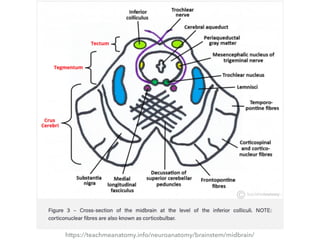
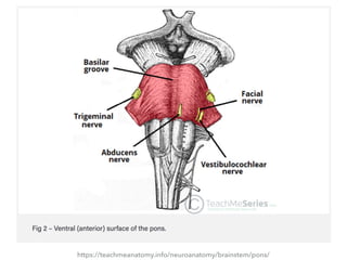
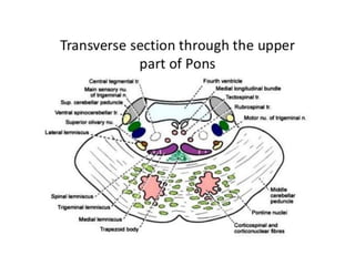
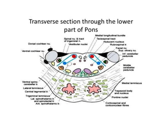
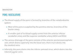
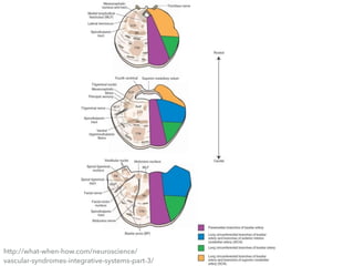
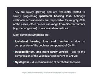
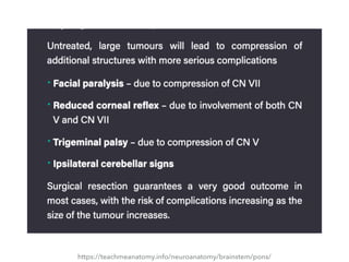
4. BRAINSTEM
MIDBRAIN: OVERVIEW
▸ The midbrain (also known as the
mesencephalon) is the most superior of
the three regions of the brainstem
▸ It acts as a conduit between the
forebrain above and the pons and
cerebellum below
6. BRAINSTEM
MIDBRAIN: EXTERNAL ANATOMY OF THE MIDBRAIN
▸ The midbrain is the smallest of the three regions of the brainstem,
measuring around 2cm in length
▸ As it ascends, the midbrain travels through the opening in the tentorium
cerebelli
▸ It can be divided into two main parts:
▸ Tectum – located posterior to the cerebral aqueduct
▸ Paired cerebral peduncles – located anteriorly and laterally
▸ Internally, the cerebral peduncles are further separated by the
substania nigra into the crus cerebri (anterior) and the tegmentum
(posterior)
7. BRAINSTEM
MIDBRAIN: EXTERNAL ANATOMY OF THE MIDBRAIN - TECTUM
▸ The tectum houses four rounded prominences named colliculi (collectively the
corpora quadrigemina) which sit directly inferior to the pineal gland
▸ The colliculi are separated by the cruciform sulcus; there are two superior and two
inferior colliculi
▸ Extending laterally from each colliculi are the quadrigeminal brachium:
▸ Superior quadrigeminal brachium forms a pathway between the superior
colliculus and the retina of the eye
▸ Inferior quadrigeminal brachium conveys fibres from the lateral lemniscus and
inferior colliculus to the medial geniculate body
▸ Inferior to the colliculi, the trochlear nerve (CN IV) emerges before sweeping
across to the anterior surface
8. BRAINSTEM
MIDBRAIN: EXTERNAL ANATOMY OF THE MIDBRAIN - CEREBRAL PEDUNCLES
▸ The paired cerebral peduncles extend from the cerebral
hemispheres to converge as they meet the pons
▸ They are separated anteriorly in the midline by
the interpeduncular fossa, the floor of which is termed the
posterior perforated substance (as many perforating blood
vessels can be identified)
▸ The oculomotor nerve (CNIII) is seen exiting from between
the peduncles while the optic tract runs around the
superior border of the midbrain
10. BRAINSTEM
MIDBRAIN: INTERNAL ANATOMY OF THE MIDBRAIN
▸ There are two transverse sections of the
midbrain:
▸ the level of the inferior colliculus
▸ the level of the superior colliculus
11. BRAINSTEM
MIDBRAIN: INTERNAL ANATOMY OF THE MIDBRAIN - INFERIOR COLLICULUS
▸ The anteriolateral surface of the midbrain houses the paired crus
cerebri
▸ Four fibre tracts run within the crus:
▸ Frontopontine fibres – located most medially
▸ Corticospinal fibres – motor fibres from the primary motor cortex
▸ Corticobulbar tracts – motor fibres from the primary motor cortex
▸ Temporopontine fibres – located posterolaterally
12. BRAINSTEM
MIDBRAIN: INTERNAL ANATOMY OF THE MIDBRAIN - INFERIOR COLLICULUS
▸ Posteriorly is the substantia nigra – a pigmented nucleus that
separates the two regions of the cerebral peduncles
▸ It is further broken down into the pars reticulata (anterior)
and pars compacta (posterior)
▸ The tegmentum is located posterior to the substantia nigra
▸ It is continuous with that found in the pons by the same name
▸ It is important to note that unlike the crus cerebri, the
tegmentum is continuous at the midline
13. BRAINSTEM
MIDBRAIN: INTERNAL ANATOMY OF THE MIDBRAIN - INFERIOR COLLICULUS
▸ The cerebral aqueduct is a midline structure surrounded
by central gray matter – the periaqueductal gray matter
▸ Within this gray matter lies the mesencephalic nucleus
of the trigeminal nerve, as well as the trochlear nucleus
with its fibres continuing around the gray matter to exit
the midbrain
▸ Anterior to this, the medial longitudinal fasciculus can
be seen
14. BRAINSTEM
MIDBRAIN: INTERNAL ANATOMY OF THE MIDBRAIN - INFERIOR COLLICULUS
▸ The decussation of the superior cerebellar peduncles can be
seen centrally at this level with some reticular formation (noted
throughout the brainstem) lying laterally
▸ Between the central gray matter and the substantia nigra are
four lemnisci
▸ Moving anterior to posterior they are the medial, spinal,
trigeminal, and lateral leminisci
▸ At the very posterior pole, we find the tectum which, at this level,
contains the inferior colliculus
16. BRAINSTEM
MIDBRAIN: INTERNAL ANATOMY OF THE MIDBRAIN - SUPERIOR COLLICULUS
▸ Much of the internal structure of the midbrain is
unchanged at this level and should be assumed present
unless mentioned differently
▸ The central portion which previously was occupied by the
decussation of the superior cerebellar peduncles now
contains the large paired red nuclei with some decussation
of the rubrospinal tract occuring anterior to this
▸ The reticular formation now fans around the posterior
borders of the red nuclei
17. BRAINSTEM
MIDBRAIN: INTERNAL ANATOMY OF THE MIDBRAIN - SUPERIOR COLLICULUS
▸ The trochlear nucleus is replaced with the
oculomotor nucleus while the oculomotor nerve
projects anteriorly
▸ The medial, spinal and trigeminal lemnisci are all
present in much the same location however the
lateral lemnisci does not reach to this level
▸ Needless to say, the tectum now contains the
superior colliculi rather than the inferior colliculi
19. BRAINSTEM
MIDBRAIN: VASCULATURE
▸ The supply to the area is derived from the basilar
artery and its branches
▸ The major suppliers are:
▸ posterior cerebral artery and its peduncular branch
▸ superior cerebellar artery
▸ posterior choroidal artery
▸ interpeduncular branches of the basilar artery
21. BRAINSTEM
REVIEW QUESTIONS: MIDBRAIN
▸ The tectum of the midbrain houses four rounded
prominences called colliculi. Where are they located in
relation to the pineal gland?
▸ Superiorly
▸ Inferiorly
▸ Laterally
▸ Medially
23. BRAINSTEM
REVIEW QUESTIONS: MIDBRAIN
▸ The colliculi of the midbrain are separated by
which structure?
▸ Substantia nigra
▸ Cruciform sulcus
▸ Oculomotor nerve
▸ Trochlear nerve
25. BRAINSTEM
REVIEW QUESTIONS: MIDBRAIN
▸ The interpeduncular fossa separates which two
structures?
▸ Cerebral peduncles
▸ Superior and inferior colliculi
▸ Cerebellar peduncles
▸ Pars reticulata and pars compacta
26. BRAINSTEM
REVIEW QUESTIONS: MIDBRAIN
▸ The paired cerebral peduncles extend
from the cerebral hemispheres to
converge as they meet the pons. They
are separated anteriorly in the midline
by the interpeduncular fossa.
29. BRAINSTEM
PONS: OVERVIEW
▸ The pons is the largest part of the brainstem, located
above the medulla and below the midbrain
▸ It is a group of nerves that function as a connection
between the cerebrum and cerebellum (pons is Latin for
bridge)
▸ The pons develops from the embryonic metencephalon
(part of the hindbrain, developed from the
rhombencephalon), alongside the cerebellum
31. BRAINSTEM
PONS: ANATOMICAL POSITION
▸ The pons is a horseshoe-shaped collection of nerve fibres
located in the anterior part of the posterior cranial fossa
▸ Its anatomical relations are as follows:
▸ Posteriorly – the cerebellum, separated by the fourth
ventricle
▸ Inferiorly – the medulla oblongata
▸ Superiorly – the midbrain lies immediately above the
pons
32. BRAINSTEM
PONS: EXTERNAL ANATOMY - ANTERIOR SURFACE
▸ The anterior or ventral surface of the pons is marked by a
bulging formed by the transverse pontocerebellar fibers
▸ These fibers wrap around the otherwise vertically oriented
brainstem which measures around 2.5 cm in adults
▸ The basilar groove demarcates the midline of the ventral surface
and is where the basilar artery is located
▸ The pontomedullary junction is an important anatomical
landmark defined by the angle between the lower border of the
pons and the superior border of the medulla
33. BRAINSTEM
PONS: EXTERNAL ANATOMY - ANTERIOR SURFACE
▸ Several cranial nerves originate from the ventral surface of the pons:
▸ Cranial nerve V: trigeminal – originates from the lateral aspect of
mid pons
▸ Cranial nerve VI: abducens – originates from the pontomedullary
junction, close to the midline
▸ Cranial nerve VII: facial – originates from the cerebellopontine
angle, the more lateral aspect of the pontomedullary junction
▸ Cranial nerve VIII: vestibulocochlear – originates laterally to the
facial nerve
35. BRAINSTEM
PONS: EXTERNAL ANATOMY - POSTERIOR SURFACE
▸ The pons is intimately related to the cerebellum, and is connected to
it by the middle cerebellar peduncles
▸ Removal of the cerebellum will reveal the underlying fourth ventricle
▸ The angle formed at the junction of the pons, medulla, and
cerebellum is another anatomical landmark and is named the
cerebellopontine angle
▸ Here, the cerebellar flocculus, the ventricular choroid plexus and the
emerging CNs VII and VIII surround the lateral apertures of the fourth
ventricle (the foramen of Luschka)
36. BRAINSTEM
PONS: EXTERNAL ANATOMY - POSTERIOR SURFACE
▸ The floor of the fourth ventricle is composed of the dorsal surface of
the pons and the medulla
▸ There are some important anatomical landmarks here:
▸ The medial eminence marks the midline of the floor
▸ The facial colliculus is a bulging formed by the fibers of the facial
nerve looping around the abducens nucleus
▸ The stria medullaris of the fourth ventricle is a bundle of nerve
fibers crossing transversely from the lateral aspect into the midline
▸ They mark the posterior border between the pons and the medulla
38. BRAINSTEM
PONS: INTERNAL ANATOMY
▸ The pons is comprised of two major components – the ventral pons and the tegmentum
▸ The Ventral Pons
▸ Contains the pontine nuclei, which are responsible for coordinating movement
▸ Fibers from the pontine nuclei cross the midline, and form the middle cerebellar
peduncles on their way to the cerebellum
▸ The Tegmentum
▸ Forms part of the reticular formation – a set of nuclei found throughout the
brainstem that are responsible for arousal and attentiveness
▸ Damage to this part of the pons may result in anosognosia for hemiplegia, where
patients are unaware of their paralysis
39. BRAINSTEM
PONS: INTERNAL ANATOMY
▸ The rest of the pons is made up of tracts passing through the pons
including:
▸ Descending corticospinal tracts – responsible for voluntary motor
control of the body
▸ Descending corticobulbar tracts – responsible for voluntary motor
control of face, head and neck
▸ Ascending medial lemniscus tracts – responsible for fine touch,
vibration and proprioception
▸ Ascending spinothalamic tracts – responsible for pain and
temperature sensation
40. BRAINSTEM
PONS: CRANIAL NERVE NUCLEI
▸ The pons houses important cranial nerve nuclei
▸ The main sensory nucleus and the trigeminal motor nucleus are located in
the midpons – at the level where the fibers originate from the lateral aspect
of the pons
▸ The main sensory nucleus receives somatosensory information from the face
▸ There are two other nuclei that receive sensory information from the
trigeminal nerve:
▸ Spinal trigeminal nucleus – extends caudally towards the medulla
▸ Mesencephalic nucleus – extends rostrally all the way to the midbrain
41. BRAINSTEM
PONS: CRANIAL NERVE NUCLEI
▸ The abducens nucleus controls the abducens nerve, which innervates the
ipsilateral lateral rectus muscle
▸ It is located in the caudal pons, on the medial aspect of its dorsal surface
▸ At the same level of the abducens nucleus, the facial nucleus is located more
anteriorly and laterally
▸ It controls the muscles of facial expression
▸ Its fibers take an unusual course and loop around the abducens nucleus
before exiting the brainstem through its ventrolateral surface
▸ The cochlear and vestibular nuclei sit dorsolaterally from the inferior pons to
the superior medulla
42.
43.
44. BRAINSTEM
PONS: VASCULATURE
▸ The blood supply of the pons is formed by branches of the vertebrobasilar
system:
▸ Most of the pons is supplied by the pontine arteries, branches of the
basilar artery
▸ A smaller part of its blood supply comes from the anterior inferior
cerebellar artery and the superior cerebellar artery (AICA and SCA)
▸ The venous drainage of the pons consists of the anterior pontomesencephalic
vein, which drains superiorly into the basal vein, that in turn drains into
the cerebral veins
▸ Inferiorly, the pons drains into the inferior petrosal sinus, which drains into the
internal jugular veins
49. BRAINSTEM
REVIEW QUESTIONS: PONS
▸ The pons develops from which part of the
embryonic brain?
▸ Telencephalon
▸ Diencephalon
▸ Mesencephalon
▸ Metencephalon
53. BRAINSTEM
REVIEW QUESTIONS: PONS
▸ Which of the following cranial nerves does NOT
originate from the pons?
▸ CN IV - Trochlear
▸ CN V - Trigeminal
▸ CN VI - Abducens
▸ CN VII - Facial
57. BRAINSTEM
MEDULLA: OVERVIEW
▸ The medulla oblongata (medulla) is one of the
three regions that make up the brainstem
▸ It is the most inferior of the three and is continuous
above with the pons and below with the spinal
cord
▸ The medulla houses essential ascending and
descending nerve tracts as well as brainstem nuclei
59. BRAINSTEM
MEDULLA: EXTERNAL ANATOMY
▸ The medulla is conical in shape, decreasing in width as it
extends inferiorly
▸ It is approximately 3cm long and 2cm wide at its largest point
▸ The superior margin of the medulla is located at the junction
between the medulla and pons, while the inferior margin is
marked by the origin of the first pair of cervical spinal nerves
▸ This occurs just as the medulla exits the skull through
the foramen magnum
60. BRAINSTEM
MEDULLA: EXTERNAL ANATOMY - ANTERIOR SURFACE
▸ There are several structures visible on the anterior surface of the medulla
▸ the three fissures/sulci
▸ the pyramids
▸ the olives
▸ five cranial nerves
▸ In the midline of the medulla is the anterior median fissure, which is continuous along the length of
the spinal cord
▸ It is interrupted temporarily by the decussation of the pyramids
▸ Moving away from the midline, two sulci are visible
▸ the ventrolateral sulcus
▸ the posterolateral sulcus
61. BRAINSTEM
MEDULLA: EXTERNAL ANATOMY - ANTERIOR SURFACE
▸ The pyramids are paired swellings found between the anterior median
fissure and the ventrolateral sulcus
▸ The olives are another pair of swellings located laterally to the pyramids –
between the ventrolateral and posterolateral sulci
▸ Arising from the junction between the pons and medulla is the abducens
nerve (CN VI)
▸ Extending out of the ventrolateral sulcus is the hypoglossal nerve (CN XII)
▸ In the posteriolateral sulcus, three more cranial nerves join the medulla
(CN IX, CN X, and CN XI)
63. BRAINSTEM
MEDULLA: EXTERNAL ANATOMY - POSTERIOR SURFACE
▸ Unlike the anterior surface of the medulla, the posterior surface is largely
obstructed from view and is relatively devoid of features
▸ In order to appreciate the posterior surface, the cerebellum must be
removed
▸ Similar to the anterior surface, the posterior surface has a midline
structure – the posterior median sulcus – which is continuous below as the
posterior median sulcus of the spinal cord
▸ Above, the sulcus ends at the point in which the fourth ventricle develops
▸ Moving laterally from the midline, the fasciculus gracilis and fasciculus
cuneatus are seen, separated by the posterior intermediate sulcus
65. BRAINSTEM
MEDULLA: INTERNAL ANATOMY
▸ The internal structures of the medulla must be viewed
in cross section to understand the layout
▸ Three levels of the medulla are typically discussed
(inferior – superior):
▸ Level of decussation of the pyramids
▸ Level of decussation of the medial lemnisci
▸ Level of the olives
66. BRAINSTEM
MEDULLA: INTERNAL ANATOMY
▸ The medulla itself is typically divided into two
regions: the open and the closed medulla
▸ This distinction is made based on whether the
CSF-containing cavities are surrounded by the
medulla (closed medulla) or not (open medulla)
▸ The medulla becomes open when the central
canal opens into the fourth ventricle
67. BRAINSTEM
MEDULLA: INTERNAL ANATOMY
▸ Some features are seen in all three cross sections
▸ Anteriorly we can see the paired lumps
representing the pyramids which are separated
by the anterior median fissure
▸ Centrally, the central canal can be seen as it rises
to form the fourth ventricle in the final cross
section
68. BRAINSTEM
MEDULLA: INTERNAL ANATOMY - DECUSSATION OF THE PYRAMIDS
▸ This is the major decussation point of the descending motor
fibres
▸ Roughly 75% of motor fibres housed within the pyramids cross
diagonally and posteriorly, and continue down the spinal
column as the lateral corticospinal tracts
▸ At this level, the central portion of the medulla contains gray
matter, while the outer portions consist of white matter
▸ The posterior white matter contains the fasiculus gracilis and the
more lateral fasiculus cuneatus
69. BRAINSTEM
MEDULLA: INTERNAL ANATOMY - DECUSSATION OF THE PYRAMIDS
▸ Corresponding portions of gray matter extend to these regions
and are the nucleus gracilis and nucleus cuneatus respectively
▸ Unchanged from the spinal cord, the spinocerebellar
tracts (posterior and anterior) are located laterally, with the
lateral spinothalamic tract situated between them
▸ The large trigeminal nucleus and tracts can be found posterior
to these tracts
▸ This is a continuation of the substantia gelatinosa of the spinal
cord
71. BRAINSTEM
MEDULLA: INTERNAL ANATOMY - DECUSSATION OF THE MEDIAL LEMNISCUS
▸ This level marks where the sensory decussation of the
medial lemniscus occurs
▸ The internal arcuate fibres run from the nucleus gracilis
and nucleus cuneatus around and anterior to the central
gray matter to form the medial lemniscus
▸ Lateral to the medial lemniscus, the trigeminal nucleus
and spinal tract can once again be seen, as can
the spinocerebellar tracts and the lateral spinothalamic
tract
72. BRAINSTEM
MEDULLA: INTERNAL ANATOMY - DECUSSATION OF THE MEDIAL LEMNISCUS
▸ Similarly, the posterior structures are much the same
at this level
▸ Centrally, the hypoglossal nucleus and medial
longitudinal fasciculus are seen
▸ Moving laterally, the nucleus ambiguous can be seen
▸ Between this structure and the pyramids is the
inferior olivary nucleus
74. BRAINSTEM
MEDULLA: INTERNAL ANATOMY - LEVEL OF THE OLIVES
▸ This level shows significant change in structure both
externally and internally when compared with previous levels
▸ The central canal has now expanded into the fourth ventricle
and as such makes this region the open medulla
▸ The large inferior olivary nucleus is responsible for the
external expansion of the olives
▸ The related medial and dorsal accessory olivary nuclei can be
seen medial and posterior to this structure respectively
75. BRAINSTEM
MEDULLA: INTERNAL ANATOMY - LEVEL OF THE OLIVES
▸ The large inferior cerebellar peduncles come into view and are
surrounded by multiple nuclei
▸ The two vestibular nuclei (medial and inferior) are both found towards
the midline while the two cochlear nuclei are found somewhat above
and below the peduncles
▸ Now a much smaller structure, the trigeminal tract and nucleus is seen
adjacent to the peduncle
▸ The nucleus ambiguous remains as it was previously, while the
hypoglossal nucleus has migrated with the central canal posteriorly,
joined by the medial longitudinal fasciulus
76. BRAINSTEM
MEDULLA: INTERNAL ANATOMY - LEVEL OF THE OLIVES
▸ An additional cranial nucleus comes into view lateral to the
hypoglossal – the dorsal vagal nucleus
▸ Moving further laterally, the nucleus of tractus solitarius comes
into view
▸ Centrally, the medial lemniscus hugs the midline posterior to the
pyramids, as does the tectospinal tract
▸ Between the peduncle and the olivary nuclei resides the lateral
spinothalamic tract and the more lateral anterior spinocerebellar
tract
78. BRAINSTEM
MEDULLA: VASCULATURE
▸ The vasculature of the medulla is complex and is dependent on the
level being viewed
▸ The vessels that supply the medulla include:
▸ the anterior spinal
▸ the posterior spinal
▸ posterior inferior cerebellar
▸ anterior inferior cerebellar
▸ vertebral arteries
79. BRAINSTEM
MEDULLA: VASCULATURE
▸ Below the level of the olives the posterior half of the
medulla is supplied by the posterior spinal artery - no
other regions are supplied by this vessel
▸ The remaining portions are supplied by the posterior
inferior cerebellar and vertebral arteries
▸ Throughout the medulla, the anterior spinal artery supplies
a region beginning at the central canal (or anterior border
of the fourth ventricle), and fans out to encompass the
pyramids
80. BRAINSTEM
MEDULLA: VASCULATURE
▸ In cross section through the olives both the posterior
inferior cerebellar and vertebral arteries take on
greater territories posterolaterally and anterolaterally
respectively
▸ They continue to do so as the medulla ascends
▸ At the highest point in the medulla, the anterior
inferior cerebellar artery supplies the outermost
portions of the posterior region
82. BRAINSTEM
REVIEW QUESTIONS: MEDULLA
▸ What are the paired swellings found between the
anterior median fissure and the ventrolateral sulcus?
▸ The pyramids
▸ The olives
▸ The tectum
▸ The colliculi
83. BRAINSTEM
REVIEW QUESTIONS: MEDULLA
▸ The pyramids are paired swellings
found between the anterior median
fissure and the ventrolateral sulcus.
(The olives are another pair of swellings
located laterally to the pyramids –
between the ventrolateral and
posterolateral sulci.)
84. BRAINSTEM
REVIEW QUESTIONS: MEDULLA
▸ The medulla becomes open when what
feature opens into the fourth ventricle?
▸ Central canal
▸ Posterior median sulcus
▸ Anterior fissure
▸ Lateral sulcus
86. BRAINSTEM
REVIEW QUESTIONS: MEDULLA
▸ Throughout the medulla, which artery supplies a region
beginning at the central canal (or anterior border of the
fourth ventricle), and fans out to encompass the pyramids?
▸ Anterior spinal artery
▸ Vertebral artery
▸ Posterior spinal artery
▸ Posterior inferior cerebellar
88. References
▸ These slide reflect a summary of the contents of
TeachMeAnatomy.info and are to be used for
educational purposes only in compliance with the
terms of use policy.
Specific portions referenced in this summary are as
follows:
▸ https://teachmeanatomy.info/neuroanatomy/
brainstem/
Additional sources are referenced on the slide
containing that specific content.