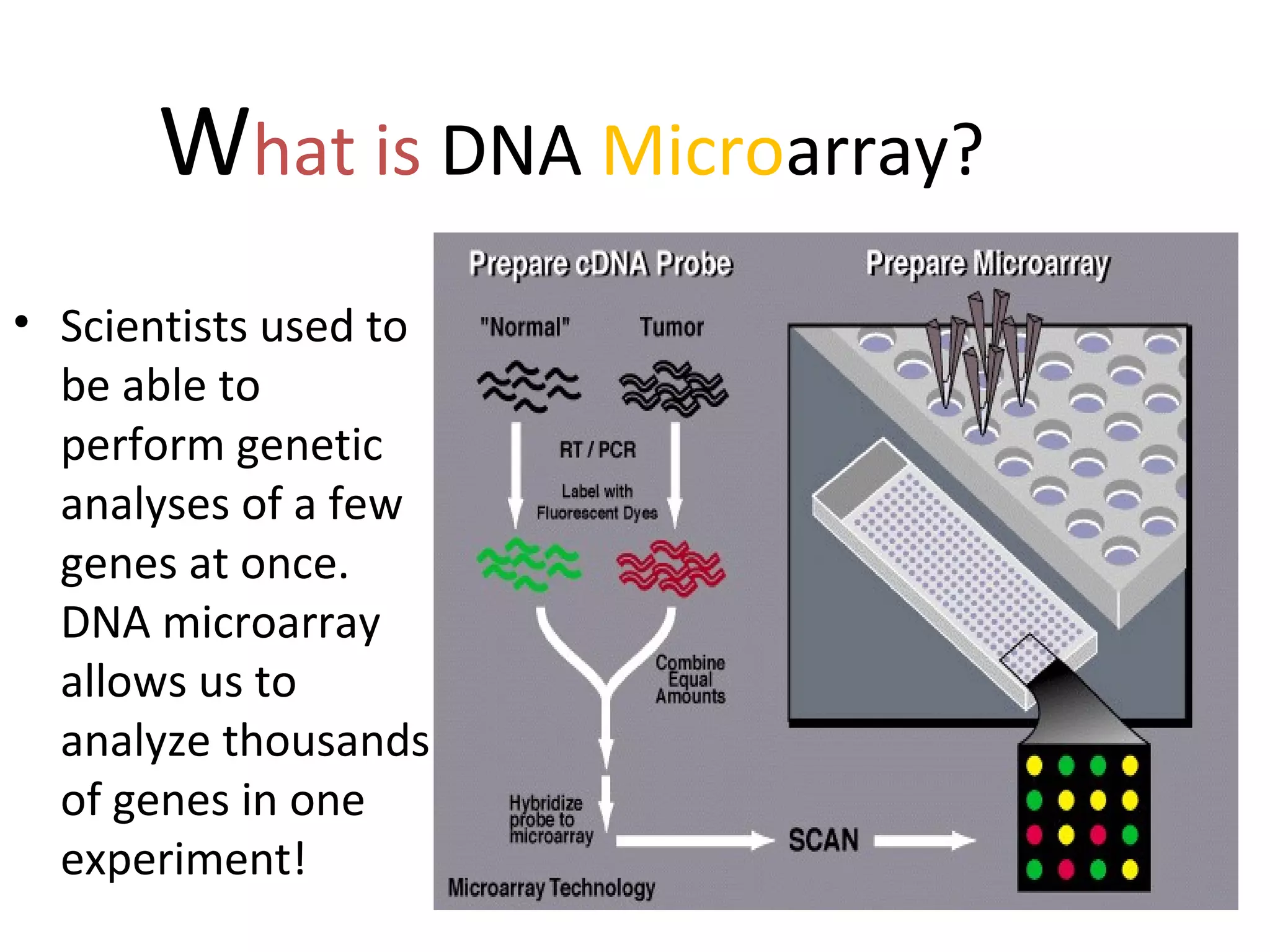
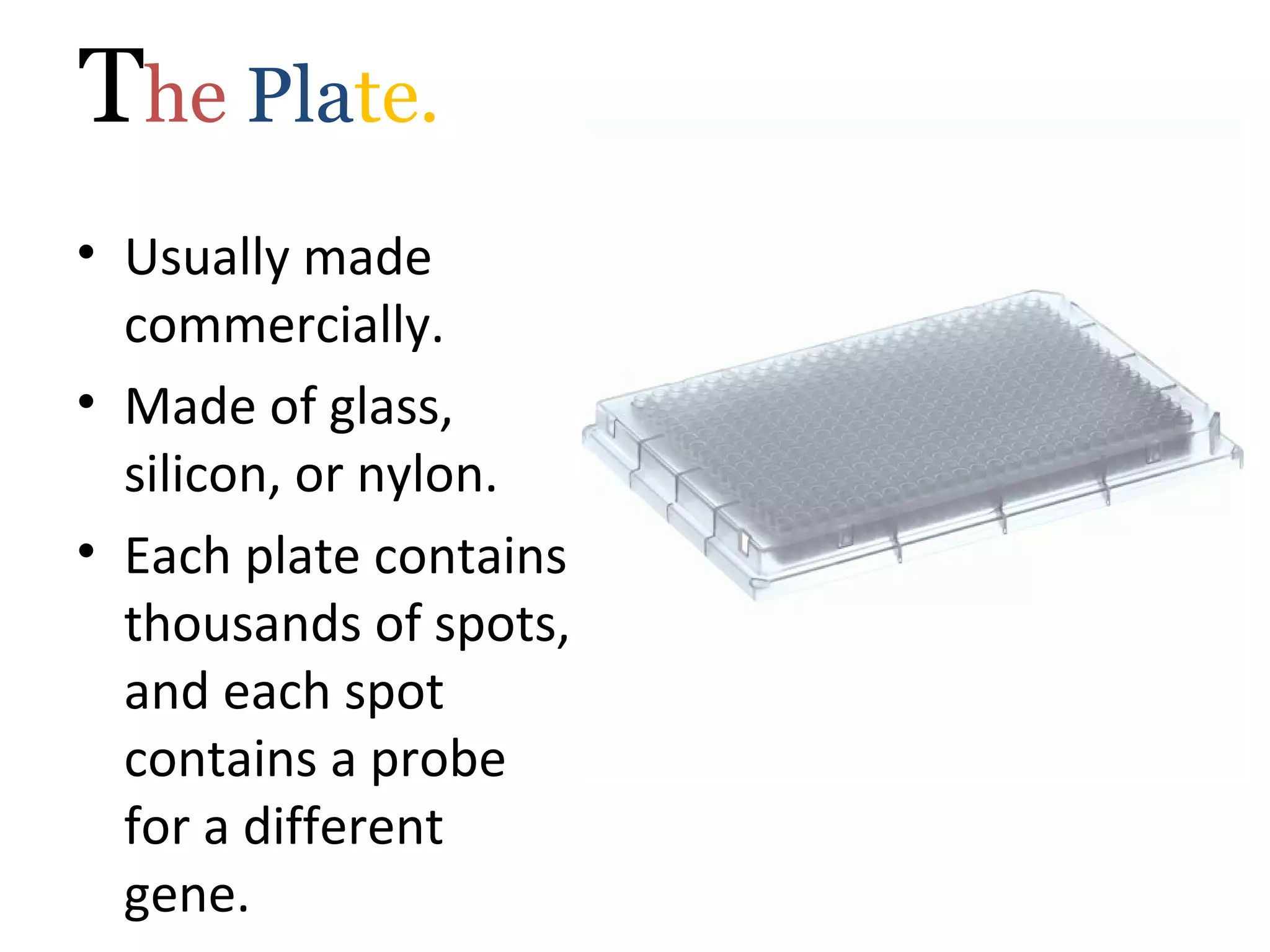
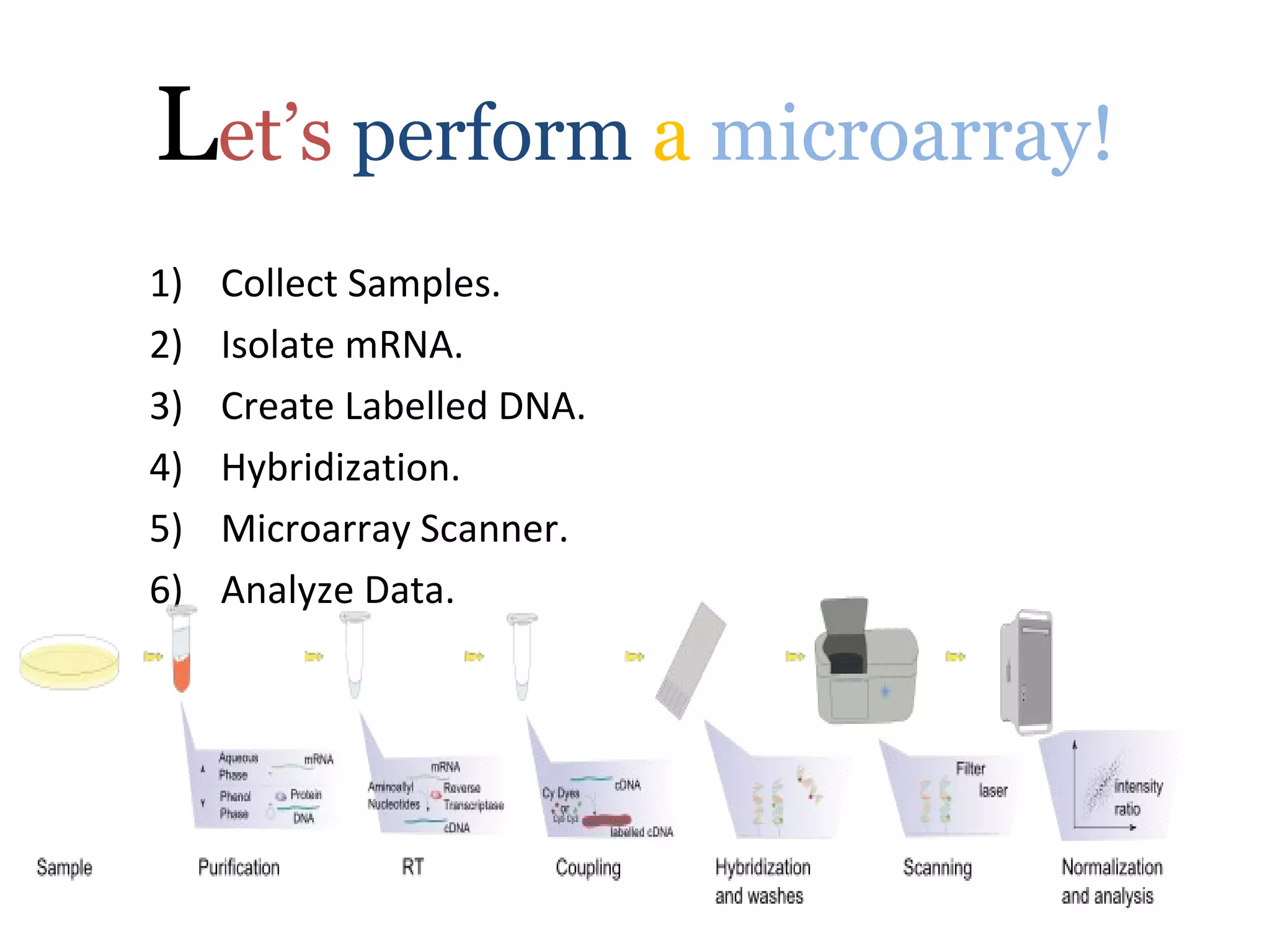
DNA Microarray.
By- Jain Hemant
What is DNA Microarray?
• Scientists used to
be able to
perform genetic
analyses of a few
genes at once.
DNA microarray
allows us to
analyze thousands
of genes in one
experiment!
Purposes.
• So why do we use DNA
microarray?
– To measure changes in
gene expression levels –
two samples’ gene
expression can be
compared from different
samples, such as from
cells of different stages of
mitosis.
– To observe genomic gains
and losses. Microarray
Comparative Genomic
Hybridization (CGH)
– To observe mutations in
DNA.
The Plate.
• Usually made
commercially.
• Made of glass,
silicon, or nylon.
• Each plate contains
thousands of spots,
and each spot
contains a probe
for a different
gene.
• A probe can be a cDNA fragment or a
synthetic oligonucleotide, such as BAC
(bacterial artificial chromosome set).
• Probes can either be attached by robotic
means, where a needle applies the cDNA to
the plate, or by a method similar to making
silicon chips for computers. The latter is
called a Gene Chip.
Let’s perform a microarray!
1) Collect Samples.
2) Isolate mRNA.
3) Create Labelled DNA.
4) Hybridization.
5) Microarray Scanner.
6) Analyze Data.
STEP 1: Collect Samples.
 This can be
from a
variety of
organisms.
We’ll use two
samples –
cancerous
human skin
tissue &
healthy
human skin
tissue
STEP 2: Isolate mRNA.
• Extract the RNA from the samples. Using either a
column, or a solvent such as phenol-chloroform.
• After isolating the RNA, we need to isolate the mRNA
from the rRNA and tRNA. mRNA has a poly-A tail, so
we can use a column containing beads with poly-T
tails to bind the mRNA.
• Rinse with buffer to release the mRNA from the
beads. The buffer disrupts the pH, disrupting the
hybrid bonds.
STEP 3: Create Labelled DNA.
Add a labeling mix to the
RNA. The labeling mix
contains poly-T (oligo
dT) primers, reverse
transcriptase (to make
cDNA), and
fluorescently dyed
nucleotides.
 We will add cyanine 3
(fluoresces green) to the
healthy cells and cyanine 5
(fluoresces red) to the
cancerous cells.
 The primer and RT bind to the
mRNA first, then add the
fluorescently dyed
nucleotides, creating a
complementary strand of DNA
STEP 4: Hybridization.
• Apply the cDNA we have
just created to a
microarray plate.
• When comparing two
samples, apply both
samples to the same
plate.
• The ssDNA will bind to
the cDNA already
present on the plate.
STEP 5: LASERS!
STEP 5: Microarray Scanner.
 The scanner has a laser, a
computer, and a camera.
 The laser causes the hybrid bonds
to fluoresce.
 The camera records the images
produced when the laser scans
the plate.
 The computer allows us to
immediately view our results and
it also stores our data.
Benefits.
• Relatively affordable (for some people!),
about $60,000 for an arrayer and scanner
setup.
• The plates are convenient to work with
because they are small.
• Fast - Thousands of genes can be analyzed at
once.
Problems.
• Oligonucleotide libraries –
redundancy and contamination.
• DNA Microarray only detects
whether a gene is turned on or
off.
• Massive amounts of data.
http://www.stuffintheair.com/very-big-problem.html
The Future of DNA Microarray.
• Gene discovery.
• Disease diagnosis: classify the types of cancer on the basis of
the patterns of gene activity in the tumor cells.
• Pharmacogenomics = is the study of correlations between
therapeutic responses to drugs and the genetic profiles of the
patients.
• Toxicogenomics – microarray technology allows us to
research the impact of toxins on cells. Some toxins can
change the genetic profiles of cells, which can be passed on to
cell progeny.
Microarray

Microarray

  • 2.
  • 3.
    What is DNAMicroarray? • Scientists used to be able to perform genetic analyses of a few genes at once. DNA microarray allows us to analyze thousands of genes in one experiment!
  • 4.
    Purposes. • So whydo we use DNA microarray? – To measure changes in gene expression levels – two samples’ gene expression can be compared from different samples, such as from cells of different stages of mitosis. – To observe genomic gains and losses. Microarray Comparative Genomic Hybridization (CGH) – To observe mutations in DNA.
  • 5.
    The Plate. • Usuallymade commercially. • Made of glass, silicon, or nylon. • Each plate contains thousands of spots, and each spot contains a probe for a different gene.
  • 6.
    • A probecan be a cDNA fragment or a synthetic oligonucleotide, such as BAC (bacterial artificial chromosome set). • Probes can either be attached by robotic means, where a needle applies the cDNA to the plate, or by a method similar to making silicon chips for computers. The latter is called a Gene Chip.
  • 7.
    Let’s perform amicroarray! 1) Collect Samples. 2) Isolate mRNA. 3) Create Labelled DNA. 4) Hybridization. 5) Microarray Scanner. 6) Analyze Data.
  • 8.
    STEP 1: CollectSamples.  This can be from a variety of organisms. We’ll use two samples – cancerous human skin tissue & healthy human skin tissue
  • 10.
    STEP 2: IsolatemRNA. • Extract the RNA from the samples. Using either a column, or a solvent such as phenol-chloroform. • After isolating the RNA, we need to isolate the mRNA from the rRNA and tRNA. mRNA has a poly-A tail, so we can use a column containing beads with poly-T tails to bind the mRNA. • Rinse with buffer to release the mRNA from the beads. The buffer disrupts the pH, disrupting the hybrid bonds.
  • 11.
    STEP 3: CreateLabelled DNA. Add a labeling mix to the RNA. The labeling mix contains poly-T (oligo dT) primers, reverse transcriptase (to make cDNA), and fluorescently dyed nucleotides.  We will add cyanine 3 (fluoresces green) to the healthy cells and cyanine 5 (fluoresces red) to the cancerous cells.  The primer and RT bind to the mRNA first, then add the fluorescently dyed nucleotides, creating a complementary strand of DNA
  • 12.
    STEP 4: Hybridization. •Apply the cDNA we have just created to a microarray plate. • When comparing two samples, apply both samples to the same plate. • The ssDNA will bind to the cDNA already present on the plate.
  • 13.
  • 14.
    STEP 5: MicroarrayScanner.  The scanner has a laser, a computer, and a camera.  The laser causes the hybrid bonds to fluoresce.  The camera records the images produced when the laser scans the plate.  The computer allows us to immediately view our results and it also stores our data.
  • 15.
    Benefits. • Relatively affordable(for some people!), about $60,000 for an arrayer and scanner setup. • The plates are convenient to work with because they are small. • Fast - Thousands of genes can be analyzed at once.
  • 16.
    Problems. • Oligonucleotide libraries– redundancy and contamination. • DNA Microarray only detects whether a gene is turned on or off. • Massive amounts of data. http://www.stuffintheair.com/very-big-problem.html
  • 17.
    The Future ofDNA Microarray. • Gene discovery. • Disease diagnosis: classify the types of cancer on the basis of the patterns of gene activity in the tumor cells. • Pharmacogenomics = is the study of correlations between therapeutic responses to drugs and the genetic profiles of the patients. • Toxicogenomics – microarray technology allows us to research the impact of toxins on cells. Some toxins can change the genetic profiles of cells, which can be passed on to cell progeny.