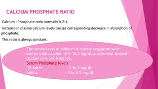
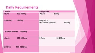
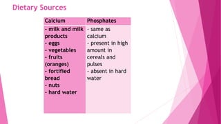
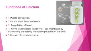
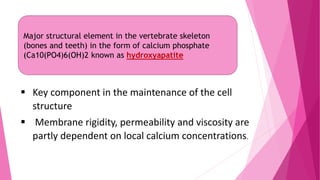
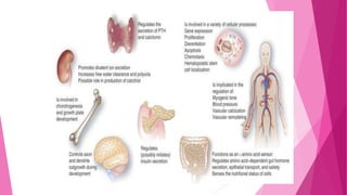
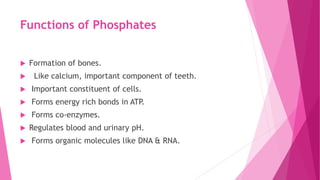
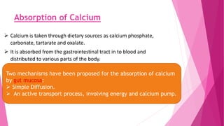
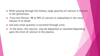
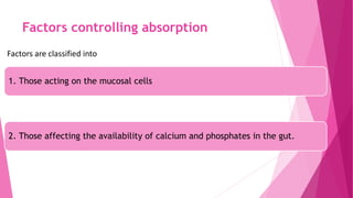
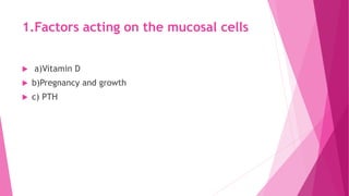
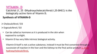
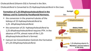
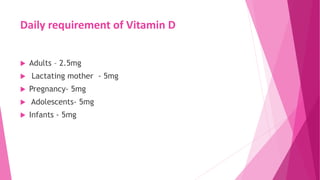
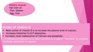
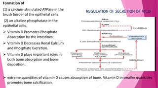
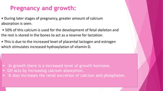
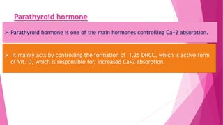
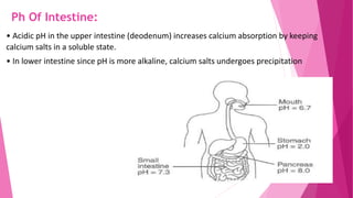
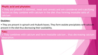
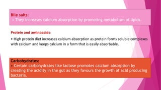
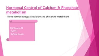
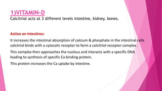
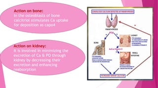
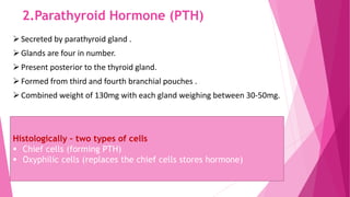
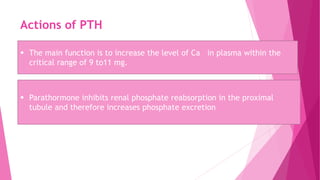
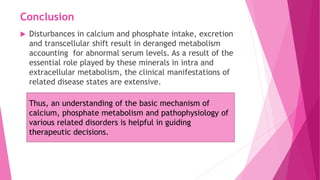
The document summarizes key information about calcium and phosphorus metabolism. It discusses their daily requirements, distribution in the body, dietary sources, functions, factors controlling absorption such as vitamin D, parathyroid hormone, and calcitonin. It also outlines hormonal control of calcium and phosphorus metabolism and clinical importance of hypo- and hypercalcemia and hyperphosphatemia. The objectives are to understand the role of calcium and phosphorus in the body and factors influencing their metabolism.
The POPPY STUDY (Preconception to post-partum cardiovascular function in prim...
Metabolism calcium @phosphorus
1. SEMINAR ON
Metabolism – Calcium and Phosphorus
Presented by:
Dr. Md. Naseem Ashraf
1st Year Postgraduate
2. Contents:
Introduction
Distribution
Daily requirements
Dietary sources
Functions
Factors controlling absorption
Hormonal control
Other hormones affecting metabolism
Clinical importance Conclusion
3. Objectives
To know the role and daily requirements of Calcium and
Phosphorous in body.
To know the various factors responsible for their
absorption
To know the hormonal influences on calcium and
phosphorus metabolism.
To know the various abnormalities related to hypo and
hyper conditions of phosphorus and calcium.
4. METABOLISM
Metabolism is the biochemical modification of
chemical compounds in living organisms and cells
that includes the biosynthesis of complex organic
molecules (anabolism) and their breakdown
(catabolism).
5. INTRODUCTION
The minerals in foods do not contribute directly to energy
needs but are important as body regulators and as
essential constituents in many vital substances within the
body.
About 25 elements have been found to be essential,
since a deficiency produces specific deficiency symptoms.
6. Principal Minerals include - Calcium, Phosphorous,
Magnesium, Sodium, Potassium and Sulphur.
Calcium and phosphorous individually have their own functions and
together they are required for the formation of hydroxyapatite and
physical strength of the skeletal tissue.
8. Calcium in the Plasma and Interstitial Fluid
The calcium in the plasma is present in three forms,
(1) About 41 per cent (1 mmol/L) of the calcium is combined with the
plasma proteins and in this form is nondiffusible through the capillary
membrane.
(2) About 9 per cent of the calcium (0.2 mmol/L) is diffusible through the
capillary membrane but is combined with anionic substances of the
plasma and interstitial fluids (citrate and phosphate, for instance) in such
a manner that it is not ionized.
(3) The remaining 50 per cent of the calcium in the plasma is both
diffusible through the capillary membrane and ionized.
9.
10. CALCIUM PHOSPHATE RATIO
Calcium : Phosphate ratio normally is 2:1.
Increase in plasma calcium levels causes corresponding decrease in absorption of
phosphate.
This ratio is always constant.
The serum level of calcium is closely regulated with
normal total calcium of 9-10.5 mg/dL and normal ionized
calcium of 4.5-5.6 mg/dL.
Serum Phosphate levels
Children - 4 to 7 mg/dL
Adults - 3 to 4.5 mg/dL
12. Dietary Sources
Calcium Phosphates
- milk and milk
products
- eggs
- vegetables
- fruits
(oranges)
- fortified
bread
- nuts
- hard water
- same as
calcium
- present in high
amount in
cereals and
pulses
- absent in hard
water
13. Functions of Calcium
1.Muscle contraction
2.Formation of bone and teeth
3. Coagulation of blood
4. Nerve transmission: Integrity of cell membrane by
maintaining the resting membrane potential of the cells
5.Release of certain hormones
14. Major structural element in the vertebrate skeleton
(bones and teeth) in the form of calcium phosphate
(Ca10(PO4)6(OH)2 known as hydroxyapatite
Key component in the maintenance of the cell
structure
Membrane rigidity, permeability and viscosity are
partly dependent on local calcium concentrations.
15.
16. Functions of Phosphates
Formation of bones.
Like calcium, important component of teeth.
Important constituent of cells.
Forms energy rich bonds in ATP.
Forms co-enzymes.
Regulates blood and urinary pH.
Forms organic molecules like DNA & RNA.
17. Absorption of Calcium
Calcium is taken through dietary sources as calcium phosphate,
carbonate, tartarate and oxalate.
It is absorbed from the gastrointestinal tract in to blood and
distributed to various parts of the body.
Two mechanisms have been proposed for the absorption of calcium
by gut mucosa:
Simple Diffusion.
An active transport process, involving energy and calcium pump.
18. While passing through the kidney, large quantity of calcium is filtered
in the glomerulus.
From the filtrate, 98 to 99% of calcium is reabsorbed in the renal
tubules in to blood
and only small quantity is excreted through urine.
In the bone, the calcium may be deposited or resorbed depending
upon the level of calcium in the plasma.
19. Factors controlling absorption
Factors are classified into
1. Those acting on the mucosal cells
2. Those affecting the availability of calcium and phosphates in the gut.
20. 1.Factors acting on the mucosal cells
a)Vitamin D
b)Pregnancy and growth
c) PTH
21. VITAMIN- D
Calcitriol (1, 25 –Dihydroxycholecalciferol(1,25-DHCC) is the
biologically active form of Vitamin D.
Synthesis of VITAMIN-D
Cholecalciferol / D3
Ergocalciferol / D2
• Can be called as hormone as it is produced in the skin when
exposed to sunlight.
• Vitamin D has very little intrinsic biological activity.
Vitamin D itself is not a active substance, instead it must be first converted through a
succession of reaction in the liver and the kidneys to the final active product 1, 25 di
hydroxycholecalciferol,
22. Cholecalciferol (Vitamin D3) Is Formed in the Skin.
Cholecalciferol Is Converted to 25-Hydroxycholecalciferol in the Liver.
Formation of 1,25-Dihydroxycholecalciferol in the
Kidneys and Its Control by Parathyroid Hormone.
the conversion in the proximal tubules of the
kidneys of 25-hydroxycholecalciferol to
1,25- dihydroxycholecalciferol.
the conversion of 25- hydroxycholecalciferol to
1,25-dihydroxycholecalciferol requires PTH. In the
absence of PTH, almost none of the 1,25-
dihydroxycholecalciferol is formed.
Calcium Ion Concentration Controls the Formation
of 1,25-Dihydroxycholecalciferol.
24. Dietary sources
fish liver oil
Fish- Salmon
Egg, liver
Action of vitamin D
Mean action of vitamin D is to increase the plasma level of calcium.
Increases intestinal Ca & P absorption.
Increases renal reabsorption of Calcium and phosphate.
1,25-Dihydroxycholecalciferol itself functions as a type of “hormone” to promote
intestinal absorption of calcium.
25. Formation of
(1) a calcium-stimulated ATPase in the
brush border of the epithelial cells
(2) an alkaline phosphatase in the
epithelial cells.
Vitamin D Promotes Phosphate
Absorption by the Intestines.
Vitamin D Decreases Renal Calcium
and Phosphate Excretion.
Vitamin D plays important roles in
both bone absorption and bone
deposition.
extreme quantities of vitamin D causes absorption of bone. Vitamin D in smaller quantities
promotes bone calcification.
REGULATION OF SECRETION OF Vit.D
26. Pregnancy and growth:
• During later stages of pregnancy, greater amount of calcium
absorption is seen.
• 50% of this calcium is used for the development of fetal skeleton and
the rest is stored in the bones to act as a reserve for lactation.
• This is due to the increased level of placental lactogen and estrogen
which stimulates increased hydroxylation of vitamin D.
In growth there is a increased level of growth hormone.
GH acts by increasing calcium absorption.
It also increases the renal excretion of calcium and phosphates.
27. Parathyroid hormone
Parathyroid hormone is one of the main hormones controlling Ca+2 absorption.
It mainly acts by controlling the formation of 1,25 DHCC, which is active form
of Vit. D, which is responsible for, increased Ca+2 absorption.
28. Factors affecting availability of Calcium and
Phosphates in gut.
pH of the intestine
Amount of dietary calcium and phosphates
Phytic acid and Phytates
Oxalates
Fats
Proteins and amino acids
Carbohydrates
Bile salts
29. Ph Of Intestine:
• Acidic pH in the upper intestine (deodenum) increases calcium absorption by keeping
calcium salts in a soluble state.
• In lower intestine since pH is more alkaline, calcium salts undergoes precipitation
30. Amount of dietary calcium and phosphates:
• Increased level of calcium and phosphate in diet increases their absorption
however up to a certain limit.
• This is because the active process of their absorption can bear with certain
amounts of load beyond which the excess would pass out into faeces
31. Phytic acid and phytates:
• They are present in oatmeal, meat and cereals and are considered anti-calcifying
factors as they combine with calcium in the diet thus forming insoluble salts of
calcium
Oxalates:
• They are present in spinach and rhubarb leaves. They form oxalate precipitates with calcium
present in the diet thus decreasing their availability.
Fats:
• They combines with calcium and form insoluble calcium , thus decreasing calcium
absorption.
32. Bile salts:
• They increases calcium absorption by promoting metabolism of lipids.
Protein and aminoacids:
• High protein diet increases calcium absorption as protein forms soluble complexes
with calcium and keeps calcium in a form that is easily absorbable.
Carbohydrates:
• Certain carbohydrates like lactose promotes calcium absorption by
creating the acidity in the gut as they favours the growth of acid producing
bacteria.
33. Hormonal Control of Calcium & Phosphate
metabolism
Three hormones regulate calcium and phosphate metabolism.
1)Vitamin D
2)PTH
3)Calcitonin
34. 1)VITAMIN-D
Calcitriol acts at 3 different levels intestine, kidney, bones.
Action on Intestines:
It increases the intestinal absorption of calcium & phosphate in the intestinal cells
calcitriol binds with a cytosolic receptor to form a calcitriol-receptor complex .
This complex then approaches the nucleus and interacts with a specific DNA
leading to synthesis of specific Ca binding protein.
This protein increases the Ca uptake by intestine.
35. Action on bone:
In the osteoblasts of bone
calcitriol stimulates Ca uptake
for deposition as capo4
Action on kidney:
It is involved in minimizing the
excretion of Ca & PO through
kidney by decreasing their
excretion and enhancing
reabsorption
36. 2.Parathyroid Hormone (PTH)
Secreted by parathyroid gland .
Glands are four in number.
Present posterior to the thyroid gland.
Formed from third and fourth branchial pouches .
Combined weight of 130mg with each gland weighing between 30-50mg.
Histologically – two types of cells
Chief cells (forming PTH)
Oxyphilic cells (replaces the chief cells stores hormone)
37.
38. Actions of PTH
The main function is to increase the level of Ca in plasma within the
critical range of 9 to11 mg.
Parathormone inhibits renal phosphate reabsorption in the proximal
tubule and therefore increases phosphate excretion
39. Parathormone increases renal Calcium reabsorption in the distal
tubule, which also increases the serum calcium.
Net effect of PTH
Increase serum calcium
Decrease serum phosphate
40. Stimulation for PTH secretion
The stimulatory effect for PTH secretion is low level of
calcium in plasma.
Maximum secretion occurs when plasma calcium level falls
below 7mg/dl.
When plasma calcium level increases to 11mg/dl there is
decreased secretion of PTH
41. 3.CALCITONIN
Minor regulator of calcium & phosphate metabolism.
Secreted by parafollicular cells or C-cells of thyroid gland.
Also called as thyrocalcitonin.
Single chain polypeptide .
Molecular weight 3400
Plasma concentration – 10-20ug/ml
42. Action of Calcitonin
Net effect of calcitonin decreases Serum Ca
Target site
Bone (osteoclasts)
decreased ability of osteoclasts to resorb bone
Osteoclasts cells
Lose their ruffled borders
Undergo cytoskeletal rearrangement
Decreased mobility
Detach from bone
43. OTHER HORMONES on CALCIUM METABOLISM
GROWTH HORMONE
INSULIN
TESTOSTERONE & OTHER HORMONES
LACTOGEN & PROLACTIN
STEROIDS
THYROID HORMONES
44. Excretion of Calcium and Phosphorous
Calcium is excreted in the urine, bile, and digestive
secretions.
The renal threshold for serum ca is 10 mg/dl.
70-90% of the calcium eliminated from the body is
excreted in the feces.
The daily loss of calcium in sweat is about 15 mg.
45. Daily turnover rates of Ca in an adult are as follows:
Intake 1000mg.
Intestinal absorption 350mg
Secretion in GI juice 250mg
Net absorption over secretion
100mg
Loss in the faeces 200mg
Excretion in the urine 80-100mg
46. Phosphorous Excretion
Phosphorous is excreted primarily through the urine.
Almost 2/3rd of total phosphorous that is excreted is
found in the urine as phosphate of various cations.
phosphorous found in the faeces is the non- absorbed
form of phosphorous.
51. Hypocalcaemia
Decreased level of calcium in the blood (<4mg/dl)
Conditions leading to hypocalcaemia
Insufficient dietary calcium
Hypoparathyroidism
Insufficient vitamin D
increase in calcitonin levels
52. Abnormal calcification
Abnormal deposits of calcium salts occur in any tissues except bones and
teeth.
Metastatic calcification Dystrophic calcification
apparently normal tissues
and is associated with
deranged calcium
metabolism and
hypercalcaemia
Characterized by deposition
of calcium salts in dead or
degenerated tissues with
normal calcium metabolism
and normal serum calcium
levels.
53. Tetany (Carpopedal spasm)
Basic feature of tetany is uncontrolled, painful, prolonged
contraction (spasm) of the voluntary muscles.
54. Chvostek’s sign
Contraction of ipsilateral facial muscles when tapping facial nerve over the angle of the
mandible.
Trousseau’s sign
• Spasm of the muscles of the
upper extremity causing flexion
of wrist and thumb and
extension of fingers.
• Clinically can be produced by
applying pressure with
sphygmomanometer cuff on the
upper arm.
Erbs sign
Hyperexitability of muscles to electrical stimulation
55. Vitamin D deficiency
Rickets
Occurs in children between 6
months to 2 years of age.
Affects long bones .
Lack of calcium causes failure
of mineralization resulting into
formation of cartilagenous
form of bone.
Most critical area that gets
affected is the center
endochondral ossification at
the epiphyseal plates.
56. Dental findings in Rickets
Developmental anomalies of enamel and dentin .
Delayed eruption .
Misalignment of teeth .
Increase caries index .
Wide predentin zone and more interglobular dentin .
Treatment
Daily administration of 1000 – 4000 units of vit.D.
57. Osteomalacia v/s Osteoporosis
Osteomalacia happens if mineralisation
doesn’t take place properly.
In osteomalacia more and more bone is
made up of collagen matrix without a
mineral covering, so the bones become
soft.
These softened bones may bend and
crack, and this can be very painful.
Osteoporosis happens when bone
density decreases and the body stops
producing as much bone as it did
before.
It can affect both males and females,
but it is most likely to occur in women
after menopause, because of the
sudden decrease in estrogen, the
hormone that normally protects
against osteoporosis.
As the bones become weaker, there is
a higher risk of a fracture during a fall
or even a fairly minor knock.
58. Osteomalacia
Clinical features
Bone pain and
tenderness
Peculiar
waddling or
“penguin”gait
Tetany
Greenstick bone
fractures
Myopathy
Oral manifestation
Severe Periodontitis
Thin or absent
trabeculae
Loosened teeth
Weakened jaw bones
59. Osteoporosis
Oral manifestation
Decreased edentulous ridge hight
Deceased posterior maxillary arch width
Progressive alveolar bone loss
Loss of attachment and gingival recession
Loss of teeth
Clinical features
Fractures
Bone pain
Change in body shape
Hight loss
60. Hyperparathyroidism
May be Primary or Secondary.
Primary hyperparathyroidism
In Primary form, a primary abnormality of the parathyroid
glands causes inappropriate, excess PTH secretion.
Caused mainly by an adenoma of parathyroid.
61. Secondary Hyperparathyroidism
In Secondary form, high levels of PTH occur as a compensation
rather than as a primary abnormality of the parathyroid glands.
It can be caused by Vitamin D deficiency or chronic renal
disease.
62. Hypoparathyroidism
Decrease level of PTH.
Due to
1.Surgical removal of parathyroid gland
2.Congenital absence of the gland
3. Atrophy of the gland
Diagnosis
◦ Decrease plasma calcium level & increase
plasma phosphate level
64. Pseudohypoparathyroidism
is the result of defective G protein in kidney and bone,
which causes end-organ resistance to PTH.
there is hypocalcemia and hyperphosphatemia that is not
correctable by administration of exogenous PTH.
circulating endogenous PTH levels are elevated.
65. HYPERPHOSPHATEMIA
Increased intake Diet containing Vit-D
Increased release of P from cells (DM, Acidaemia,
Starvation)
Increased release of P from bone (malignancy, Renal
failure, Increased PTH)
Decreased excretion (Renal failure, Hyperparathyroidism,
Increased growth hormone)
68. HYPOPHOSPHATASIA
The basic disorder is a deficiency of the enzyme
alkaline phosphatase in serum or tissues and
excretion of phosphoethanolamine in the urine.
Oral manifestations:
Loosening and premature loss of deciduous
teeth, chiefly the incisors.
69. PSEUDOHYPOPHOSPHATASIA:
A disease resembling classic hypophosphatasia
but with a normal serum alkaline phosphatase level.
Patients exhibit osteopathy of the long bones and skull,
premature loss of deciduous teeth,
hypotonia, hypercalcemia and phosphoethanolaminauria.
70. DENTAL IMPLICATIONS
It has often been supposed that a low intake of calcium,
or phosphorous, or both might lead to poor calcification of
teeth and possibly, therefore, to an increased risk of
dental caries.
the calcification of the teeth could be affected if calcium
was very low in the diet.
71. deficiencies in calcium and phosphate intake do not affect
tooth calcification they do reduce that of bone, and they
result in mobilization of calcium from already formed
bone.
In pregnancy, when the dietary need of the mother for
calcium and phosphate are increased by the demands
imposed by the growing fetus, there is mobilization of
bone calcium if the dietary supply is insufficient.
72. Conclusion
Disturbances in calcium and phosphate intake, excretion
and transcellular shift result in deranged metabolism
accounting for abnormal serum levels. As a result of the
essential role played by these minerals in intra and
extracellular metabolism, the clinical manifestations of
related disease states are extensive.
Thus, an understanding of the basic mechanism of
calcium, phosphate metabolism and pathophysiology of
various related disorders is helpful in guiding
therapeutic decisions.
73. References:
1. Textbook of Biochemistry by U. Satyanarayana, second
edition.
2. Essentials of Medical Physiology by K. Sambulingam,
third edition.
3. Textbook of Medical Physiology by Guyton and Hall,
tenth edition.
4. Shafer’s textbook of oral pathology, Fifth edition
5. Burkets oral medicine 11th edition