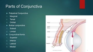
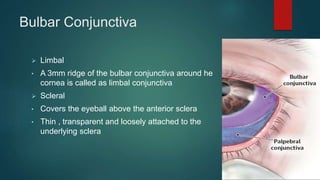
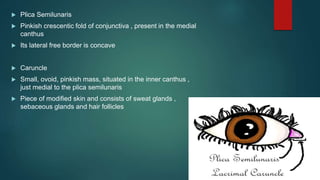
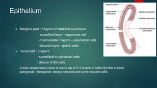
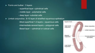
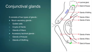
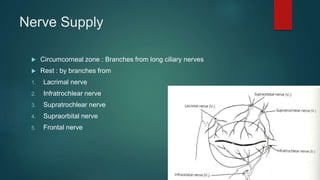
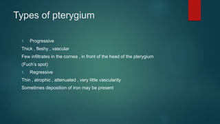
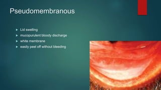
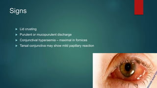
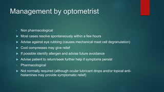
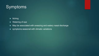
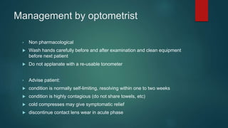
SlideShare a Scribd company logo
Conjunctiva
SIMRAN PAHUJA
B.OPTOM 4TH YEAR
Introduction:
 It is a Translucent mucous membrane which
lines the posterior surface of the eyelids and
anterior aspect of the eyeball
 It stretches from the lid margin to the limbus
Parts of Conjunctiva
 Palpebral Conjunctiva
1. Marginal
2. Tarsal
3. Orbital
 Bulbar conjunctiva
1. Scleral
2. Limbal
 Conjunctival fornix
1. Superior
2. Inferior
3. Lateral
4. Medial
Palpebral Conjunctival
 Marginal conjunctiva
• It extends from the lid margin to about 2mm on the
back of the lid upto a shallow groove – Subtarsal
sulcus .
• The sulcus is the commonest site for lodgement of a
conjunctival foreign body .
 Tarsal Conjunctiva
• Thin, transparent, highly vascular
• It is firmly adherent to the whole tarsal
plate in the upper lid and in lower lid only
half width of the tarsus.
 Orbital part
• It lies loose between the tarsal plate and
fornix
Bulbar Conjunctiva
 Limbal
• A 3mm ridge of the bulbar conjunctiva around he
cornea is called as limbal conjunctiva
 Scleral
• Covers the eyeball above the anterior sclera
• Thin , transparent and loosely attached to the
underlying sclera
Conjunctival Fornix
 It joins the bulbar conjunctiva with the palpebral conjunctiva
 It is broken only at the medial side by the caruncle and plica
semilunaris
 Plica Semilunaris
 Pinkish crescentic fold of conjunctiva , present in the medial
canthus
 Its lateral free border is concave
 Caruncle
 Small, ovoid, pinkish mass, situated in the inner canthus ,
just medial to the plica semilunaris
 Piece of modified skin and consists of sweat glands ,
sebaceous glands and hair follicles
Structure Of Conjunctiva
 Conjunctiva consists of 3 layers
1. Epithelium
2. Adenoid layer
3. Fibrous layer
Epithelium
 Marginal part : 5 layers of stratified squamous
- superficial layer –squamous cell
-intermediate 3 layers – polyhedral cells
-deepest layer –goblet cells
 Tarsal part : 2 layers
- superficial is cylindrical cells
- deeper is flat cells
• Lower tarsal conjunctiva is made up of 3-4 layers of cells like the cubical,
polygonal , elongated ,wedge shaped and cone shaped cells
 Fornix and bulbar : 3 layers
- superficial layer -cylindrical cells
- middle layer - polyhedral cells
- deep layer -cuboidal cells
 Limbal conjunctiva : 8-10 layer of stratified squamous epithelium
-Most superficial 1-2 layers – squamous cells
-Intermediate several layers – polygonal cells
-Basal layer – cylindrical or cubical cells
Adenoid layer
 Lymphoid layer
 It is not present since birth but develops after 3-4 months of life .
 For this reason, conjunctival inflammation in an infant does not produce
follicular reactions.
Fibrous layer
 Meshwork of collagenous and elastic fibers
 Thicker than adenoid layer except in the Tarsal conjunctiva
 Consists of nerves and vessels of conjunctiva
Conjunctival glands
It consists of two types of glands :
 Mucin secretory glands :
1. Goblet cells
2. Crypts of Henle
3. Glands of Manz
 Accessory lacrimal glands :
1. Glands of Krause
2. Glands of Wolfring
Blood Supply
 Arteries :
- Derived from 3 sources
1. Peripheral arterial arcade of eyelids
2. Marginal arterial arcade of eyelids
3. Anterior ciliary arteries
 Palpebral and fornix part : Peripheral and Marginal arterial arcade of
eyelid
 Bulbar part : Anterior and posterior conjunctival arteries
Nerve Supply
 Circumcorneal zone : Branches from long ciliary nerves
 Rest : by branches from
1. Lacrimal nerve
2. Infratrochlear nerve
3. Supratrochlear nerve
4. Supraorbital nerve
5. Frontal nerve
Lymphatic Drainage
 Lymphatics of conjunctiva are arranged in 2 layers
- superficial
- deep
 Lymphatics from the lateral side drain into preauricular lymph nodes
 Lymphatics from the medial side drain into the submandibular lymph
nodes.
Diseases Of Conjunctiva
1. Degenerative conditions
2. Inflammatory conditions
3. Symptomatic conditions
4. Cysts and Tumours
1.Degenerative conditions
1. Pinguecula
2. Pterygium
3. Concretions
4. Amyloid degenerations
Pinguecula
 Extremely common degenerative change
 Yellowish white path on bulbar conjunctiva near the limbus ,
nasal or temporal .
Etiology
 Age related change
 Strong sunlight exposure
 Dusty , windy and smoky working environment
 ?Considered as a precursor of pterygium
Symptoms :
 Usually asymptomatic
 Possible mild foreign body sensation and redness when inflamed
 Occasional cosmetic concern
Signs :
 Area of conjunctival thickening adjoining the limbus
 In the palpebral aperture, usually at 3 & 9 o’clock positions
 More common nasally
 Usually bilateral
 Elevated and less transparent than normal conjunctiva
 White to yellow colour, fat like appearance
 May become inflamed causing mild ocular irritation
 Decreased TBUT
Management by Optometrist
 Non pharmacological
• Reassure patient about benign nature of the lesion (no threat to health or
sight)
• Advise on UV protection to minimise risk of inflammation
-brimmed hat, sunglasses in wrap-around style for side protection
• Cold compresses when inflamed
 Pharmacological
• Ocular lubricants for symptomatic relief
Pterygium
 Pterygium is a wing shaped fold of conjunctiva encroaching upon the
cornea from either side within the interpalpebral fissure.
 Etiology ;
1. More common in people living in hot climates .
2. Exposure to sun
3. Dry heat
4. High wind
5. Abundance of dust
Signs
 Usually bilateral; often asymmetrical.
 More common nasally
 Starts with scarring, thickening and distortion of the bulbar conjunctiva
 Slow insidious growth on to cornea (or may become stable)
 Destroys Bowman’s membrane and superficial stroma lamellae
 Epithelial iron deposit (Stocker’s line) ahead of advancing pterygium
 Relatively rich surface vascularisation
 Flattening of cornea in horizontal meridian
Symptoms
 Mild irritation (redness, dryness, foreign body sensation)
 May be exacerbated by incidents of acute inflammation
 Cosmetic concern
Types of pterygium
1. Progressive
Thick , fleshy , vascular
Few infiltrates in the cornea , in front of the head of the pterygium
(Fuch’s spot)
1. Regressive
Thin , atrophic , attenuated , very little vascularity
Sometimes deposition of iron may be present
Treatment
Surgical excision is the only satisfactory treatment .
 Indications :
1. Cosmetic disfigurement
2. Visual impairment
3. Continued progression
4. Diplopia due to interference in ocular movement
Management by Optometrist
 Non pharmacological
• Advise on UV protection:
-brimmed hat, tinted lenses, wrap-around style for side protection
-reduces risk of progression and of becoming inflamed and irritated
• Measure and draw diagram (photo document if possible)
• Cold compress when inflamed
 Pharmacological
Ocular lubricants for symptomatic relief
Concretions
Concretions are formed due to accumulation of inspisated
mucus and dead epithelial cell debris into the conjunctival
depressions called loops of henle .
 Symptoms :
1. Usually none
2. May erode through the epithelium
3. foreign body sensation
 Signs :
1. Small white/yellow-white bodies with distinct edges in tarsal conjunctiva
2. Single or multiple Usually <1mm dia, sometimes up to 3mm
3. Usually low profile but may be raised if large.
Management by optometrist
 Non pharmacological
• Treatment rarely required
• Artificial tears and lubricating ointments
• Eroded concretions leading to irritation can be removed at the slit lamp
-topical anaesthetic
- with sterile hypodermic needle
 Pharmacological
• No specific drug treatment available
Amyloid Degeneration Of Conjunctiva
 Rare
 Occurs in 2 forms :
1. Primary conjunctival amyloid
2. Secondary conjunctival amyloid
 Clinical Features :
1. Deposition of yellowish , well demarcated, irregular amyloid
material in the conjunctiva
2. Subconjunctival haemorrhage may be associated with amyloid
deposition in blood vessels
 Treatment :
1. Lubricating drops for mild symptoms .
2. Excision can be performed with marked irritation due to raised
lesions .
Symptomology
• Non-Specific
 Lacrimation
 Irritation
 Stinging
 Photophobia
 Burning
 Redness
• Specific
 Pain and FB sensation in Corneal Involvement
 Itching in allergic, Blepharitis and dry eyes
Conjunctival Signs
 Type of Discharge
 Type of conjunctival reaction
 Presence of membrane/ Pseudo-membrane
 Lymphadenopathy
 Pigmentations
Discharge :
It is exudation from abnormally dilated blood vessels filtered
through conjunctival epithelium .
Types of discharge :
1. Watery Discharge – Viral infection
2. Mucinous Discharge – keratoconjunctivitis sicca
3. Purulent Discharge – severe bacterial infection
4. Mucopurulent Discharge – mild bacterial infection
Conjunctival Reactions :
1. Hyperaemia
2. Papillae
3. Follicles
4. Concretions
5. Chemosis / Conjunctival Oedema
Papillae
 It is response of conjunctiva to inflammation
 It consists of small capillary network surrounded by fibrous
network
 hyperplasia of epithelium
 usually more discrete and more red than follicles
 side walls of papillae appear perpendicular to tarsal plate
Follicles
 Follicles appear as multiple discrete slightly elevated lesions
 They may be 0.5 to 5 mm in size depending upon severity and duration
 Follicles present lymphocytic response
 Commonly seen in lower palpebral conjunctiva
 Acute Follicular Response- <4 weeks
 Chronic Follicular Response- >4 weeks
 hyperplasia of lymphoid tissue
 generally seen in viral conditions
 smooth, pale, pink-to-yellow, elevated lesions
 surrounded by displaced vessels
Chemosis
 It is seen when conjunctiva is inflamed and hyperemic due to transdation
of fibrin and protein rich fluid through damaged blood vessels .
 Due to laxity of bulbar conjunctiva the fluid commonly gets collected there
 Ballooning of conjunctiva occurs when fluid is in large quantity it is labelled
as chemosis
Pseudomembranous
 Lid swelling
 mucopurulent bloody discharge
 white membrane
 easily peel off without bleeding
Membranous
 Acute inflammation of the conjunctiva
 Lids are hardened
 semisolid exudates: result in necrosis of conjunctiva and cornea
 difficult to peel off
 associated with bleeding from the under surface
Lymphadenopathy-Swelling of Lymph
nodes
 Pre auricular and sub mandibular.
1. Viral infection.
2. Chlamydial infection.
3. Severe bacterial infections. (Gonococcal)
4. Parinaud oculo-glandular syndrome.
Bacterial Conjunctivitis
• Predisposing factors
 contamination of the conjunctival surface
 superficial trauma
 contact lens wear
 secondary to viral conjunctivitis
 recent cold, upper respiratory tract infection or sinusitis
 Diabetes
 Steroids
 Blepharitis
Symptoms
Acute onset of:
• redness
• discomfort, usually described as burning
• discharge
• Usually bilateral – one eye may be affected before the other (by one or two
days)
Signs
 Lid crusting
 Purulent or mucopurulent discharge
 Conjunctival hyperaemia – maximal in fornices
 Tarsal conjunctiva may show mild papillary reaction
Management by optometrist
• Non pharmacological
 Often resolves in 5-7 days without treatment
 Bathe/clean the eyelids with lint or cotton wool dipped in sterile saline or
boiled (cooled) water to remove crusting
 Advise patient that condition is contagious (do not share towels, etc.)
• Pharmacological
 Treatment with topical antibiotic may improve short-term outcome and
render patient less infectious to others
CL-associated Papillary Conjunctivitis (CLAPC),
Giant Papillary Conjunctivitis (GPC)
• Predisposing factors
 Common in soft compared to rigid lenses
 reported in silicone hydrogel, as well as hydrogel, lens wearers
 Lens deposits
 Thick or poorly designed or manufactured lens edges
 Meibomian gland dysfunction
Symptoms
 Itching and non-specific irritation
 may increase after lens removal Mucus discharge
 Increased lens movement
 Loss of lens tolerance
 Decreasing comfort
 Blurred vision
Signs
 Almost always bilateral
 Upper tarsal conjunctiva (lower usually not affected)
 papillae
 macro papillae (diameter between 0.3 and 1 mm) or giant papillae
(diameter > 1 mm)
 hyperaemia
 stringy mucus in tear film and on conjunctival surfaces
 conjunctival oedema
Management by optometrist
• Non pharmacological
 Removal of lens deposits
 Reduce exposure time
 Optimize lens fit, material and wearing regime
o change soft lens material to one with improved deposit resistance
o change to daily disposable soft lenses
Acute Allergic Conjunctivitis
• Aetiology
 A self-limiting reaction to an allergen (often unidentified) that comes into
contact with the conjunctiva provoking an immediate response
 Common in children
 Allergens include: grass pollen, animal dander , dust
• Predisposing factors
 History of allergic disease; can also occur without such history
• Symptoms
 Eyelid swelling
 Itching
 May be unilateral (if a direct contact response)
• Signs
 Lid oedema
 Conjunctival chemosis
 Mild watery or mucoid discharge
Management by optometrist
• Non pharmacological
 Most cases resolve spontaneously within a few hours
 Advise against eye rubbing (causes mechanical mast cell degranulation)
 Cool compresses may give relief
 If possible identify allergen and advise future avoidance
 Advise patient to return/seek further help if symptoms persist
• Pharmacological
 Not normally required (although ocular lubricant drops and/or topical anti-
histamines may provide symptomatic relief)
Seasonal Allergic Conjunctivitis
• Predisposing factors
 Atopic disposition
 Personal history of allergic disease (hay fever, asthma, eczema, food or
drug allergy)
 Family history of allergic disease
 Exposure to allergens
Symptoms
 Itching
 Watering of eye
 May be associated with sneezing and watery nasal discharge
 symptoms seasonal with climatic variations
Signs
Mild to moderate lid oedema
Bulbar and tarsal conjunctiva: chemosis , hyperaemia and diffuse papillary
reaction
Management by optometrist
 Identify allergens
 Advise avoidance of allergens
 Cool compresses for symptomatic relief
 Advise against eye rubbing
Viral Conjunctivitis
• Predisposing factors
 Recent cold or other upper respiratory tract infection
 Low standards of hygiene
 Crowded conditions (schools, camps, clinics)
 Eye clinics (transmission by clinicians’ fingers, tonometer , etc.)
Symptoms
Acute onset of :
 redness
 Discomfort
 watering
 Eyelids may be stuck together in the morning
 Often unilateral at first, becoming bilateral
 Blurred vision if central cornea involved
Signs
 Watery discharge
 Hyperemia
 Follicles
 Subconjunctival haemorrhages
 Pseudo membranes
 Pre-auricular lymphadenopathy which may be tender (not present in every
case)
 Corneal involvement in some cases
Management by optometrist
• Non pharmacological
 Wash hands carefully before and after examination and clean equipment
before next patient
 Do not applanate with a re-usable tonometer
• Advise patient:
 condition is normally self-limiting, resolving within one to two weeks
 condition is highly contagious (do not share towels, etc)
 cold compresses may give symptomatic relief
 discontinue contact lens wear in acute phase
Thank You !

More Related Content

What's hot

Sturm's conoid
Sturm's conoidSturm's conoid
Sturm's conoid
Dr Samarth Mishra
 
Anatomy Of Cornea
Anatomy Of  CorneaAnatomy Of  Cornea
Anatomy Of Cornea
Husain Patanwala
 
Tear film Dr Ferdous
Tear film Dr Ferdous  Tear film Dr Ferdous
Tear film Dr Ferdous
Ferdous101531
 
Sclera
ScleraSclera
Sclera
kamal thakur
 
Tear film and dynamics
Tear film and dynamics Tear film and dynamics
Tear film and dynamics
SSSIHMS-PG
 
Limbus
LimbusLimbus
disorders of lashes
disorders of lashesdisorders of lashes
disorders of lashesSiva Wurity
 
Pterygium and its management
Pterygium and its managementPterygium and its management
Pterygium and its management
Jyoti Prakash
 
Coloboma
ColobomaColoboma
Anatomy of macula
Anatomy of maculaAnatomy of macula
Anatomy of macula
Dr.Siddharth Gautam
 
Anatomy and physiology of cornea
Anatomy and physiology of corneaAnatomy and physiology of cornea
Anatomy and physiology of cornea
SSSIHMS-PG
 
Anatomy of uvea
Anatomy of uveaAnatomy of uvea
Anatomy of uvea
Barun Garg
 
Aqueous humor dynamics
Aqueous humor dynamicsAqueous humor dynamics
Aqueous humor dynamics
Laxmi Eye Institute
 
Eyelid anatomy and physiology
Eyelid anatomy and physiologyEyelid anatomy and physiology
Eyelid anatomy and physiology
Najara Thapa
 
Primary open angle glaucoma (poag)
Primary open angle glaucoma (poag)Primary open angle glaucoma (poag)
Primary open angle glaucoma (poag)
Om Patel
 
eyelid anatomy slideshare
eyelid anatomy slideshareeyelid anatomy slideshare
eyelid anatomy slideshare
University of Gondar
 
Corneal anatomy and physiology 2
Corneal anatomy and physiology 2Corneal anatomy and physiology 2
Corneal anatomy and physiology 2
Om Patel
 
Keratoconus
KeratoconusKeratoconus
Keratoconus
ikramdr01
 

What's hot (20)

Sturm's conoid
Sturm's conoidSturm's conoid
Sturm's conoid
 
Anatomy Of Cornea
Anatomy Of  CorneaAnatomy Of  Cornea
Anatomy Of Cornea
 
Tear film Dr Ferdous
Tear film Dr Ferdous  Tear film Dr Ferdous
Tear film Dr Ferdous
 
Sclera
ScleraSclera
Sclera
 
Tear film and dynamics
Tear film and dynamics Tear film and dynamics
Tear film and dynamics
 
Limbus
LimbusLimbus
Limbus
 
disorders of lashes
disorders of lashesdisorders of lashes
disorders of lashes
 
Pterygium and its management
Pterygium and its managementPterygium and its management
Pterygium and its management
 
Coloboma
ColobomaColoboma
Coloboma
 
Anatomy of macula
Anatomy of maculaAnatomy of macula
Anatomy of macula
 
Anatomy and physiology of cornea
Anatomy and physiology of corneaAnatomy and physiology of cornea
Anatomy and physiology of cornea
 
Anatomy of uvea
Anatomy of uveaAnatomy of uvea
Anatomy of uvea
 
Aqueous humor dynamics
Aqueous humor dynamicsAqueous humor dynamics
Aqueous humor dynamics
 
Eyelid anatomy and physiology
Eyelid anatomy and physiologyEyelid anatomy and physiology
Eyelid anatomy and physiology
 
Anterior Uveitis
Anterior UveitisAnterior Uveitis
Anterior Uveitis
 
Primary open angle glaucoma (poag)
Primary open angle glaucoma (poag)Primary open angle glaucoma (poag)
Primary open angle glaucoma (poag)
 
Tear film test
Tear film testTear film test
Tear film test
 
eyelid anatomy slideshare
eyelid anatomy slideshareeyelid anatomy slideshare
eyelid anatomy slideshare
 
Corneal anatomy and physiology 2
Corneal anatomy and physiology 2Corneal anatomy and physiology 2
Corneal anatomy and physiology 2
 
Keratoconus
KeratoconusKeratoconus
Keratoconus
 

Similar to Conjunctiva and its Disorders

Conjunctiva
ConjunctivaConjunctiva
CONJUCTIVA-2.pptx
CONJUCTIVA-2.pptxCONJUCTIVA-2.pptx
CONJUCTIVA-2.pptx
Harshika Malik
 
Eyelid infection
Eyelid infectionEyelid infection
Eyelid infection
Dipali Dumbre
 
Layers of skin of the Eye Lids, Eye Lashes, Eye Lid Pathology, Anatomy of Con...
Layers of skin of the Eye Lids, Eye Lashes, Eye Lid Pathology, Anatomy of Con...Layers of skin of the Eye Lids, Eye Lashes, Eye Lid Pathology, Anatomy of Con...
Layers of skin of the Eye Lids, Eye Lashes, Eye Lid Pathology, Anatomy of Con...Nina Ko
 
Eyelid infections ppt
Eyelid infections pptEyelid infections ppt
Eyelid infections ppt
OM VERMA
 
opthalmolgy.The conjunctiva lecture 1.(dr.ali)
opthalmolgy.The conjunctiva lecture 1.(dr.ali)opthalmolgy.The conjunctiva lecture 1.(dr.ali)
opthalmolgy.The conjunctiva lecture 1.(dr.ali)student
 
L-2 Eye lid disorder.pdf
L-2 Eye lid disorder.pdfL-2 Eye lid disorder.pdf
L-2 Eye lid disorder.pdf
Ahad412190
 
Degenerative condition of eye
Degenerative condition of eyeDegenerative condition of eye
Degenerative condition of eye
OPTOM FASLU MUHAMMED
 
Conjuctival diseases
Conjuctival diseasesConjuctival diseases
Conjuctival diseases
mohammed Qazzaz
 
Eyelid disorders
Eyelid disordersEyelid disorders
Eyelid disorders
SOUMYA SUBRAMANI
 
Diseases of Cornea Keratitis
Diseases of Cornea KeratitisDiseases of Cornea Keratitis
Diseases of Cornea Keratitis
Arslan Chaudhry
 
anomalies of eyemargin ophthalmology.pptx
anomalies of eyemargin ophthalmology.pptxanomalies of eyemargin ophthalmology.pptx
anomalies of eyemargin ophthalmology.pptx
sarathrajum17
 
conjunctiva.pptx
conjunctiva.pptxconjunctiva.pptx
conjunctiva.pptx
ShashiBhushan44431
 
Allergic conjunctivitis
Allergic conjunctivitisAllergic conjunctivitis
Allergic conjunctivitis
University of Gondar
 
Conjonctivite allergiqueeeeeeeeeeee.pptx
Conjonctivite allergiqueeeeeeeeeeee.pptxConjonctivite allergiqueeeeeeeeeeee.pptx
Conjonctivite allergiqueeeeeeeeeeee.pptx
miaorned
 
EYELID DISORDER
EYELID DISORDEREYELID DISORDER
EYELID DISORDER
ParameshP4
 
CLICK THIS LINK TO OPEN PRESENTATION
CLICK THIS LINK TO  OPEN PRESENTATIONCLICK THIS LINK TO  OPEN PRESENTATION
CLICK THIS LINK TO OPEN PRESENTATIONqumberabbas10
 

Similar to Conjunctiva and its Disorders (20)

Conjunctiva
ConjunctivaConjunctiva
Conjunctiva
 
CONJUCTIVA-2.pptx
CONJUCTIVA-2.pptxCONJUCTIVA-2.pptx
CONJUCTIVA-2.pptx
 
Eyelid infection
Eyelid infectionEyelid infection
Eyelid infection
 
Layers of skin of the Eye Lids, Eye Lashes, Eye Lid Pathology, Anatomy of Con...
Layers of skin of the Eye Lids, Eye Lashes, Eye Lid Pathology, Anatomy of Con...Layers of skin of the Eye Lids, Eye Lashes, Eye Lid Pathology, Anatomy of Con...
Layers of skin of the Eye Lids, Eye Lashes, Eye Lid Pathology, Anatomy of Con...
 
Eyelid infections ppt
Eyelid infections pptEyelid infections ppt
Eyelid infections ppt
 
opthalmolgy.The conjunctiva lecture 1.(dr.ali)
opthalmolgy.The conjunctiva lecture 1.(dr.ali)opthalmolgy.The conjunctiva lecture 1.(dr.ali)
opthalmolgy.The conjunctiva lecture 1.(dr.ali)
 
L-2 Eye lid disorder.pdf
L-2 Eye lid disorder.pdfL-2 Eye lid disorder.pdf
L-2 Eye lid disorder.pdf
 
Degenerative condition of eye
Degenerative condition of eyeDegenerative condition of eye
Degenerative condition of eye
 
Conjuctival diseases
Conjuctival diseasesConjuctival diseases
Conjuctival diseases
 
Allergic conjunctivitis
Allergic conjunctivitisAllergic conjunctivitis
Allergic conjunctivitis
 
Eyelid disorders
Eyelid disordersEyelid disorders
Eyelid disorders
 
Diseases of Cornea Keratitis
Diseases of Cornea KeratitisDiseases of Cornea Keratitis
Diseases of Cornea Keratitis
 
anomalies of eyemargin ophthalmology.pptx
anomalies of eyemargin ophthalmology.pptxanomalies of eyemargin ophthalmology.pptx
anomalies of eyemargin ophthalmology.pptx
 
conjunctiva.pptx
conjunctiva.pptxconjunctiva.pptx
conjunctiva.pptx
 
Allergic conjunctivitis
Allergic conjunctivitisAllergic conjunctivitis
Allergic conjunctivitis
 
Conjonctivite allergiqueeeeeeeeeeee.pptx
Conjonctivite allergiqueeeeeeeeeeee.pptxConjonctivite allergiqueeeeeeeeeeee.pptx
Conjonctivite allergiqueeeeeeeeeeee.pptx
 
EYELID DISORDER
EYELID DISORDEREYELID DISORDER
EYELID DISORDER
 
CLICK THIS LINK TO OPEN PRESENTATION
CLICK THIS LINK TO  OPEN PRESENTATIONCLICK THIS LINK TO  OPEN PRESENTATION
CLICK THIS LINK TO OPEN PRESENTATION
 
Uveitis 2lectures by dr. khaled
Uveitis 2lectures by dr. khaledUveitis 2lectures by dr. khaled
Uveitis 2lectures by dr. khaled
 
Uveitis 2lectures by dr. khaled
Uveitis 2lectures by dr. khaledUveitis 2lectures by dr. khaled
Uveitis 2lectures by dr. khaled
 

Recently uploaded

Nursing Care of Client With Acute And Chronic Renal Failure.ppt
Nursing Care of Client With Acute And Chronic Renal Failure.pptNursing Care of Client With Acute And Chronic Renal Failure.ppt
Nursing Care of Client With Acute And Chronic Renal Failure.ppt
Rommel Luis III Israel
 
Demystifying-Gene-Editing-The-Promise-and-Peril-of-CRISPR.pdf
Demystifying-Gene-Editing-The-Promise-and-Peril-of-CRISPR.pdfDemystifying-Gene-Editing-The-Promise-and-Peril-of-CRISPR.pdf
Demystifying-Gene-Editing-The-Promise-and-Peril-of-CRISPR.pdf
SasikiranMarri
 
Contact ME {89011**83002} Haridwar ℂall Girls By Full Service Call Girl In Ha...
Contact ME {89011**83002} Haridwar ℂall Girls By Full Service Call Girl In Ha...Contact ME {89011**83002} Haridwar ℂall Girls By Full Service Call Girl In Ha...
Contact ME {89011**83002} Haridwar ℂall Girls By Full Service Call Girl In Ha...
ranishasharma67
 
Myopia Management & Control Strategies.pptx
Myopia Management & Control Strategies.pptxMyopia Management & Control Strategies.pptx
Myopia Management & Control Strategies.pptx
RitonDeb1
 
Dimensions of Healthcare Quality
Dimensions of Healthcare QualityDimensions of Healthcare Quality
Dimensions of Healthcare Quality
Naeemshahzad51
 
.Metabolic.disordersYYSSSFFSSSSSSSSSSDDD
.Metabolic.disordersYYSSSFFSSSSSSSSSSDDD.Metabolic.disordersYYSSSFFSSSSSSSSSSDDD
.Metabolic.disordersYYSSSFFSSSSSSSSSSDDD
samahesh1
 
GURGAON Call Girls ❤8901183002❤ #ℂALL# #gIRLS# In GURGAON ₹,2500 Cash Payment...
GURGAON Call Girls ❤8901183002❤ #ℂALL# #gIRLS# In GURGAON ₹,2500 Cash Payment...GURGAON Call Girls ❤8901183002❤ #ℂALL# #gIRLS# In GURGAON ₹,2500 Cash Payment...
GURGAON Call Girls ❤8901183002❤ #ℂALL# #gIRLS# In GURGAON ₹,2500 Cash Payment...
ranishasharma67
 
Jaipur ❤cALL gIRLS 89O1183002 ❤ℂall Girls IN JaiPuR ESCORT SERVICE
Jaipur ❤cALL gIRLS 89O1183002 ❤ℂall Girls IN JaiPuR ESCORT SERVICEJaipur ❤cALL gIRLS 89O1183002 ❤ℂall Girls IN JaiPuR ESCORT SERVICE
Jaipur ❤cALL gIRLS 89O1183002 ❤ℂall Girls IN JaiPuR ESCORT SERVICE
ranishasharma67
 
CANCER CANCER CANCER CANCER CANCER CANCER
CANCER  CANCER  CANCER  CANCER  CANCER CANCERCANCER  CANCER  CANCER  CANCER  CANCER CANCER
CANCER CANCER CANCER CANCER CANCER CANCER
KRISTELLEGAMBOA2
 
POLYCYSTIC OVARIAN SYNDROME (PCOS)......
POLYCYSTIC OVARIAN SYNDROME (PCOS)......POLYCYSTIC OVARIAN SYNDROME (PCOS)......
POLYCYSTIC OVARIAN SYNDROME (PCOS)......
Ameena Kadar
 
A Community health , health for prisoners
A Community health  , health for prisonersA Community health  , health for prisoners
A Community health , health for prisoners
Ahmed Elmi
 
The Impact of Meeting: How It Can Change Your Life
The Impact of Meeting: How It Can Change Your LifeThe Impact of Meeting: How It Can Change Your Life
The Impact of Meeting: How It Can Change Your Life
ranishasharma67
 
India Clinical Trials Market: Industry Size and Growth Trends [2030] Analyzed...
India Clinical Trials Market: Industry Size and Growth Trends [2030] Analyzed...India Clinical Trials Market: Industry Size and Growth Trends [2030] Analyzed...
India Clinical Trials Market: Industry Size and Growth Trends [2030] Analyzed...
Kumar Satyam
 
Antibiotic Stewardship by Anushri Srivastava.pptx
Antibiotic Stewardship by Anushri Srivastava.pptxAntibiotic Stewardship by Anushri Srivastava.pptx
Antibiotic Stewardship by Anushri Srivastava.pptx
AnushriSrivastav
 
Introduction to Forensic Pathology course
Introduction to Forensic Pathology courseIntroduction to Forensic Pathology course
Introduction to Forensic Pathology course
fprxsqvnz5
 
一比一原版纽约大学毕业证(NYU毕业证)成绩单留信认证
一比一原版纽约大学毕业证(NYU毕业证)成绩单留信认证一比一原版纽约大学毕业证(NYU毕业证)成绩单留信认证
一比一原版纽约大学毕业证(NYU毕业证)成绩单留信认证
o6ov5dqmf
 
Neuro Saphirex Cranial Brochure
Neuro Saphirex Cranial BrochureNeuro Saphirex Cranial Brochure
Neuro Saphirex Cranial Brochure
RXOOM Healthcare Pvt. Ltd. ​
 
Yemen National Tuberculosis Program .ppt
Yemen National Tuberculosis Program .pptYemen National Tuberculosis Program .ppt
Yemen National Tuberculosis Program .ppt
Esam43
 
Navigating Challenges: Mental Health, Legislation, and the Prison System in B...
Navigating Challenges: Mental Health, Legislation, and the Prison System in B...Navigating Challenges: Mental Health, Legislation, and the Prison System in B...
Navigating Challenges: Mental Health, Legislation, and the Prison System in B...
Guillermo Rivera
 
How many patients does case series should have In comparison to case reports.pdf
How many patients does case series should have In comparison to case reports.pdfHow many patients does case series should have In comparison to case reports.pdf
How many patients does case series should have In comparison to case reports.pdf
pubrica101
 

Recently uploaded (20)

Nursing Care of Client With Acute And Chronic Renal Failure.ppt
Nursing Care of Client With Acute And Chronic Renal Failure.pptNursing Care of Client With Acute And Chronic Renal Failure.ppt
Nursing Care of Client With Acute And Chronic Renal Failure.ppt
 
Demystifying-Gene-Editing-The-Promise-and-Peril-of-CRISPR.pdf
Demystifying-Gene-Editing-The-Promise-and-Peril-of-CRISPR.pdfDemystifying-Gene-Editing-The-Promise-and-Peril-of-CRISPR.pdf
Demystifying-Gene-Editing-The-Promise-and-Peril-of-CRISPR.pdf
 
Contact ME {89011**83002} Haridwar ℂall Girls By Full Service Call Girl In Ha...
Contact ME {89011**83002} Haridwar ℂall Girls By Full Service Call Girl In Ha...Contact ME {89011**83002} Haridwar ℂall Girls By Full Service Call Girl In Ha...
Contact ME {89011**83002} Haridwar ℂall Girls By Full Service Call Girl In Ha...
 
Myopia Management & Control Strategies.pptx
Myopia Management & Control Strategies.pptxMyopia Management & Control Strategies.pptx
Myopia Management & Control Strategies.pptx
 
Dimensions of Healthcare Quality
Dimensions of Healthcare QualityDimensions of Healthcare Quality
Dimensions of Healthcare Quality
 
.Metabolic.disordersYYSSSFFSSSSSSSSSSDDD
.Metabolic.disordersYYSSSFFSSSSSSSSSSDDD.Metabolic.disordersYYSSSFFSSSSSSSSSSDDD
.Metabolic.disordersYYSSSFFSSSSSSSSSSDDD
 
GURGAON Call Girls ❤8901183002❤ #ℂALL# #gIRLS# In GURGAON ₹,2500 Cash Payment...
GURGAON Call Girls ❤8901183002❤ #ℂALL# #gIRLS# In GURGAON ₹,2500 Cash Payment...GURGAON Call Girls ❤8901183002❤ #ℂALL# #gIRLS# In GURGAON ₹,2500 Cash Payment...
GURGAON Call Girls ❤8901183002❤ #ℂALL# #gIRLS# In GURGAON ₹,2500 Cash Payment...
 
Jaipur ❤cALL gIRLS 89O1183002 ❤ℂall Girls IN JaiPuR ESCORT SERVICE
Jaipur ❤cALL gIRLS 89O1183002 ❤ℂall Girls IN JaiPuR ESCORT SERVICEJaipur ❤cALL gIRLS 89O1183002 ❤ℂall Girls IN JaiPuR ESCORT SERVICE
Jaipur ❤cALL gIRLS 89O1183002 ❤ℂall Girls IN JaiPuR ESCORT SERVICE
 
CANCER CANCER CANCER CANCER CANCER CANCER
CANCER  CANCER  CANCER  CANCER  CANCER CANCERCANCER  CANCER  CANCER  CANCER  CANCER CANCER
CANCER CANCER CANCER CANCER CANCER CANCER
 
POLYCYSTIC OVARIAN SYNDROME (PCOS)......
POLYCYSTIC OVARIAN SYNDROME (PCOS)......POLYCYSTIC OVARIAN SYNDROME (PCOS)......
POLYCYSTIC OVARIAN SYNDROME (PCOS)......
 
A Community health , health for prisoners
A Community health  , health for prisonersA Community health  , health for prisoners
A Community health , health for prisoners
 
The Impact of Meeting: How It Can Change Your Life
The Impact of Meeting: How It Can Change Your LifeThe Impact of Meeting: How It Can Change Your Life
The Impact of Meeting: How It Can Change Your Life
 
India Clinical Trials Market: Industry Size and Growth Trends [2030] Analyzed...
India Clinical Trials Market: Industry Size and Growth Trends [2030] Analyzed...India Clinical Trials Market: Industry Size and Growth Trends [2030] Analyzed...
India Clinical Trials Market: Industry Size and Growth Trends [2030] Analyzed...
 
Antibiotic Stewardship by Anushri Srivastava.pptx
Antibiotic Stewardship by Anushri Srivastava.pptxAntibiotic Stewardship by Anushri Srivastava.pptx
Antibiotic Stewardship by Anushri Srivastava.pptx
 
Introduction to Forensic Pathology course
Introduction to Forensic Pathology courseIntroduction to Forensic Pathology course
Introduction to Forensic Pathology course
 
一比一原版纽约大学毕业证(NYU毕业证)成绩单留信认证
一比一原版纽约大学毕业证(NYU毕业证)成绩单留信认证一比一原版纽约大学毕业证(NYU毕业证)成绩单留信认证
一比一原版纽约大学毕业证(NYU毕业证)成绩单留信认证
 
Neuro Saphirex Cranial Brochure
Neuro Saphirex Cranial BrochureNeuro Saphirex Cranial Brochure
Neuro Saphirex Cranial Brochure
 
Yemen National Tuberculosis Program .ppt
Yemen National Tuberculosis Program .pptYemen National Tuberculosis Program .ppt
Yemen National Tuberculosis Program .ppt
 
Navigating Challenges: Mental Health, Legislation, and the Prison System in B...
Navigating Challenges: Mental Health, Legislation, and the Prison System in B...Navigating Challenges: Mental Health, Legislation, and the Prison System in B...
Navigating Challenges: Mental Health, Legislation, and the Prison System in B...
 
How many patients does case series should have In comparison to case reports.pdf
How many patients does case series should have In comparison to case reports.pdfHow many patients does case series should have In comparison to case reports.pdf
How many patients does case series should have In comparison to case reports.pdf
 

Conjunctiva and its Disorders

  • 2. Introduction:  It is a Translucent mucous membrane which lines the posterior surface of the eyelids and anterior aspect of the eyeball  It stretches from the lid margin to the limbus
  • 3. Parts of Conjunctiva  Palpebral Conjunctiva 1. Marginal 2. Tarsal 3. Orbital  Bulbar conjunctiva 1. Scleral 2. Limbal  Conjunctival fornix 1. Superior 2. Inferior 3. Lateral 4. Medial
  • 4. Palpebral Conjunctival  Marginal conjunctiva • It extends from the lid margin to about 2mm on the back of the lid upto a shallow groove – Subtarsal sulcus . • The sulcus is the commonest site for lodgement of a conjunctival foreign body .
  • 5.  Tarsal Conjunctiva • Thin, transparent, highly vascular • It is firmly adherent to the whole tarsal plate in the upper lid and in lower lid only half width of the tarsus.  Orbital part • It lies loose between the tarsal plate and fornix
  • 6. Bulbar Conjunctiva  Limbal • A 3mm ridge of the bulbar conjunctiva around he cornea is called as limbal conjunctiva  Scleral • Covers the eyeball above the anterior sclera • Thin , transparent and loosely attached to the underlying sclera
  • 7. Conjunctival Fornix  It joins the bulbar conjunctiva with the palpebral conjunctiva  It is broken only at the medial side by the caruncle and plica semilunaris
  • 8.  Plica Semilunaris  Pinkish crescentic fold of conjunctiva , present in the medial canthus  Its lateral free border is concave  Caruncle  Small, ovoid, pinkish mass, situated in the inner canthus , just medial to the plica semilunaris  Piece of modified skin and consists of sweat glands , sebaceous glands and hair follicles
  • 9. Structure Of Conjunctiva  Conjunctiva consists of 3 layers 1. Epithelium 2. Adenoid layer 3. Fibrous layer
  • 10. Epithelium  Marginal part : 5 layers of stratified squamous - superficial layer –squamous cell -intermediate 3 layers – polyhedral cells -deepest layer –goblet cells  Tarsal part : 2 layers - superficial is cylindrical cells - deeper is flat cells • Lower tarsal conjunctiva is made up of 3-4 layers of cells like the cubical, polygonal , elongated ,wedge shaped and cone shaped cells
  • 11.  Fornix and bulbar : 3 layers - superficial layer -cylindrical cells - middle layer - polyhedral cells - deep layer -cuboidal cells  Limbal conjunctiva : 8-10 layer of stratified squamous epithelium -Most superficial 1-2 layers – squamous cells -Intermediate several layers – polygonal cells -Basal layer – cylindrical or cubical cells
  • 12. Adenoid layer  Lymphoid layer  It is not present since birth but develops after 3-4 months of life .  For this reason, conjunctival inflammation in an infant does not produce follicular reactions.
  • 13. Fibrous layer  Meshwork of collagenous and elastic fibers  Thicker than adenoid layer except in the Tarsal conjunctiva  Consists of nerves and vessels of conjunctiva
  • 14. Conjunctival glands It consists of two types of glands :  Mucin secretory glands : 1. Goblet cells 2. Crypts of Henle 3. Glands of Manz  Accessory lacrimal glands : 1. Glands of Krause 2. Glands of Wolfring
  • 15. Blood Supply  Arteries : - Derived from 3 sources 1. Peripheral arterial arcade of eyelids 2. Marginal arterial arcade of eyelids 3. Anterior ciliary arteries  Palpebral and fornix part : Peripheral and Marginal arterial arcade of eyelid  Bulbar part : Anterior and posterior conjunctival arteries
  • 16. Nerve Supply  Circumcorneal zone : Branches from long ciliary nerves  Rest : by branches from 1. Lacrimal nerve 2. Infratrochlear nerve 3. Supratrochlear nerve 4. Supraorbital nerve 5. Frontal nerve
  • 17. Lymphatic Drainage  Lymphatics of conjunctiva are arranged in 2 layers - superficial - deep  Lymphatics from the lateral side drain into preauricular lymph nodes  Lymphatics from the medial side drain into the submandibular lymph nodes.
  • 18. Diseases Of Conjunctiva 1. Degenerative conditions 2. Inflammatory conditions 3. Symptomatic conditions 4. Cysts and Tumours
  • 19. 1.Degenerative conditions 1. Pinguecula 2. Pterygium 3. Concretions 4. Amyloid degenerations
  • 20. Pinguecula  Extremely common degenerative change  Yellowish white path on bulbar conjunctiva near the limbus , nasal or temporal .
  • 21. Etiology  Age related change  Strong sunlight exposure  Dusty , windy and smoky working environment  ?Considered as a precursor of pterygium
  • 22. Symptoms :  Usually asymptomatic  Possible mild foreign body sensation and redness when inflamed  Occasional cosmetic concern
  • 23. Signs :  Area of conjunctival thickening adjoining the limbus  In the palpebral aperture, usually at 3 & 9 o’clock positions  More common nasally  Usually bilateral  Elevated and less transparent than normal conjunctiva  White to yellow colour, fat like appearance  May become inflamed causing mild ocular irritation  Decreased TBUT
  • 24. Management by Optometrist  Non pharmacological • Reassure patient about benign nature of the lesion (no threat to health or sight) • Advise on UV protection to minimise risk of inflammation -brimmed hat, sunglasses in wrap-around style for side protection • Cold compresses when inflamed  Pharmacological • Ocular lubricants for symptomatic relief
  • 25. Pterygium  Pterygium is a wing shaped fold of conjunctiva encroaching upon the cornea from either side within the interpalpebral fissure.  Etiology ; 1. More common in people living in hot climates . 2. Exposure to sun 3. Dry heat 4. High wind 5. Abundance of dust
  • 26. Signs  Usually bilateral; often asymmetrical.  More common nasally  Starts with scarring, thickening and distortion of the bulbar conjunctiva  Slow insidious growth on to cornea (or may become stable)  Destroys Bowman’s membrane and superficial stroma lamellae  Epithelial iron deposit (Stocker’s line) ahead of advancing pterygium  Relatively rich surface vascularisation  Flattening of cornea in horizontal meridian
  • 27. Symptoms  Mild irritation (redness, dryness, foreign body sensation)  May be exacerbated by incidents of acute inflammation  Cosmetic concern
  • 28. Types of pterygium 1. Progressive Thick , fleshy , vascular Few infiltrates in the cornea , in front of the head of the pterygium (Fuch’s spot) 1. Regressive Thin , atrophic , attenuated , very little vascularity Sometimes deposition of iron may be present
  • 29. Treatment Surgical excision is the only satisfactory treatment .  Indications : 1. Cosmetic disfigurement 2. Visual impairment 3. Continued progression 4. Diplopia due to interference in ocular movement
  • 30. Management by Optometrist  Non pharmacological • Advise on UV protection: -brimmed hat, tinted lenses, wrap-around style for side protection -reduces risk of progression and of becoming inflamed and irritated • Measure and draw diagram (photo document if possible) • Cold compress when inflamed  Pharmacological Ocular lubricants for symptomatic relief
  • 31. Concretions Concretions are formed due to accumulation of inspisated mucus and dead epithelial cell debris into the conjunctival depressions called loops of henle .
  • 32.  Symptoms : 1. Usually none 2. May erode through the epithelium 3. foreign body sensation  Signs : 1. Small white/yellow-white bodies with distinct edges in tarsal conjunctiva 2. Single or multiple Usually <1mm dia, sometimes up to 3mm 3. Usually low profile but may be raised if large.
  • 33. Management by optometrist  Non pharmacological • Treatment rarely required • Artificial tears and lubricating ointments • Eroded concretions leading to irritation can be removed at the slit lamp -topical anaesthetic - with sterile hypodermic needle  Pharmacological • No specific drug treatment available
  • 34. Amyloid Degeneration Of Conjunctiva  Rare  Occurs in 2 forms : 1. Primary conjunctival amyloid 2. Secondary conjunctival amyloid
  • 35.  Clinical Features : 1. Deposition of yellowish , well demarcated, irregular amyloid material in the conjunctiva 2. Subconjunctival haemorrhage may be associated with amyloid deposition in blood vessels  Treatment : 1. Lubricating drops for mild symptoms . 2. Excision can be performed with marked irritation due to raised lesions .
  • 36. Symptomology • Non-Specific  Lacrimation  Irritation  Stinging  Photophobia  Burning  Redness • Specific  Pain and FB sensation in Corneal Involvement  Itching in allergic, Blepharitis and dry eyes
  • 37. Conjunctival Signs  Type of Discharge  Type of conjunctival reaction  Presence of membrane/ Pseudo-membrane  Lymphadenopathy  Pigmentations
  • 38. Discharge : It is exudation from abnormally dilated blood vessels filtered through conjunctival epithelium . Types of discharge : 1. Watery Discharge – Viral infection 2. Mucinous Discharge – keratoconjunctivitis sicca 3. Purulent Discharge – severe bacterial infection 4. Mucopurulent Discharge – mild bacterial infection
  • 39. Conjunctival Reactions : 1. Hyperaemia 2. Papillae 3. Follicles 4. Concretions 5. Chemosis / Conjunctival Oedema
  • 40. Papillae  It is response of conjunctiva to inflammation  It consists of small capillary network surrounded by fibrous network  hyperplasia of epithelium  usually more discrete and more red than follicles  side walls of papillae appear perpendicular to tarsal plate
  • 41. Follicles  Follicles appear as multiple discrete slightly elevated lesions  They may be 0.5 to 5 mm in size depending upon severity and duration  Follicles present lymphocytic response  Commonly seen in lower palpebral conjunctiva  Acute Follicular Response- <4 weeks  Chronic Follicular Response- >4 weeks  hyperplasia of lymphoid tissue  generally seen in viral conditions  smooth, pale, pink-to-yellow, elevated lesions  surrounded by displaced vessels
  • 42. Chemosis  It is seen when conjunctiva is inflamed and hyperemic due to transdation of fibrin and protein rich fluid through damaged blood vessels .  Due to laxity of bulbar conjunctiva the fluid commonly gets collected there  Ballooning of conjunctiva occurs when fluid is in large quantity it is labelled as chemosis
  • 43. Pseudomembranous  Lid swelling  mucopurulent bloody discharge  white membrane  easily peel off without bleeding
  • 44. Membranous  Acute inflammation of the conjunctiva  Lids are hardened  semisolid exudates: result in necrosis of conjunctiva and cornea  difficult to peel off  associated with bleeding from the under surface
  • 45. Lymphadenopathy-Swelling of Lymph nodes  Pre auricular and sub mandibular. 1. Viral infection. 2. Chlamydial infection. 3. Severe bacterial infections. (Gonococcal) 4. Parinaud oculo-glandular syndrome.
  • 46. Bacterial Conjunctivitis • Predisposing factors  contamination of the conjunctival surface  superficial trauma  contact lens wear  secondary to viral conjunctivitis  recent cold, upper respiratory tract infection or sinusitis  Diabetes  Steroids  Blepharitis
  • 47. Symptoms Acute onset of: • redness • discomfort, usually described as burning • discharge • Usually bilateral – one eye may be affected before the other (by one or two days)
  • 48. Signs  Lid crusting  Purulent or mucopurulent discharge  Conjunctival hyperaemia – maximal in fornices  Tarsal conjunctiva may show mild papillary reaction
  • 49. Management by optometrist • Non pharmacological  Often resolves in 5-7 days without treatment  Bathe/clean the eyelids with lint or cotton wool dipped in sterile saline or boiled (cooled) water to remove crusting  Advise patient that condition is contagious (do not share towels, etc.) • Pharmacological  Treatment with topical antibiotic may improve short-term outcome and render patient less infectious to others
  • 50. CL-associated Papillary Conjunctivitis (CLAPC), Giant Papillary Conjunctivitis (GPC) • Predisposing factors  Common in soft compared to rigid lenses  reported in silicone hydrogel, as well as hydrogel, lens wearers  Lens deposits  Thick or poorly designed or manufactured lens edges  Meibomian gland dysfunction
  • 51. Symptoms  Itching and non-specific irritation  may increase after lens removal Mucus discharge  Increased lens movement  Loss of lens tolerance  Decreasing comfort  Blurred vision
  • 52. Signs  Almost always bilateral  Upper tarsal conjunctiva (lower usually not affected)  papillae  macro papillae (diameter between 0.3 and 1 mm) or giant papillae (diameter > 1 mm)  hyperaemia  stringy mucus in tear film and on conjunctival surfaces  conjunctival oedema
  • 53. Management by optometrist • Non pharmacological  Removal of lens deposits  Reduce exposure time  Optimize lens fit, material and wearing regime o change soft lens material to one with improved deposit resistance o change to daily disposable soft lenses
  • 54. Acute Allergic Conjunctivitis • Aetiology  A self-limiting reaction to an allergen (often unidentified) that comes into contact with the conjunctiva provoking an immediate response  Common in children  Allergens include: grass pollen, animal dander , dust • Predisposing factors  History of allergic disease; can also occur without such history
  • 55. • Symptoms  Eyelid swelling  Itching  May be unilateral (if a direct contact response) • Signs  Lid oedema  Conjunctival chemosis  Mild watery or mucoid discharge
  • 56. Management by optometrist • Non pharmacological  Most cases resolve spontaneously within a few hours  Advise against eye rubbing (causes mechanical mast cell degranulation)  Cool compresses may give relief  If possible identify allergen and advise future avoidance  Advise patient to return/seek further help if symptoms persist • Pharmacological  Not normally required (although ocular lubricant drops and/or topical anti- histamines may provide symptomatic relief)
  • 57. Seasonal Allergic Conjunctivitis • Predisposing factors  Atopic disposition  Personal history of allergic disease (hay fever, asthma, eczema, food or drug allergy)  Family history of allergic disease  Exposure to allergens
  • 58. Symptoms  Itching  Watering of eye  May be associated with sneezing and watery nasal discharge  symptoms seasonal with climatic variations
  • 59. Signs Mild to moderate lid oedema Bulbar and tarsal conjunctiva: chemosis , hyperaemia and diffuse papillary reaction
  • 60. Management by optometrist  Identify allergens  Advise avoidance of allergens  Cool compresses for symptomatic relief  Advise against eye rubbing
  • 61. Viral Conjunctivitis • Predisposing factors  Recent cold or other upper respiratory tract infection  Low standards of hygiene  Crowded conditions (schools, camps, clinics)  Eye clinics (transmission by clinicians’ fingers, tonometer , etc.)
  • 62. Symptoms Acute onset of :  redness  Discomfort  watering  Eyelids may be stuck together in the morning  Often unilateral at first, becoming bilateral  Blurred vision if central cornea involved
  • 63. Signs  Watery discharge  Hyperemia  Follicles  Subconjunctival haemorrhages  Pseudo membranes  Pre-auricular lymphadenopathy which may be tender (not present in every case)  Corneal involvement in some cases
  • 64. Management by optometrist • Non pharmacological  Wash hands carefully before and after examination and clean equipment before next patient  Do not applanate with a re-usable tonometer • Advise patient:  condition is normally self-limiting, resolving within one to two weeks  condition is highly contagious (do not share towels, etc)  cold compresses may give symptomatic relief  discontinue contact lens wear in acute phase