SlideShare a Scribd company logo
1 of 26
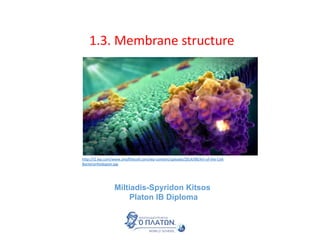
1.3. Membrane structure
Miltiadis-Spyridon Kitsos
Platon IB Diploma
http://i2.wp.com/www.artofthecell.com/wp-content/uploads/2014/08/Art-of-the-Cell-
Bacteriorhodopsin.jpg
The official IB Diploma Biology guide
https://ibpublishing.ibo.org/server2/rest/app/tsm.xql?doc=d_4_biolo_gui_1402_1_e&part=3
&chapter=1
Phospholipids
Phospholipids form bilayers in water due to the amphipathic properties of phospholipid
molecules.
http://carpinteriavalleyassociation.org/wp-
content/uploads/2013/07/Oil-and-Water-multi-racial-
churches2.jpg
This is what happens when water mixes with oil. Can you describe it?
Oil molecules are non-polar while water molecules are polar.
http://www.whatischemistry.unina.it/idrofobica.jpg
https://www.oliveoilsource.com/sites/default/files/basic/formul
as.png
Polar molecules are usually hydrophilic (may mix or dissolve in
water), while non-polar molecules are hydrophobic (the
opposite)
Structure of phospholipids
Phospholipids form bilayers in water due to the amphipathic properties of phospholipid
molecules.
It is an extraordinary molecule since
the:
Phospholipid head (choline,
phosphate group and glycerol) is
hydrophilic (attracts water) while the
phospholipid tail (fatty acid) is
hydrophobic (repels water).
Substances being hydrophilic and
hydrophobic simultaneously are
called amphipathic
How to draw a phospholipid. Individual phospholipid
molecules should be shown using the symbol of a circle
with two parallel lines attached. Don’t forget to indicate
the hydrophilic head and the hydrophobic tail.
hydrophilic head
hydrophobic tail
http://bio1151.nicerweb.com/Locked/media/ch05/05_13PhospholipidStructur.jpg
Micelle and liposomes
Phospholipids form bilayers in water due to the amphipathic properties of phospholipid
molecules.
The amphipathic properties of
phospholipids are responsible for the
development of phospholipid
monolayers, when placed in water
The orientation is simple: the
hydrophilic heads towards water,
hydrophobic tails away from water
These structures are called micelle
How is this related with the emerging
properties of the different levels of
organisation?
http://eng.thesaurus.rusnano.com/upload/iblock/383/micelle1.jpg
Micelle and liposomes
Phospholipids form bilayers in water due to the amphipathic properties of phospholipid
molecules.
Phospholipids may also form bilayers
called liposomes
Liposomes have many applications
in drug delivery. Read more here
http://www.sciencedirect.com/science/article/pii/S0169409X120029
80
In liposomes phospholipids may
exhibit lateral movement within the
layer but not vertical.
https://upload.wikimedia.org/wikipedia/en
/2/28/Liposome.jpg
http://www.mdpi.com/marinedrugs/marinedrugs-12-
06014/article_deploy/html/images/marinedrugs-12-06014-g001-1024.png
http://cancurecancer.org/wp-
content/uploads/2015/09/liposome.png
Theories on the structure of membranes
Using models as representations of the real world—there are alternative models of membrane structure.
1920. Gorter and Grendel hypothesized quite simply that if a plasma membrane was a bilayer then its
surface area should be half that occupied by all its amphipathic lipids spread out in a monolayer.
To prove that they used a Langmuir–Blodgett trough a laboratory apparatus that is able to compress
monolayers of molecules on the surface of water and measures surface phenomena due to this
compression.
https://upload.wikimedia.org/wikipedia/commons/b/b1/LBcompression.jpg
A schematic of a Langmuir Blodgett trough: 1. Amphiphile
monolayer 2. Liquid subphase 3. LB Trough 4. Solid substrate 5.
Dipping mechanism 6. Wilhelmy Plate 7. Electrobalance 8. Barrier
9. Barrier Mechanism 10. Vibration reduction system 11. Clean
room enclosure
https://upload.wikimedia.org/wikipedia/commons/9/9f/LB_trough.jpg
http://cr.middlebury.edu/biology/labbook/membranes/frap/membranes/chap4b.htm
Their initial hypothesis was not falsified. The
plasma membrane of erythrocytes proved to be
a bilayer.
Bilayers (2 layers) vs monolayer (1 layer).
https://upload.wikimedia.org/wikipedia/commons/thumb/c/c8/Lipid_bilayer_and_micelle.sv
g/2000px-Lipid_bilayer_and_micelle.svg.png
Membrane proteins
Membrane proteins are diverse in terms of structure, position in the membrane and function
Proteins play an important role in the
structure of the membrane.
Membrane proteins are divided
according to their position in the
membrane
https://youtu.be/0emD1AmfdjY
http://www.mun.ca/biology/desmid/brian/BIOL2060/BIOL2060-07-08/07_19.jpg
Glycoproteins
Membrane proteins are diverse in terms of structure, position in the membrane and function
Glycoproteins contain
oligosaccharide chains covalently
attached to polypeptide side-chains
[(oligo (few) and saccharide (sugar)]
Important in cell to cell interactions
and as hormone receptors.
https://upload.wikimedia.org/wikipedia/commons/thumb/e/ed/Glicoprot
ein.svg/900px-Glicoprotein.svg.png
https://classconnection.s3.amazonaws.com/811/flashcards/141811/jpg/cellmembrane213270
83769742.jpg
http://www.erin.utoronto.ca/~w3bio315/picts/lectures/lecture2/MembraneGlycoprotein1.jp
g
Transport: Protein channels (facilitated) and protein pumps (active)
Receptors: Peptide-based hormones (insulin, glucagon, etc.)
Anchorage: Cytoskeleton attachments and extracellular matrix
Cell recognition: MHC proteins and antigens
Intercellular joinings: Tight junctions and plasmodesmata
Enzymatic activity: Metabolic pathways (e.g. electron transport chain)
http://www.ib.bioninja.com.au/standard-level/topic-2-cells/24-membranes.html
Slide from
Functions of proteins
Membrane proteins are diverse in terms of structure, position in the membrane and function
Cholesterol
Cholesterol is a component of animal cell membranes
Cholesterol is a steroid molecule (type of
lipid) which plays a significant role in
membrane integrity and fluidity.
http://www.raw-milk-facts.com/images/Cholesterol2.gif
-OH group is polar and
hydrophilic. Attracted by
phosphate heads of bilayer.
Chain of methyl groups
creates a hydrophobic
tail attracted to the
Hydrophobic fatty acid
chains of the
phospholipids
http://biology4ibdp.weebly.com/uploads/9/0/8/0/9080078/608539464.jpg
Cholesterol
Cholesterol in mammalian membranes reduces membrane fluidity and permeability to some
solutes.
http://ib.bioninja.com.au/_Media/membrane-fluidity_med.jpeg
Membrane fluidity refers to the degree of
membrane viscosity and it reflects the
ability of membrane components to move
freely.
There are many factors that determine
Membrane fluidity such as temperature
and the type of lipids
Construct your answers in IB Biology as a series of logical arguments
Example: Outline the role of cholesterol in membrane fluidity
1. In any membrane, phospholipid head usually behave as solids whereas, hydrophobic tails as
liquids.
2. Cholesterol disrupts the packing of tails and thus, prevents them behaving as solid.
3. However, it also limits the movement of components and thus, has a negative effect on
membrane fluidity.
4. Furthermore, reduced the permeability of the membrane to polar molecules (sodium and
hydrogen ions)
Cholesterol
Cholesterol in mammalian membranes reduces membrane fluidity and permeability to some
solutes.
Example 2: Explain why membrane fluidity, needs to be controlled.
1. Increased fluidity would mean less control in membrane permeability.
2. Decreased fluidity would mean decreased ability for
1. cell movement
2. control of substance transfer inside and outside the cell.
https://upload.wikimedia.org/wikipedia/commons/thumb/d/da/Cell_membrane_detailed_di
agram_en.svg/2000px-Cell_membrane_detailed_diagram_en.svg.png
https://www.wisc-online.com//LearningContent/ap1101/index.html
http://www.phschool.com/science/biology_place/biocoach/biomembrane1/regi
ons.html
http://www.bio.davidson.edu/people/macampbell/111/memb-
swf/membranes.swf
Use the tutorials to learn and review membrane
structure
Slide from
Media
Drawing of the fluid mosaic model.
(1) Draw part of the phospholipid bilayer and
label
hydrophilic
heads
Hydrophobic
tails
(2) Draw an integral protein and annotate
hydrophilic
heads
Hydrophobic
tails
Integral protein
(3) Draw another channel protein and
annotate.
(4) Add a peripheral protein. Keep
annotating
Chanel protein
Peripheral protein
Drawing of the fluid mosaic model.
(5) Finally, add a glycoprotein and the cholesterol molecules. Keep annotating
hydrophilic
heads
Hydropho
bic tails
Phospholipids
cholesterol Integral protein Channel protein
Peripheral
protein
glycoprotein
http://www.ib.bioninja.com.au/_Media/phospholipid_bilayer_med.jpeg
• Good use of space
• Clear strong lines
• Label lines are straight
• Labels clearly written
• (Scale bar if appropriate)
• Lines touch the labeled
structure
• No unnecessary shading
or colouring
Reminder of features that make good diagrams:
Slide from
Drawing of the fluid mosaic model.
The concept of the model – The Davson-Danielli model
In science, a model is a representation of an idea, an object or even a process or a
system that is used to describe and explain phenomena that cannot be experienced
directly. Can you think of models in our everyday lives?
In the case of membranes, the extremely
small size does not allow its direct
observation. Thus, the structure of the
membranes is studied through models
which rely on scientific evidence.
Analysis of the falsification of the Davson-Danielli model that led to the Singer-Nicolson model
In the case of membranes, the extremely small size does not allow its direct
observation. Thus, the structure of the membranes is studied through models which
rely on scientific evidence.
Analysis of the falsification of the Davson-Danielli model that led to the Singer-Nicolson
model
The Davson-Danielli Model
Evidence for the development of the method
relied on electron micrographs showing:
(a) two distinct black lines
(b) a lighter band in-between the two black ones.
Interpretation: The two black lines were
considered to be showing proteins and the lighter
band the phospholipid bilayer.
https://natureofscienceib.files.wordpress.com/2015/01/screen
-shot-2015-01-27-at-10-23-00-am.png
Analysis of the falsification of the Davson-Danielli model that led to the Singer-Nicolson
model
This explains: Despite being very thin membranes are an
effective barrier to the movement of certain substances.
The Davson-Danielli Model
The model was developed by 1935 by Hugh Davson and James Danielli.
Basic concepts
• There is a phospholipid bilayer with the
phosphate heads outside and the tails
inside
• The proteins are found as two separate
layers coating the internal and the
external surface of the bilayer
Slide edited from
Analysis of the falsification of the Davson-Danielli model that led to the Singer-Nicolson
model
Evidence falsifying the Davson-Danielli Model
The freeze fracture technique
In the freeze fracturing process, a
sample is frozen and cracked on a
plane through the tissue.
http://www1.udel.edu/biology/Wags/histopage/empage/ecu/ecu14.gif
https://cmrf.research.uiowa.edu/sites/cmrf.research.uiowa.edu/
files/styles/large/public/freezefrac1.gif?itok=7S_MSNoy
Interpreting the image:
• The fracture occurs along
lines of weakness, including
the centre of membranes.
• The fracture reveals an
irregular rough surface
inside the phospholipid
bilayer
• The globular structures
were interpreted as trans-
membrane proteins.
The Davson –Danielli model can not explain the
presence of trans-membrane proteins
Analysis of the falsification of the Davson-Danielli model that led to the Singer-Nicolson
model
Evidence falsifying the Davson-Danielli Model
http://www1.udel.edu/biology/Wags/histopage/empage/ecu/ecu14.gif
Slide edited from
The current model on the membrane structure is the model developed by
Singer and Nicolson in 1972
Key features:
• Phospholipid molecules form a bilayer - phospholipids are fluid and move laterally.
• Peripheral proteins are bound to either the inner or outer surface of the
membrane.
• Integral proteins - permeate the surface of the membrane.
• The membrane is a fluid mosaic of phospholipids and proteins, meaning the
proteins in the bilayer are placed like smaill piecies in a mosaic.
• Proteins can move laterally along membrane.
Analysis of the falsification of the Davson-Danielli model that led to the Singer-Nicolson
model
The Singer-Nicholson fluid mosaic model
Biochemical
techniques
• Membrane proteins
were found to be
very varied in size
and globular in
shape
• Such proteins
would be unable to
form continuous
layers on the
periphery of the
membrane.
• The membrane proteins had hydrophobic regions and
therefore would embed in the membrane not layer the
outside
Slide edited from
Hydrophilic
Hydrophilic Hydrophobic
Analysis of the falsification of the Davson-Danielli model that led to the Singer-Nicolson
model
Evidence supporting the Singer-Nicolson model
http://biochemistry.utoronto.ca/wp-content/uploads/2014/10/Bch422-Moraes-Lecture1-2-
Sept30-2014-21-e1417034958906-670x378.jpg
Fluorescent
antibody tagging
• Within 40 minutes the red and green markers
were mixed throughout the membrane of the
fused cell.
• This showed that membrane proteins are
free to move within the membrane rather
than being fixed in a peripheral layer.
• red or green
fluorescent
markers attached
to antibodies which
would bind to
membrane proteins
• The membrane proteins
of some cells were
tagged with red
markers and other cells
with green markers.
• The cells were fused together.
Analysis of the falsification of the Davson-Danielli model that led to the Singer-Nicolson
model
Evidence supporting the Singer-Nicolson model
http://www.mun.ca/biology/desmid/brian/BIOL2060/BIOL2060-07/07_28.jpg
www.stepsinbiology.com

More Related Content

What's hot

Lipid rafts an overview
Lipid rafts an overviewLipid rafts an overview
Lipid rafts an overviewJaiKumar377
 
types of cleavage, blastula formation, gastrula , morphogenetic movements etc
types of cleavage, blastula formation, gastrula , morphogenetic movements etc types of cleavage, blastula formation, gastrula , morphogenetic movements etc
types of cleavage, blastula formation, gastrula , morphogenetic movements etc Veena Dongare
 
Determination of amphibian axes
Determination of amphibian axesDetermination of amphibian axes
Determination of amphibian axesGauri Haval
 
2.8 cellular respiration
2.8 cellular respiration 2.8 cellular respiration
2.8 cellular respiration Bob Smullen
 
Cell fate (fate maps)
Cell fate (fate maps)Cell fate (fate maps)
Cell fate (fate maps)Shashank Patel
 
Vulva development - Caenorhabditis elegans
Vulva development - Caenorhabditis elegansVulva development - Caenorhabditis elegans
Vulva development - Caenorhabditis elegansnaren
 
Morphogens, induction and cytoplasmic determinants
Morphogens, induction and cytoplasmic determinantsMorphogens, induction and cytoplasmic determinants
Morphogens, induction and cytoplasmic determinantsSakshi Saxena
 
Development of tetrapod limb
Development of tetrapod limbDevelopment of tetrapod limb
Development of tetrapod limbMerlyn Denesia
 
Evolution of photosynthesis
Evolution of photosynthesis Evolution of photosynthesis
Evolution of photosynthesis Bishnu Adhikari
 
Fertilization 1
Fertilization 1Fertilization 1
Fertilization 1Shoeb Ahmad
 
Sponge reproduction notes
Sponge reproduction notesSponge reproduction notes
Sponge reproduction notesericchapman81
 

What's hot (20)

Morphogen gradient
Morphogen gradientMorphogen gradient
Morphogen gradient
 
Lipid rafts an overview
Lipid rafts an overviewLipid rafts an overview
Lipid rafts an overview
 
Cleavage
CleavageCleavage
Cleavage
 
types of cleavage, blastula formation, gastrula , morphogenetic movements etc
types of cleavage, blastula formation, gastrula , morphogenetic movements etc types of cleavage, blastula formation, gastrula , morphogenetic movements etc
types of cleavage, blastula formation, gastrula , morphogenetic movements etc
 
Determination of amphibian axes
Determination of amphibian axesDetermination of amphibian axes
Determination of amphibian axes
 
2.8 cellular respiration
2.8 cellular respiration 2.8 cellular respiration
2.8 cellular respiration
 
Evolution of Urinogenital Ducts
Evolution of Urinogenital DuctsEvolution of Urinogenital Ducts
Evolution of Urinogenital Ducts
 
Cell fate (fate maps)
Cell fate (fate maps)Cell fate (fate maps)
Cell fate (fate maps)
 
FATE Map
FATE MapFATE Map
FATE Map
 
Vulva development - Caenorhabditis elegans
Vulva development - Caenorhabditis elegansVulva development - Caenorhabditis elegans
Vulva development - Caenorhabditis elegans
 
BT 4.1 La Celula en Accion CLEAN.pptx
BT 4.1 La Celula en Accion CLEAN.pptxBT 4.1 La Celula en Accion CLEAN.pptx
BT 4.1 La Celula en Accion CLEAN.pptx
 
Nervous systems in vertebrates: T.Y.B.Sc. Sem VI Notes
Nervous systems in vertebrates: T.Y.B.Sc. Sem VI NotesNervous systems in vertebrates: T.Y.B.Sc. Sem VI Notes
Nervous systems in vertebrates: T.Y.B.Sc. Sem VI Notes
 
Morphogens, induction and cytoplasmic determinants
Morphogens, induction and cytoplasmic determinantsMorphogens, induction and cytoplasmic determinants
Morphogens, induction and cytoplasmic determinants
 
organogenesis ,vulva formation in Caenorhabditis elegans
 organogenesis ,vulva formation in Caenorhabditis elegans  organogenesis ,vulva formation in Caenorhabditis elegans
organogenesis ,vulva formation in Caenorhabditis elegans
 
Development of tetrapod limb
Development of tetrapod limbDevelopment of tetrapod limb
Development of tetrapod limb
 
Fungi botany
Fungi botanyFungi botany
Fungi botany
 
Evolution of photosynthesis
Evolution of photosynthesis Evolution of photosynthesis
Evolution of photosynthesis
 
Aortic arches in vertebrates
Aortic arches in vertebratesAortic arches in vertebrates
Aortic arches in vertebrates
 
Fertilization 1
Fertilization 1Fertilization 1
Fertilization 1
 
Sponge reproduction notes
Sponge reproduction notesSponge reproduction notes
Sponge reproduction notes
 

Viewers also liked

Exercises on microscope magnifications and conversions
Exercises on microscope magnifications and conversionsExercises on microscope magnifications and conversions
Exercises on microscope magnifications and conversionsMiltiadis Kitsos
 
1.4. Sustainability
1.4. Sustainability 1.4. Sustainability
1.4. Sustainability Miltiadis Kitsos
 
Topic 4.2. Energy flow
Topic 4.2. Energy flowTopic 4.2. Energy flow
Topic 4.2. Energy flowMiltiadis Kitsos
 
9.3 Growth in plants
 9.3 Growth in plants 9.3 Growth in plants
9.3 Growth in plantsMiltiadis Kitsos
 
IB Biology Notes on the Individual investigation assessment
IB Biology Notes on the Individual investigation assessment IB Biology Notes on the Individual investigation assessment
IB Biology Notes on the Individual investigation assessment Miltiadis Kitsos
 
Revision questions on 1.3. structure of membranes
Revision questions on 1.3. structure of membranesRevision questions on 1.3. structure of membranes
Revision questions on 1.3. structure of membranesMiltiadis Kitsos
 
1.5. Humans and pollution
1.5. Humans and pollution1.5. Humans and pollution
1.5. Humans and pollutionMiltiadis Kitsos
 
1.3. Energy and equilibria
1.3. Energy and equilibria 1.3. Energy and equilibria
1.3. Energy and equilibria Miltiadis Kitsos
 
2.1. Molecules to Metabolism
 2.1. Molecules to Metabolism 2.1. Molecules to Metabolism
2.1. Molecules to MetabolismMiltiadis Kitsos
 
1.2. Ultrastructure of cells - practise questions
1.2. Ultrastructure of cells - practise questions1.2. Ultrastructure of cells - practise questions
1.2. Ultrastructure of cells - practise questionsMiltiadis Kitsos
 

Viewers also liked (11)

Exercises on microscope magnifications and conversions
Exercises on microscope magnifications and conversionsExercises on microscope magnifications and conversions
Exercises on microscope magnifications and conversions
 
1.4. Sustainability
1.4. Sustainability 1.4. Sustainability
1.4. Sustainability
 
Topic 4.2. Energy flow
Topic 4.2. Energy flowTopic 4.2. Energy flow
Topic 4.2. Energy flow
 
9.3 Growth in plants
 9.3 Growth in plants 9.3 Growth in plants
9.3 Growth in plants
 
IB Biology Notes on the Individual investigation assessment
IB Biology Notes on the Individual investigation assessment IB Biology Notes on the Individual investigation assessment
IB Biology Notes on the Individual investigation assessment
 
Revision questions on 1.3. structure of membranes
Revision questions on 1.3. structure of membranesRevision questions on 1.3. structure of membranes
Revision questions on 1.3. structure of membranes
 
1.5. Humans and pollution
1.5. Humans and pollution1.5. Humans and pollution
1.5. Humans and pollution
 
1.3. Energy and equilibria
1.3. Energy and equilibria 1.3. Energy and equilibria
1.3. Energy and equilibria
 
2.1. Molecules to Metabolism
 2.1. Molecules to Metabolism 2.1. Molecules to Metabolism
2.1. Molecules to Metabolism
 
2.2. Water
2.2. Water 2.2. Water
2.2. Water
 
1.2. Ultrastructure of cells - practise questions
1.2. Ultrastructure of cells - practise questions1.2. Ultrastructure of cells - practise questions
1.2. Ultrastructure of cells - practise questions
 

Similar to 1.3 Membrane Structure

membrane structure
membrane structuremembrane structure
membrane structureWayne Goodwin
 
2-150804182721-lva1-app6891.pdf
2-150804182721-lva1-app6891.pdf2-150804182721-lva1-app6891.pdf
2-150804182721-lva1-app6891.pdfOcquliVictor
 
2.1 How to teach a concept. e
2.1 How to teach a concept. e2.1 How to teach a concept. e
2.1 How to teach a concept. eBelghith Abidi
 
1.3 membrane structure
1.3 membrane structure1.3 membrane structure
1.3 membrane structureBob Smullen
 
IB Biology 1.3 Slides: Membrane Structure
IB Biology 1.3 Slides: Membrane StructureIB Biology 1.3 Slides: Membrane Structure
IB Biology 1.3 Slides: Membrane StructureJacob Cedarbaum
 
1.3 membrane structure
1.3 membrane structure1.3 membrane structure
1.3 membrane structurelucascw
 
IB Biology Topic 1.3 - Cell Membrane Structure
IB Biology Topic 1.3 - Cell Membrane StructureIB Biology Topic 1.3 - Cell Membrane Structure
IB Biology Topic 1.3 - Cell Membrane StructureEran Earland
 
1.3 Cell Membrane
1.3 Cell Membrane1.3 Cell Membrane
1.3 Cell MembraneEran Earland
 
IB Biology Core 2.4: Cell Membranes
IB Biology Core 2.4: Cell MembranesIB Biology Core 2.4: Cell Membranes
IB Biology Core 2.4: Cell MembranesJason de Nys
 
HMP01 02Membrane structure of excitable cell (1).pdf
HMP01 02Membrane structure of excitable cell (1).pdfHMP01 02Membrane structure of excitable cell (1).pdf
HMP01 02Membrane structure of excitable cell (1).pdfkennedymaweu032
 
A and P Mod. #1 Plasma membrane
A and P Mod. #1 Plasma membraneA and P Mod. #1 Plasma membrane
A and P Mod. #1 Plasma membraneKelley Crawford
 
Structure of plasma membrane
Structure of plasma membraneStructure of plasma membrane
Structure of plasma membraneshilpa nair
 
Structure and function of plasma membrane 2
Structure and function of plasma membrane 2Structure and function of plasma membrane 2
Structure and function of plasma membrane 2ICHHA PURAK
 
B9-10 Cell Membranes
B9-10 Cell MembranesB9-10 Cell Membranes
B9-10 Cell Membranessciencechris
 
Unit B9 10 Cell Membranes
Unit B9 10 Cell MembranesUnit B9 10 Cell Membranes
Unit B9 10 Cell Membranessciencechris
 
Bio practise questions unit_1_module_1
Bio practise questions unit_1_module_1Bio practise questions unit_1_module_1
Bio practise questions unit_1_module_1Taj Taylor
 

Similar to 1.3 Membrane Structure (20)

membrane structure
membrane structuremembrane structure
membrane structure
 
2-150804182721-lva1-app6891.pdf
2-150804182721-lva1-app6891.pdf2-150804182721-lva1-app6891.pdf
2-150804182721-lva1-app6891.pdf
 
2.1 How to teach a concept. e
2.1 How to teach a concept. e2.1 How to teach a concept. e
2.1 How to teach a concept. e
 
1.3 membrane structure
1.3 membrane structure1.3 membrane structure
1.3 membrane structure
 
Cell membrane
Cell membraneCell membrane
Cell membrane
 
IB Biology 1.3 Slides: Membrane Structure
IB Biology 1.3 Slides: Membrane StructureIB Biology 1.3 Slides: Membrane Structure
IB Biology 1.3 Slides: Membrane Structure
 
1.3 membrane structure
1.3 membrane structure1.3 membrane structure
1.3 membrane structure
 
IB Biology Topic 1.3 - Cell Membrane Structure
IB Biology Topic 1.3 - Cell Membrane StructureIB Biology Topic 1.3 - Cell Membrane Structure
IB Biology Topic 1.3 - Cell Membrane Structure
 
1.3 Cell Membrane
1.3 Cell Membrane1.3 Cell Membrane
1.3 Cell Membrane
 
MEMBRANE STRUCTURE
MEMBRANE STRUCTUREMEMBRANE STRUCTURE
MEMBRANE STRUCTURE
 
IB Biology Core 2.4: Cell Membranes
IB Biology Core 2.4: Cell MembranesIB Biology Core 2.4: Cell Membranes
IB Biology Core 2.4: Cell Membranes
 
Cell mem ppt
Cell mem pptCell mem ppt
Cell mem ppt
 
HMP01 02Membrane structure of excitable cell (1).pdf
HMP01 02Membrane structure of excitable cell (1).pdfHMP01 02Membrane structure of excitable cell (1).pdf
HMP01 02Membrane structure of excitable cell (1).pdf
 
A and P Mod. #1 Plasma membrane
A and P Mod. #1 Plasma membraneA and P Mod. #1 Plasma membrane
A and P Mod. #1 Plasma membrane
 
Structure of plasma membrane
Structure of plasma membraneStructure of plasma membrane
Structure of plasma membrane
 
Structure and function of plasma membrane 2
Structure and function of plasma membrane 2Structure and function of plasma membrane 2
Structure and function of plasma membrane 2
 
B9-10 Cell Membranes
B9-10 Cell MembranesB9-10 Cell Membranes
B9-10 Cell Membranes
 
Unit B9 10 Cell Membranes
Unit B9 10 Cell MembranesUnit B9 10 Cell Membranes
Unit B9 10 Cell Membranes
 
Cell Membarne ppt.pptx
Cell Membarne ppt.pptxCell Membarne ppt.pptx
Cell Membarne ppt.pptx
 
Bio practise questions unit_1_module_1
Bio practise questions unit_1_module_1Bio practise questions unit_1_module_1
Bio practise questions unit_1_module_1
 

More from Miltiadis Kitsos

Topic 3.1. An introduction to Biodiversity
Topic 3.1. An introduction to Biodiversity Topic 3.1. An introduction to Biodiversity
Topic 3.1. An introduction to Biodiversity Miltiadis Kitsos
 
D.5. hormones and metabolism
 D.5. hormones and metabolism D.5. hormones and metabolism
D.5. hormones and metabolismMiltiadis Kitsos
 
2.1. Species and populations
2.1. Species and populations 2.1. Species and populations
2.1. Species and populations Miltiadis Kitsos
 
2.2. Communities and ecosystems
2.2. Communities and ecosystems 2.2. Communities and ecosystems
2.2. Communities and ecosystems Miltiadis Kitsos
 
Revision questions 2.2. Water
Revision questions 2.2. Water Revision questions 2.2. Water
Revision questions 2.2. Water Miltiadis Kitsos
 
Revision questions on Topic 2.1. From Molecules to Metabolism
Revision questions on Topic 2.1. From Molecules to MetabolismRevision questions on Topic 2.1. From Molecules to Metabolism
Revision questions on Topic 2.1. From Molecules to MetabolismMiltiadis Kitsos
 
Revision questions on 1.6. Cell division of IB Biology
Revision questions on 1.6. Cell division of IB Biology Revision questions on 1.6. Cell division of IB Biology
Revision questions on 1.6. Cell division of IB Biology Miltiadis Kitsos
 
1.5. - The origin of cells
1.5. - The origin of cells 1.5. - The origin of cells
1.5. - The origin of cells Miltiadis Kitsos
 
9.4. Reproduction in plants
 9.4. Reproduction in plants  9.4. Reproduction in plants
9.4. Reproduction in plants Miltiadis Kitsos
 
4.1 Species, communities and ecosystems
 4.1 Species, communities and ecosystems 4.1 Species, communities and ecosystems
4.1 Species, communities and ecosystemsMiltiadis Kitsos
 
1.2. Cell ultrastructure
1.2. Cell ultrastructure1.2. Cell ultrastructure
1.2. Cell ultrastructureMiltiadis Kitsos
 
3.5. Genetic modification and cloning
3.5. Genetic modification and cloning3.5. Genetic modification and cloning
3.5. Genetic modification and cloningMiltiadis Kitsos
 
1.1. Cell theory and microscopy
1.1. Cell theory and microscopy 1.1. Cell theory and microscopy
1.1. Cell theory and microscopy Miltiadis Kitsos
 
Introduction to IB Biology
Introduction to IB BiologyIntroduction to IB Biology
Introduction to IB BiologyMiltiadis Kitsos
 

More from Miltiadis Kitsos (17)

Topic 3.1. An introduction to Biodiversity
Topic 3.1. An introduction to Biodiversity Topic 3.1. An introduction to Biodiversity
Topic 3.1. An introduction to Biodiversity
 
D.5. hormones and metabolism
 D.5. hormones and metabolism D.5. hormones and metabolism
D.5. hormones and metabolism
 
2.1. Species and populations
2.1. Species and populations 2.1. Species and populations
2.1. Species and populations
 
2.2. Communities and ecosystems
2.2. Communities and ecosystems 2.2. Communities and ecosystems
2.2. Communities and ecosystems
 
Revision questions 2.2. Water
Revision questions 2.2. Water Revision questions 2.2. Water
Revision questions 2.2. Water
 
Revision questions on Topic 2.1. From Molecules to Metabolism
Revision questions on Topic 2.1. From Molecules to MetabolismRevision questions on Topic 2.1. From Molecules to Metabolism
Revision questions on Topic 2.1. From Molecules to Metabolism
 
Revision questions on 1.6. Cell division of IB Biology
Revision questions on 1.6. Cell division of IB Biology Revision questions on 1.6. Cell division of IB Biology
Revision questions on 1.6. Cell division of IB Biology
 
1.5. - The origin of cells
1.5. - The origin of cells 1.5. - The origin of cells
1.5. - The origin of cells
 
4.3 Carbon cycling
 4.3 Carbon cycling 4.3 Carbon cycling
4.3 Carbon cycling
 
9.4. Reproduction in plants
 9.4. Reproduction in plants  9.4. Reproduction in plants
9.4. Reproduction in plants
 
4.1 Species, communities and ecosystems
 4.1 Species, communities and ecosystems 4.1 Species, communities and ecosystems
4.1 Species, communities and ecosystems
 
1.2. Cell ultrastructure
1.2. Cell ultrastructure1.2. Cell ultrastructure
1.2. Cell ultrastructure
 
Stem cells
Stem cellsStem cells
Stem cells
 
3.5. Genetic modification and cloning
3.5. Genetic modification and cloning3.5. Genetic modification and cloning
3.5. Genetic modification and cloning
 
1.1. Cell theory and microscopy
1.1. Cell theory and microscopy 1.1. Cell theory and microscopy
1.1. Cell theory and microscopy
 
Introduction to IB Biology
Introduction to IB BiologyIntroduction to IB Biology
Introduction to IB Biology
 
Cas presentation
Cas presentationCas presentation
Cas presentation
 

Recently uploaded

Sanyam Choudhary Chemistry practical.pdf
Sanyam Choudhary Chemistry practical.pdfSanyam Choudhary Chemistry practical.pdf
Sanyam Choudhary Chemistry practical.pdfsanyamsingh5019
 
Introduction to AI in Higher Education_draft.pptx
Introduction to AI in Higher Education_draft.pptxIntroduction to AI in Higher Education_draft.pptx
Introduction to AI in Higher Education_draft.pptxpboyjonauth
 
Mastering the Unannounced Regulatory Inspection
Mastering the Unannounced Regulatory InspectionMastering the Unannounced Regulatory Inspection
Mastering the Unannounced Regulatory InspectionSafetyChain Software
 
Interactive Powerpoint_How to Master effective communication
Interactive Powerpoint_How to Master effective communicationInteractive Powerpoint_How to Master effective communication
Interactive Powerpoint_How to Master effective communicationnomboosow
 
Crayon Activity Handout For the Crayon A
Crayon Activity Handout For the Crayon ACrayon Activity Handout For the Crayon A
Crayon Activity Handout For the Crayon AUnboundStockton
 
How to Configure Email Server in Odoo 17
How to Configure Email Server in Odoo 17How to Configure Email Server in Odoo 17
How to Configure Email Server in Odoo 17Celine George
 
A Critique of the Proposed National Education Policy Reform
A Critique of the Proposed National Education Policy ReformA Critique of the Proposed National Education Policy Reform
A Critique of the Proposed National Education Policy ReformChameera Dedduwage
 
Solving Puzzles Benefits Everyone (English).pptx
Solving Puzzles Benefits Everyone (English).pptxSolving Puzzles Benefits Everyone (English).pptx
Solving Puzzles Benefits Everyone (English).pptxOH TEIK BIN
 
Paris 2024 Olympic Geographies - an activity
Paris 2024 Olympic Geographies - an activityParis 2024 Olympic Geographies - an activity
Paris 2024 Olympic Geographies - an activityGeoBlogs
 
Alper Gobel In Media Res Media Component
Alper Gobel In Media Res Media ComponentAlper Gobel In Media Res Media Component
Alper Gobel In Media Res Media ComponentInMediaRes1
 
Enzyme, Pharmaceutical Aids, Miscellaneous Last Part of Chapter no 5th.pdf
Enzyme, Pharmaceutical Aids, Miscellaneous Last Part of Chapter no 5th.pdfEnzyme, Pharmaceutical Aids, Miscellaneous Last Part of Chapter no 5th.pdf
Enzyme, Pharmaceutical Aids, Miscellaneous Last Part of Chapter no 5th.pdfSumit Tiwari
 
Employee wellbeing at the workplace.pptx
Employee wellbeing at the workplace.pptxEmployee wellbeing at the workplace.pptx
Employee wellbeing at the workplace.pptxNirmalaLoungPoorunde1
 
Accessible design: Minimum effort, maximum impact
Accessible design: Minimum effort, maximum impactAccessible design: Minimum effort, maximum impact
Accessible design: Minimum effort, maximum impactdawncurless
 
Concept of Vouching. B.Com(Hons) /B.Compdf
Concept of Vouching. B.Com(Hons) /B.CompdfConcept of Vouching. B.Com(Hons) /B.Compdf
Concept of Vouching. B.Com(Hons) /B.CompdfUmakantAnnand
 
Kisan Call Centre - To harness potential of ICT in Agriculture by answer farm...
Kisan Call Centre - To harness potential of ICT in Agriculture by answer farm...Kisan Call Centre - To harness potential of ICT in Agriculture by answer farm...
Kisan Call Centre - To harness potential of ICT in Agriculture by answer farm...Krashi Coaching
 
How to Make a Pirate ship Primary Education.pptx
How to Make a Pirate ship Primary Education.pptxHow to Make a Pirate ship Primary Education.pptx
How to Make a Pirate ship Primary Education.pptxmanuelaromero2013
 
KSHARA STURA .pptx---KSHARA KARMA THERAPY (CAUSTIC THERAPY)————IMP.OF KSHARA ...
KSHARA STURA .pptx---KSHARA KARMA THERAPY (CAUSTIC THERAPY)————IMP.OF KSHARA ...KSHARA STURA .pptx---KSHARA KARMA THERAPY (CAUSTIC THERAPY)————IMP.OF KSHARA ...
KSHARA STURA .pptx---KSHARA KARMA THERAPY (CAUSTIC THERAPY)————IMP.OF KSHARA ...M56BOOKSTORE PRODUCT/SERVICE
 

Recently uploaded (20)

TataKelola dan KamSiber Kecerdasan Buatan v022.pdf
TataKelola dan KamSiber Kecerdasan Buatan v022.pdfTataKelola dan KamSiber Kecerdasan Buatan v022.pdf
TataKelola dan KamSiber Kecerdasan Buatan v022.pdf
 
Sanyam Choudhary Chemistry practical.pdf
Sanyam Choudhary Chemistry practical.pdfSanyam Choudhary Chemistry practical.pdf
Sanyam Choudhary Chemistry practical.pdf
 
Introduction to AI in Higher Education_draft.pptx
Introduction to AI in Higher Education_draft.pptxIntroduction to AI in Higher Education_draft.pptx
Introduction to AI in Higher Education_draft.pptx
 
Mastering the Unannounced Regulatory Inspection
Mastering the Unannounced Regulatory InspectionMastering the Unannounced Regulatory Inspection
Mastering the Unannounced Regulatory Inspection
 
Interactive Powerpoint_How to Master effective communication
Interactive Powerpoint_How to Master effective communicationInteractive Powerpoint_How to Master effective communication
Interactive Powerpoint_How to Master effective communication
 
Crayon Activity Handout For the Crayon A
Crayon Activity Handout For the Crayon ACrayon Activity Handout For the Crayon A
Crayon Activity Handout For the Crayon A
 
How to Configure Email Server in Odoo 17
How to Configure Email Server in Odoo 17How to Configure Email Server in Odoo 17
How to Configure Email Server in Odoo 17
 
A Critique of the Proposed National Education Policy Reform
A Critique of the Proposed National Education Policy ReformA Critique of the Proposed National Education Policy Reform
A Critique of the Proposed National Education Policy Reform
 
Solving Puzzles Benefits Everyone (English).pptx
Solving Puzzles Benefits Everyone (English).pptxSolving Puzzles Benefits Everyone (English).pptx
Solving Puzzles Benefits Everyone (English).pptx
 
Paris 2024 Olympic Geographies - an activity
Paris 2024 Olympic Geographies - an activityParis 2024 Olympic Geographies - an activity
Paris 2024 Olympic Geographies - an activity
 
Alper Gobel In Media Res Media Component
Alper Gobel In Media Res Media ComponentAlper Gobel In Media Res Media Component
Alper Gobel In Media Res Media Component
 
Enzyme, Pharmaceutical Aids, Miscellaneous Last Part of Chapter no 5th.pdf
Enzyme, Pharmaceutical Aids, Miscellaneous Last Part of Chapter no 5th.pdfEnzyme, Pharmaceutical Aids, Miscellaneous Last Part of Chapter no 5th.pdf
Enzyme, Pharmaceutical Aids, Miscellaneous Last Part of Chapter no 5th.pdf
 
Model Call Girl in Tilak Nagar Delhi reach out to us at 🔝9953056974🔝
Model Call Girl in Tilak Nagar Delhi reach out to us at 🔝9953056974🔝Model Call Girl in Tilak Nagar Delhi reach out to us at 🔝9953056974🔝
Model Call Girl in Tilak Nagar Delhi reach out to us at 🔝9953056974🔝
 
Employee wellbeing at the workplace.pptx
Employee wellbeing at the workplace.pptxEmployee wellbeing at the workplace.pptx
Employee wellbeing at the workplace.pptx
 
Accessible design: Minimum effort, maximum impact
Accessible design: Minimum effort, maximum impactAccessible design: Minimum effort, maximum impact
Accessible design: Minimum effort, maximum impact
 
Concept of Vouching. B.Com(Hons) /B.Compdf
Concept of Vouching. B.Com(Hons) /B.CompdfConcept of Vouching. B.Com(Hons) /B.Compdf
Concept of Vouching. B.Com(Hons) /B.Compdf
 
Staff of Color (SOC) Retention Efforts DDSD
Staff of Color (SOC) Retention Efforts DDSDStaff of Color (SOC) Retention Efforts DDSD
Staff of Color (SOC) Retention Efforts DDSD
 
Kisan Call Centre - To harness potential of ICT in Agriculture by answer farm...
Kisan Call Centre - To harness potential of ICT in Agriculture by answer farm...Kisan Call Centre - To harness potential of ICT in Agriculture by answer farm...
Kisan Call Centre - To harness potential of ICT in Agriculture by answer farm...
 
How to Make a Pirate ship Primary Education.pptx
How to Make a Pirate ship Primary Education.pptxHow to Make a Pirate ship Primary Education.pptx
How to Make a Pirate ship Primary Education.pptx
 
KSHARA STURA .pptx---KSHARA KARMA THERAPY (CAUSTIC THERAPY)————IMP.OF KSHARA ...
KSHARA STURA .pptx---KSHARA KARMA THERAPY (CAUSTIC THERAPY)————IMP.OF KSHARA ...KSHARA STURA .pptx---KSHARA KARMA THERAPY (CAUSTIC THERAPY)————IMP.OF KSHARA ...
KSHARA STURA .pptx---KSHARA KARMA THERAPY (CAUSTIC THERAPY)————IMP.OF KSHARA ...
 

1.3 Membrane Structure

  • 1. 1.3. Membrane structure Miltiadis-Spyridon Kitsos Platon IB Diploma http://i2.wp.com/www.artofthecell.com/wp-content/uploads/2014/08/Art-of-the-Cell- Bacteriorhodopsin.jpg
  • 2. The official IB Diploma Biology guide https://ibpublishing.ibo.org/server2/rest/app/tsm.xql?doc=d_4_biolo_gui_1402_1_e&part=3 &chapter=1
  • 3. Phospholipids Phospholipids form bilayers in water due to the amphipathic properties of phospholipid molecules. http://carpinteriavalleyassociation.org/wp- content/uploads/2013/07/Oil-and-Water-multi-racial- churches2.jpg This is what happens when water mixes with oil. Can you describe it? Oil molecules are non-polar while water molecules are polar. http://www.whatischemistry.unina.it/idrofobica.jpg https://www.oliveoilsource.com/sites/default/files/basic/formul as.png Polar molecules are usually hydrophilic (may mix or dissolve in water), while non-polar molecules are hydrophobic (the opposite)
  • 4. Structure of phospholipids Phospholipids form bilayers in water due to the amphipathic properties of phospholipid molecules. It is an extraordinary molecule since the: Phospholipid head (choline, phosphate group and glycerol) is hydrophilic (attracts water) while the phospholipid tail (fatty acid) is hydrophobic (repels water). Substances being hydrophilic and hydrophobic simultaneously are called amphipathic How to draw a phospholipid. Individual phospholipid molecules should be shown using the symbol of a circle with two parallel lines attached. Don’t forget to indicate the hydrophilic head and the hydrophobic tail. hydrophilic head hydrophobic tail http://bio1151.nicerweb.com/Locked/media/ch05/05_13PhospholipidStructur.jpg http://bio1151.nicerweb.com/Locked/media/ch05/05_13PhospholipidStructur.jpg
  • 5. Micelle and liposomes Phospholipids form bilayers in water due to the amphipathic properties of phospholipid molecules. The amphipathic properties of phospholipids are responsible for the development of phospholipid monolayers, when placed in water The orientation is simple: the hydrophilic heads towards water, hydrophobic tails away from water These structures are called micelle How is this related with the emerging properties of the different levels of organisation? http://eng.thesaurus.rusnano.com/upload/iblock/383/micelle1.jpg
  • 6. Micelle and liposomes Phospholipids form bilayers in water due to the amphipathic properties of phospholipid molecules. Phospholipids may also form bilayers called liposomes Liposomes have many applications in drug delivery. Read more here http://www.sciencedirect.com/science/article/pii/S0169409X120029 80 In liposomes phospholipids may exhibit lateral movement within the layer but not vertical. https://upload.wikimedia.org/wikipedia/en /2/28/Liposome.jpg http://www.mdpi.com/marinedrugs/marinedrugs-12- 06014/article_deploy/html/images/marinedrugs-12-06014-g001-1024.png http://cancurecancer.org/wp- content/uploads/2015/09/liposome.png
  • 7. Theories on the structure of membranes Using models as representations of the real world—there are alternative models of membrane structure. 1920. Gorter and Grendel hypothesized quite simply that if a plasma membrane was a bilayer then its surface area should be half that occupied by all its amphipathic lipids spread out in a monolayer. To prove that they used a Langmuir–Blodgett trough a laboratory apparatus that is able to compress monolayers of molecules on the surface of water and measures surface phenomena due to this compression. https://upload.wikimedia.org/wikipedia/commons/b/b1/LBcompression.jpg A schematic of a Langmuir Blodgett trough: 1. Amphiphile monolayer 2. Liquid subphase 3. LB Trough 4. Solid substrate 5. Dipping mechanism 6. Wilhelmy Plate 7. Electrobalance 8. Barrier 9. Barrier Mechanism 10. Vibration reduction system 11. Clean room enclosure https://upload.wikimedia.org/wikipedia/commons/9/9f/LB_trough.jpg http://cr.middlebury.edu/biology/labbook/membranes/frap/membranes/chap4b.htm Their initial hypothesis was not falsified. The plasma membrane of erythrocytes proved to be a bilayer. Bilayers (2 layers) vs monolayer (1 layer). https://upload.wikimedia.org/wikipedia/commons/thumb/c/c8/Lipid_bilayer_and_micelle.sv g/2000px-Lipid_bilayer_and_micelle.svg.png
  • 8. Membrane proteins Membrane proteins are diverse in terms of structure, position in the membrane and function Proteins play an important role in the structure of the membrane. Membrane proteins are divided according to their position in the membrane https://youtu.be/0emD1AmfdjY http://www.mun.ca/biology/desmid/brian/BIOL2060/BIOL2060-07-08/07_19.jpg
  • 9. Glycoproteins Membrane proteins are diverse in terms of structure, position in the membrane and function Glycoproteins contain oligosaccharide chains covalently attached to polypeptide side-chains [(oligo (few) and saccharide (sugar)] Important in cell to cell interactions and as hormone receptors. https://upload.wikimedia.org/wikipedia/commons/thumb/e/ed/Glicoprot ein.svg/900px-Glicoprotein.svg.png https://classconnection.s3.amazonaws.com/811/flashcards/141811/jpg/cellmembrane213270 83769742.jpg http://www.erin.utoronto.ca/~w3bio315/picts/lectures/lecture2/MembraneGlycoprotein1.jp g
  • 10. Transport: Protein channels (facilitated) and protein pumps (active) Receptors: Peptide-based hormones (insulin, glucagon, etc.) Anchorage: Cytoskeleton attachments and extracellular matrix Cell recognition: MHC proteins and antigens Intercellular joinings: Tight junctions and plasmodesmata Enzymatic activity: Metabolic pathways (e.g. electron transport chain) http://www.ib.bioninja.com.au/standard-level/topic-2-cells/24-membranes.html Slide from Functions of proteins Membrane proteins are diverse in terms of structure, position in the membrane and function
  • 11. Cholesterol Cholesterol is a component of animal cell membranes Cholesterol is a steroid molecule (type of lipid) which plays a significant role in membrane integrity and fluidity. http://www.raw-milk-facts.com/images/Cholesterol2.gif -OH group is polar and hydrophilic. Attracted by phosphate heads of bilayer. Chain of methyl groups creates a hydrophobic tail attracted to the Hydrophobic fatty acid chains of the phospholipids http://biology4ibdp.weebly.com/uploads/9/0/8/0/9080078/608539464.jpg
  • 12. Cholesterol Cholesterol in mammalian membranes reduces membrane fluidity and permeability to some solutes. http://ib.bioninja.com.au/_Media/membrane-fluidity_med.jpeg Membrane fluidity refers to the degree of membrane viscosity and it reflects the ability of membrane components to move freely. There are many factors that determine Membrane fluidity such as temperature and the type of lipids Construct your answers in IB Biology as a series of logical arguments Example: Outline the role of cholesterol in membrane fluidity 1. In any membrane, phospholipid head usually behave as solids whereas, hydrophobic tails as liquids. 2. Cholesterol disrupts the packing of tails and thus, prevents them behaving as solid. 3. However, it also limits the movement of components and thus, has a negative effect on membrane fluidity. 4. Furthermore, reduced the permeability of the membrane to polar molecules (sodium and hydrogen ions)
  • 13. Cholesterol Cholesterol in mammalian membranes reduces membrane fluidity and permeability to some solutes. Example 2: Explain why membrane fluidity, needs to be controlled. 1. Increased fluidity would mean less control in membrane permeability. 2. Decreased fluidity would mean decreased ability for 1. cell movement 2. control of substance transfer inside and outside the cell. https://upload.wikimedia.org/wikipedia/commons/thumb/d/da/Cell_membrane_detailed_di agram_en.svg/2000px-Cell_membrane_detailed_diagram_en.svg.png
  • 15. Drawing of the fluid mosaic model. (1) Draw part of the phospholipid bilayer and label hydrophilic heads Hydrophobic tails (2) Draw an integral protein and annotate hydrophilic heads Hydrophobic tails Integral protein (3) Draw another channel protein and annotate. (4) Add a peripheral protein. Keep annotating Chanel protein Peripheral protein
  • 16. Drawing of the fluid mosaic model. (5) Finally, add a glycoprotein and the cholesterol molecules. Keep annotating hydrophilic heads Hydropho bic tails Phospholipids cholesterol Integral protein Channel protein Peripheral protein glycoprotein
  • 17. http://www.ib.bioninja.com.au/_Media/phospholipid_bilayer_med.jpeg • Good use of space • Clear strong lines • Label lines are straight • Labels clearly written • (Scale bar if appropriate) • Lines touch the labeled structure • No unnecessary shading or colouring Reminder of features that make good diagrams: Slide from Drawing of the fluid mosaic model.
  • 18. The concept of the model – The Davson-Danielli model In science, a model is a representation of an idea, an object or even a process or a system that is used to describe and explain phenomena that cannot be experienced directly. Can you think of models in our everyday lives? In the case of membranes, the extremely small size does not allow its direct observation. Thus, the structure of the membranes is studied through models which rely on scientific evidence. Analysis of the falsification of the Davson-Danielli model that led to the Singer-Nicolson model In the case of membranes, the extremely small size does not allow its direct observation. Thus, the structure of the membranes is studied through models which rely on scientific evidence.
  • 19. Analysis of the falsification of the Davson-Danielli model that led to the Singer-Nicolson model The Davson-Danielli Model Evidence for the development of the method relied on electron micrographs showing: (a) two distinct black lines (b) a lighter band in-between the two black ones. Interpretation: The two black lines were considered to be showing proteins and the lighter band the phospholipid bilayer. https://natureofscienceib.files.wordpress.com/2015/01/screen -shot-2015-01-27-at-10-23-00-am.png
  • 20. Analysis of the falsification of the Davson-Danielli model that led to the Singer-Nicolson model This explains: Despite being very thin membranes are an effective barrier to the movement of certain substances. The Davson-Danielli Model The model was developed by 1935 by Hugh Davson and James Danielli. Basic concepts • There is a phospholipid bilayer with the phosphate heads outside and the tails inside • The proteins are found as two separate layers coating the internal and the external surface of the bilayer Slide edited from
  • 21. Analysis of the falsification of the Davson-Danielli model that led to the Singer-Nicolson model Evidence falsifying the Davson-Danielli Model The freeze fracture technique In the freeze fracturing process, a sample is frozen and cracked on a plane through the tissue. http://www1.udel.edu/biology/Wags/histopage/empage/ecu/ecu14.gif https://cmrf.research.uiowa.edu/sites/cmrf.research.uiowa.edu/ files/styles/large/public/freezefrac1.gif?itok=7S_MSNoy
  • 22. Interpreting the image: • The fracture occurs along lines of weakness, including the centre of membranes. • The fracture reveals an irregular rough surface inside the phospholipid bilayer • The globular structures were interpreted as trans- membrane proteins. The Davson –Danielli model can not explain the presence of trans-membrane proteins Analysis of the falsification of the Davson-Danielli model that led to the Singer-Nicolson model Evidence falsifying the Davson-Danielli Model http://www1.udel.edu/biology/Wags/histopage/empage/ecu/ecu14.gif Slide edited from
  • 23. The current model on the membrane structure is the model developed by Singer and Nicolson in 1972 Key features: • Phospholipid molecules form a bilayer - phospholipids are fluid and move laterally. • Peripheral proteins are bound to either the inner or outer surface of the membrane. • Integral proteins - permeate the surface of the membrane. • The membrane is a fluid mosaic of phospholipids and proteins, meaning the proteins in the bilayer are placed like smaill piecies in a mosaic. • Proteins can move laterally along membrane. Analysis of the falsification of the Davson-Danielli model that led to the Singer-Nicolson model The Singer-Nicholson fluid mosaic model
  • 24. Biochemical techniques • Membrane proteins were found to be very varied in size and globular in shape • Such proteins would be unable to form continuous layers on the periphery of the membrane. • The membrane proteins had hydrophobic regions and therefore would embed in the membrane not layer the outside Slide edited from Hydrophilic Hydrophilic Hydrophobic Analysis of the falsification of the Davson-Danielli model that led to the Singer-Nicolson model Evidence supporting the Singer-Nicolson model http://biochemistry.utoronto.ca/wp-content/uploads/2014/10/Bch422-Moraes-Lecture1-2- Sept30-2014-21-e1417034958906-670x378.jpg
  • 25. Fluorescent antibody tagging • Within 40 minutes the red and green markers were mixed throughout the membrane of the fused cell. • This showed that membrane proteins are free to move within the membrane rather than being fixed in a peripheral layer. • red or green fluorescent markers attached to antibodies which would bind to membrane proteins • The membrane proteins of some cells were tagged with red markers and other cells with green markers. • The cells were fused together. Analysis of the falsification of the Davson-Danielli model that led to the Singer-Nicolson model Evidence supporting the Singer-Nicolson model http://www.mun.ca/biology/desmid/brian/BIOL2060/BIOL2060-07/07_28.jpg