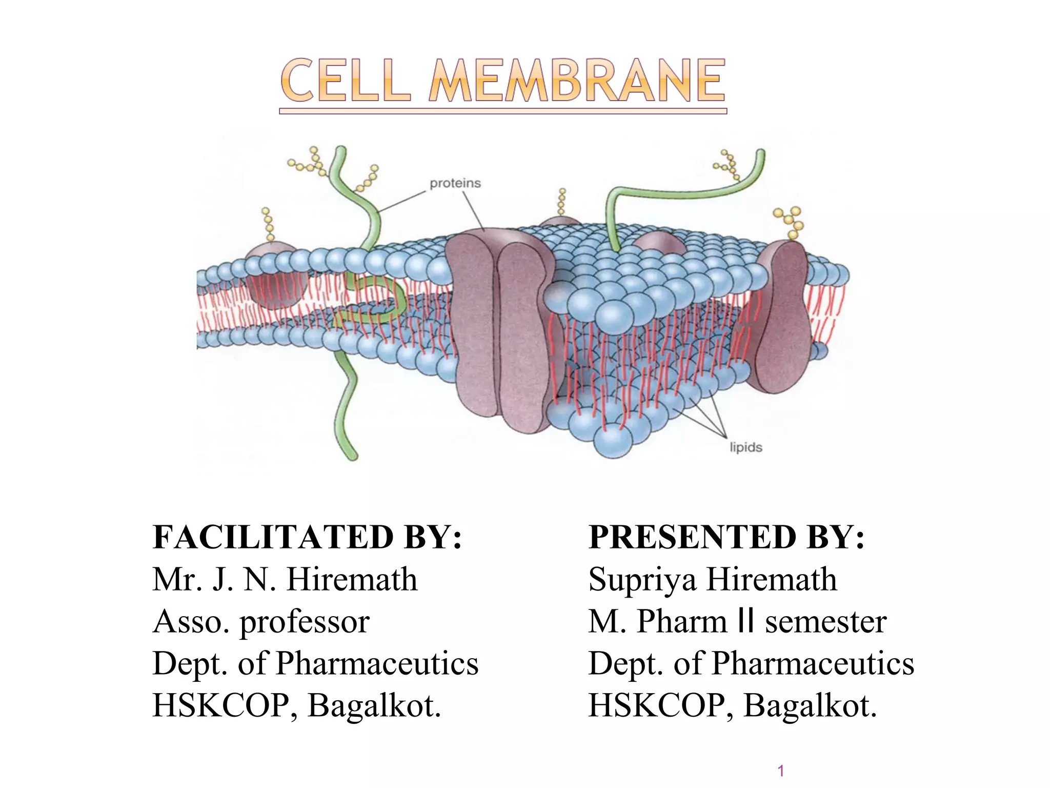
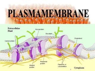
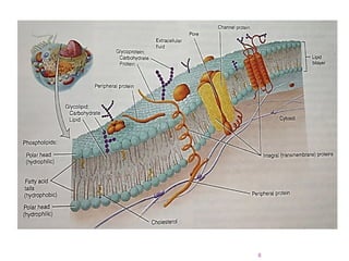
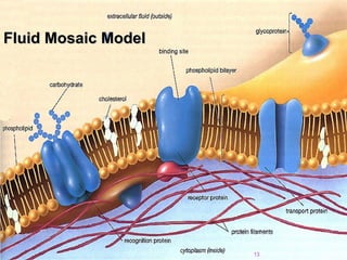
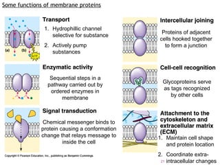
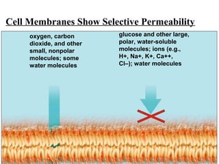
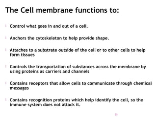
FACILITATED BY:
Mr. J. N. Hiremath
Asso. professor
Dept. of Pharmaceutics
HSKCOP, Bagalkot.
PRESENTED BY:
Supriya Hiremath
M. Pharm semesterⅡ
Dept. of Pharmaceutics
HSKCOP, Bagalkot.
1
 Structure of cell
 Plasma membrane
 Lipid bilayer
 Fluid mosaic model
 Membrane protein
 Functions of membrane protein
 Functions of cell membrane.
2
3
Plasma MembranePlasma Membrane
cytoplasm
plasma
membrane
4
 For a drug to be absorbed and distributed into organs
and tissues and eliminated from the body, it must pass
through one or more biological membranes/barriers at
various locations.
 Such a movement of drug across the membrane is
called as ‘drug transport’.
 The membrane is like a ‘mayonnaise sandwich’.
Structure of cell membrane
5
6
 The cellular membrane consists of a double layer of
amphiphilic phospholipids molecules arranged in such a
fashion that their hydrocarbon chains are oriented
inwards to form the hydrophobic or lipophilic phase and
their polar heads oriented to form the outer and inner
hydrophilic boundaries of the cellular membrane that
face the surrounding aq. Environment.
 Globular protein molecules are associated on either side
of these hydrophilic boundaries.
 The bio-membrane acts like a semipermeable barrier
permitting rapid and limited passage of some compounds
while restricting others.
7
8
The basic structural framework of the plasma membrane is the lipid
bilayer, two back to back layers made up of three types of lipid
molecules-----
# Phospholipids (75% of the membrane lipid)
# Cholesterol ( About 20%)
# Glycolipid (About 5%)
The bilayer arrangement occurs because the lipids are
Amphipathic molecule which means they have both polar and
nonpolar parts.
The polar head is hydrophilic.
The nonpolar parts is hydrophobic.
9
Phospholipids
 Has a hydrophilic
(water loving head)
 Hydrophobic (water
fearing) Tail
 Made up mostly of
carbon (black)&
Hydrogen (light blue)
10
Phospholipid bi-layer
 The cell membrane is made
up of 2 layers of
phospholipids.
 The Hydrophilic heads face
out.
 The Hydrophobic tails face
inward
11
Other Membrane components
Cholesterol makes the cell membrane more stiff and
less permeable.
12
Fluid Mosaic ModelFluid Mosaic Model
13
 Singer and Nicolson developed the fluid mosaic model
 The phospholipids molecules orient themselves in the
bilayer with their hydrophilic heads facing outward. In this
way the heads face a watery fluid on either side – cytosol on
the inside and extracellular fluid on the outside.
 Cholesterol molecules are weakly amphipathic. It is
attached with the polar heads of the phospholipids and
glycolipids.
 Cholesterol may increase or decrease fluidity depending
on other factors, such as the fatty acid composition of the
other lipids found in the membrane.
14
MEMBRANE PROTEIN
It categorized into:
1) Integral protein.
2) Peripheral protein.
3) Glycoprotein.
Integral Protein: It extends into or through the lipid bilayer
among the fatty acid tails and are firmly embedded in it. Most
integral proteins are transmembrane proteins, which means
that they span the entire lipid bilayer and protrude into both the
cytosol and extracellular fluid.
They are Amphipathic. The hydrophilic ends protrude into either
the watery extracellular fluid or the cytosol.
The hydrophobic regions extends among the fatty acid tails.
15
Peripheral Protein:
Are firmly embedded in the membrane. They are
attached more loosely with the polar heads of the
membrane lipids or with the integral Proteins.
Glycoprotein:
It acts as cell identity marker.
16
17
TRANSPORT PROTEINS – regulate the movement of
water-soluble molecules through the cell membrane.
RECEPTOR PROTEINS –recognizes and binds to specific
molecules. Often trigger some response by the cell such as
endocytosis or cell division.
RECOGNITION PROTEINS – identifies the cell as
belonging to a specific species or organ. Helps immune
system tell invading cells from cells belonging to the
organism.
18
Inside of cell
Outside of cell
Some proteins penetrate the
bilayer only partially.
Some proteins adhere one cell
to another.
Some proteins cross
the entire membrane.
Peripheral proteins do not penetrate the
bilayer at all.
19
1.Integral membrane proteins acts as ion channels.
2.Integral membrane proteins acts as transporters.
3.Integral membrane proteins acts as receptors.
4.Integral membrane proteins acts as Linkers.
5.Integral membrane proteins acts as enzymes.
6.Glycoproteins acts as cell identity marker.
7.pheripheral proteins acts as enzymes and linkers.
20
Some functions of membrane proteins
1. Hydrophillic channel
selective for substance
2. Actively pump
substances
Sequential steps in a
pathway carried out by
ordered enzymes in
membrane
Chemical messenger binds to
protein causing a conformation
change that relays message to
inside the cell
Proteins of adjacent
cells hooked together
to form a junction
Glycoproteins serve
as tags recognized
by other cells
1. Maintain cell shape
and protein location
2. Coordinate extra-
intracellular changes21
Cell Membranes Show Selective Permeability
oxygen, carbon
dioxide, and other
small, nonpolar
molecules; some
water molecules
glucose and other large,
polar, water-soluble
molecules; ions (e.g.,
H+, Na+, K+, Ca++,
Cl–); water molecules
22
The Cell membrane functions to:
 Control what goes in and out of a cell.
 Anchors the cytoskeleton to help provide shape.
 Attaches to a substrate outside of the cell or to other cells to help
form tissues
 Controls the transportation of substances across the membrane by
using proteins as carriers and channels
 Contains receptors that allow cells to communicate through chemical
messages
 Contains recognition proteins which help identify the cell, so the
immune system does not attack it.
23
 Principles of anatomy and physiology 12th
edition by G.J.TORTORA & B.H.DERRICKSON
page no: 61 to 100
 https://en.wikibooks.org/wiki/Structural_Biochemistry/
 https://en.wikipedia.org/wiki/Cell_membrane
24
25

cell membrane supriya

  • 1.
    FACILITATED BY: Mr. J.N. Hiremath Asso. professor Dept. of Pharmaceutics HSKCOP, Bagalkot. PRESENTED BY: Supriya Hiremath M. Pharm semesterⅡ Dept. of Pharmaceutics HSKCOP, Bagalkot. 1
  • 2.
     Structure ofcell  Plasma membrane  Lipid bilayer  Fluid mosaic model  Membrane protein  Functions of membrane protein  Functions of cell membrane. 2
  • 3.
  • 4.
  • 5.
     For adrug to be absorbed and distributed into organs and tissues and eliminated from the body, it must pass through one or more biological membranes/barriers at various locations.  Such a movement of drug across the membrane is called as ‘drug transport’.  The membrane is like a ‘mayonnaise sandwich’. Structure of cell membrane 5
  • 6.
  • 7.
     The cellularmembrane consists of a double layer of amphiphilic phospholipids molecules arranged in such a fashion that their hydrocarbon chains are oriented inwards to form the hydrophobic or lipophilic phase and their polar heads oriented to form the outer and inner hydrophilic boundaries of the cellular membrane that face the surrounding aq. Environment.  Globular protein molecules are associated on either side of these hydrophilic boundaries.  The bio-membrane acts like a semipermeable barrier permitting rapid and limited passage of some compounds while restricting others. 7
  • 8.
  • 9.
    The basic structuralframework of the plasma membrane is the lipid bilayer, two back to back layers made up of three types of lipid molecules----- # Phospholipids (75% of the membrane lipid) # Cholesterol ( About 20%) # Glycolipid (About 5%) The bilayer arrangement occurs because the lipids are Amphipathic molecule which means they have both polar and nonpolar parts. The polar head is hydrophilic. The nonpolar parts is hydrophobic. 9
  • 10.
    Phospholipids  Has ahydrophilic (water loving head)  Hydrophobic (water fearing) Tail  Made up mostly of carbon (black)& Hydrogen (light blue) 10
  • 11.
    Phospholipid bi-layer  Thecell membrane is made up of 2 layers of phospholipids.  The Hydrophilic heads face out.  The Hydrophobic tails face inward 11
  • 12.
    Other Membrane components Cholesterolmakes the cell membrane more stiff and less permeable. 12
  • 13.
    Fluid Mosaic ModelFluidMosaic Model 13
  • 14.
     Singer andNicolson developed the fluid mosaic model  The phospholipids molecules orient themselves in the bilayer with their hydrophilic heads facing outward. In this way the heads face a watery fluid on either side – cytosol on the inside and extracellular fluid on the outside.  Cholesterol molecules are weakly amphipathic. It is attached with the polar heads of the phospholipids and glycolipids.  Cholesterol may increase or decrease fluidity depending on other factors, such as the fatty acid composition of the other lipids found in the membrane. 14
  • 15.
    MEMBRANE PROTEIN It categorizedinto: 1) Integral protein. 2) Peripheral protein. 3) Glycoprotein. Integral Protein: It extends into or through the lipid bilayer among the fatty acid tails and are firmly embedded in it. Most integral proteins are transmembrane proteins, which means that they span the entire lipid bilayer and protrude into both the cytosol and extracellular fluid. They are Amphipathic. The hydrophilic ends protrude into either the watery extracellular fluid or the cytosol. The hydrophobic regions extends among the fatty acid tails. 15
  • 16.
    Peripheral Protein: Are firmlyembedded in the membrane. They are attached more loosely with the polar heads of the membrane lipids or with the integral Proteins. Glycoprotein: It acts as cell identity marker. 16
  • 17.
  • 18.
    TRANSPORT PROTEINS –regulate the movement of water-soluble molecules through the cell membrane. RECEPTOR PROTEINS –recognizes and binds to specific molecules. Often trigger some response by the cell such as endocytosis or cell division. RECOGNITION PROTEINS – identifies the cell as belonging to a specific species or organ. Helps immune system tell invading cells from cells belonging to the organism. 18
  • 19.
    Inside of cell Outsideof cell Some proteins penetrate the bilayer only partially. Some proteins adhere one cell to another. Some proteins cross the entire membrane. Peripheral proteins do not penetrate the bilayer at all. 19
  • 20.
    1.Integral membrane proteinsacts as ion channels. 2.Integral membrane proteins acts as transporters. 3.Integral membrane proteins acts as receptors. 4.Integral membrane proteins acts as Linkers. 5.Integral membrane proteins acts as enzymes. 6.Glycoproteins acts as cell identity marker. 7.pheripheral proteins acts as enzymes and linkers. 20
  • 21.
    Some functions ofmembrane proteins 1. Hydrophillic channel selective for substance 2. Actively pump substances Sequential steps in a pathway carried out by ordered enzymes in membrane Chemical messenger binds to protein causing a conformation change that relays message to inside the cell Proteins of adjacent cells hooked together to form a junction Glycoproteins serve as tags recognized by other cells 1. Maintain cell shape and protein location 2. Coordinate extra- intracellular changes21
  • 22.
    Cell Membranes ShowSelective Permeability oxygen, carbon dioxide, and other small, nonpolar molecules; some water molecules glucose and other large, polar, water-soluble molecules; ions (e.g., H+, Na+, K+, Ca++, Cl–); water molecules 22
  • 23.
    The Cell membranefunctions to:  Control what goes in and out of a cell.  Anchors the cytoskeleton to help provide shape.  Attaches to a substrate outside of the cell or to other cells to help form tissues  Controls the transportation of substances across the membrane by using proteins as carriers and channels  Contains receptors that allow cells to communicate through chemical messages  Contains recognition proteins which help identify the cell, so the immune system does not attack it. 23
  • 24.
     Principles ofanatomy and physiology 12th edition by G.J.TORTORA & B.H.DERRICKSON page no: 61 to 100  https://en.wikibooks.org/wiki/Structural_Biochemistry/  https://en.wikipedia.org/wiki/Cell_membrane 24
  • 25.