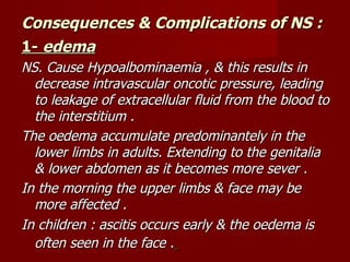
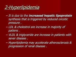
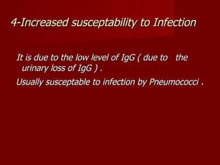
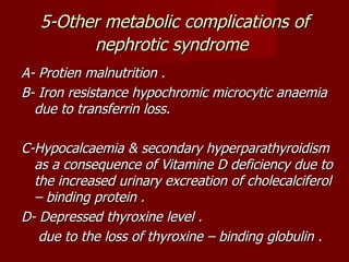
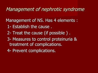
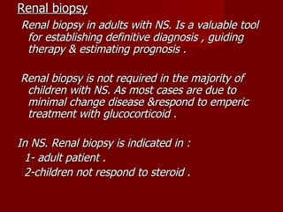
Nephrotic syndrome
Nephrotic syndrome
Nephrotic syndrome is a clinical complex
charecterized by a number of renal and extrarenal
features :
1- protien urea of > 3.5 gram /24 hr.
2- hypoalbominaemia (usually <30 gm/L ).
3- oedema .
4- hyperlipidemia , hyperlipiduria .
5- hypercoagulability .
   The key point for nephrotic syndrom is proteinuria
    (>3.5 gm/24 hr ) .

   The proteinuria result from altered permiability of
    the glumerular filtration barrier for protein (namely
    the GBM & podocytes )

   The other components & metabolic complications of
    nephrotic syndrome are all secondary to protein loss
    in urine .
Consequences & Complications of NS :
1- edema
NS. Cause Hypoalbominaemia , & this results in
  decrease intravascular oncotic pressure, leading
  to leakage of extracellular fluid from the blood to
  the interstitium .
The oedema accumulate predominantely in the
  lower limbs in adults. Extending to the genitalia
  & lower abdomen as it becomes more sever .
In the morning the upper limbs & face may be
  more affected .
In children : ascitis occurs early & the oedema is
  often seen in the face .
2-Hyperlipidemia
– It is due to the increased hepatic lipoprotein
  synthesis that is triggered by reduced oncotic
  pressure .
– LDL & cholestrol are increase in majority of
  patiens .
– VLDL & triglyceride are increase in patients with
  sever disease .
– Hyperlipidemia may accelerate atherosclerosis &
  progression of renal disease .
3-hypercoagulability
   It is multifactorial in origin & caused by :
  1-Increased urinary loss of antithrombin 3 .
  2-Altered level &lor activity of protein C & S .
 3-Hyperfibrinogenemia (due to increased hepatic
  synthesis ) .
4-Impaired fibrinolysis .
5-Increased platelet aggregability .

The patient may develop peripheral arterial or
  venous thrombosis , renal vein thrombosis or
  pulmonary embolism .
Venous thrombosis is common & some time fatal.
4-Increased susceptability to Infection


 It is due to the low level of IgG ( due to the
   urinary loss of IgG ) .
 Usually susceptable to infection by Pneumococci .
5-Other metabolic complications of
         nephrotic syndrome
A- Protien malnutrition .
B- Iron resistance hypochromic microcytic anaemia
  due to transferrin loss.

C-Hypocalcaemia & secondary hyperparathyroidism
  as a consequence of Vitamine D deficiency due to
  the increased urinary excreation of cholecalciferol
  – binding protein .
D- Depressed thyroxine level .
   due to the loss of thyroxine – binding globulin .
Causes of nephrotic syndrome(types)
A- Nephrotic range proteinuria(NS.) with
  bland urine sidement (non-inflammatory)
      (Pure nephrotic )

 1- Primary glomerular disease :
    - minimal change NS.
    - membranous glomerulopathy.
    - focal glomerulosclerosis .

2- Secondary glomerular disease :
   - diabetic nephropathy .
   - amyloidosis .
B- Nephrotic range proteinuria with active
  urine sidement
  (inflammatory,proliferative)
   (mixed nephrotic / nephritic

 1- Primary glomerular disease :
    - membranoproliferative glomerulopathy.

 2- Secondary glomerular disease :
    - membranoproliferative glomerulopathy.
    - SLE .
    - Henoch-Schonlein purpara .
    - Mixed essential cryoglobulinemia .
Management of nephrotic syndrome

Management of NS. Has 4 elements :
1- Establish the cause .
2- Treat the cause (if possible ) .
3- Measures to control proteinuria &
 treatment of complications.
4- Prevent complications.
Renal biopsy
Renal biopsy in adults with NS. Is a valuable tool
 for establishing definitive diagnosis , guiding
 therapy & estimating prognosis .

Renal biopsy is not required in the majority of
 children with NS. As most cases are due to
 minimal change disease &respond to emperic
 treatment with glucocorticoid .

In NS. Renal biopsy is indicated in :
 1- adult patient .
 2-children not respond to steroid .
Lines of management of NS
A- Treatment of proteinuria:
  non specific measure that may reduce
 proteinuria is Angiotensin-converting enzyme
 inhibitors like lisinopril .

 ACE-inhibitors reduce proteinuria & slow the rate
  of progression of renal failure by lowering
  intraglomerular pressure & preventing the
  development of hemodynamically mediated focal
  segmental glomerulosclerosis .
 ACE-inhibitors are renoprotective in diabetic
  nephropathy.
B-Treatment of complications of NS
1-Oedema
-Moderate salt restriction ( 1-2 g/day)

-Diuretics : usually we use loop diuretic (frusemide) . But
 in sever NS & sever oedema combination of diuretics can
 be used acting on different parts of the nephron
 (frusemide , thiazide , amiloride )

We should not remove > 1 Kg. of the oedema per day ,
 as more aggressive diuretics may precipitate
 intravascular volume depletion & pre-renal azotemia .
If the patient have sever hypoalbuminaemia , the
  oedema may be resistant to diuretics & may
  need to give IV albumine by infusion.

Giving albumine in NS. Is some times harmful ,
  because the more albumine you give the more
  will be loss in the urine & this may cause more
  glomerular damage .
2-Hypercholestrolemia
Treated by lipid lowering drugs ( statin
 group ) to prevent accelerated
 atherosclerosis & slow the decline in GFR.
3- Thromboembolism
Anticoagulation is indicated for patients with
 NS. developed DVT , arterial thrombosis or
 pulmonary embolism.
  Also anticoagulation indicated routinly in
 all patients with chronic or sever NS.

There may be a resistance to heparin
 because of antithrombine III deficiency .
4-Infection
Risk of infection can be reduced by immunization
  ( particularly pneumococci).
5- Vit. D deficiency
Vitamin D supplementation is advisable in
 patients with clinical or biochemical
 evidence of vit. D deficiency .
C-Steroid & other immunosupressant
                 drugs
In adults :
  renal biopsy should be done before giving steroid &
   other immunosupressant drugs ( as cyclophosphamide ).
  giving steroid & other immunosupressant drugs depend
   on the result of renal biopsy

In children :
  we can start steroid without renal biops because most
   cases are minimal change type which have a good
   response to steroid .
 renal biopsy is indicated if the child with NS. have no
   response to steroid.

medicine.Renal 2.(dr.kawa)

  • 1.
  • 2.
    Nephrotic syndrome Nephrotic syndromeis a clinical complex charecterized by a number of renal and extrarenal features : 1- protien urea of > 3.5 gram /24 hr. 2- hypoalbominaemia (usually <30 gm/L ). 3- oedema . 4- hyperlipidemia , hyperlipiduria . 5- hypercoagulability .
  • 3.
    The key point for nephrotic syndrom is proteinuria (>3.5 gm/24 hr ) .  The proteinuria result from altered permiability of the glumerular filtration barrier for protein (namely the GBM & podocytes )  The other components & metabolic complications of nephrotic syndrome are all secondary to protein loss in urine .
  • 4.
    Consequences & Complicationsof NS : 1- edema NS. Cause Hypoalbominaemia , & this results in decrease intravascular oncotic pressure, leading to leakage of extracellular fluid from the blood to the interstitium . The oedema accumulate predominantely in the lower limbs in adults. Extending to the genitalia & lower abdomen as it becomes more sever . In the morning the upper limbs & face may be more affected . In children : ascitis occurs early & the oedema is often seen in the face .
  • 5.
    2-Hyperlipidemia – It isdue to the increased hepatic lipoprotein synthesis that is triggered by reduced oncotic pressure . – LDL & cholestrol are increase in majority of patiens . – VLDL & triglyceride are increase in patients with sever disease . – Hyperlipidemia may accelerate atherosclerosis & progression of renal disease .
  • 6.
    3-hypercoagulability  It is multifactorial in origin & caused by : 1-Increased urinary loss of antithrombin 3 . 2-Altered level &lor activity of protein C & S . 3-Hyperfibrinogenemia (due to increased hepatic synthesis ) . 4-Impaired fibrinolysis . 5-Increased platelet aggregability . The patient may develop peripheral arterial or venous thrombosis , renal vein thrombosis or pulmonary embolism . Venous thrombosis is common & some time fatal.
  • 7.
    4-Increased susceptability toInfection It is due to the low level of IgG ( due to the urinary loss of IgG ) . Usually susceptable to infection by Pneumococci .
  • 8.
    5-Other metabolic complicationsof nephrotic syndrome A- Protien malnutrition . B- Iron resistance hypochromic microcytic anaemia due to transferrin loss. C-Hypocalcaemia & secondary hyperparathyroidism as a consequence of Vitamine D deficiency due to the increased urinary excreation of cholecalciferol – binding protein . D- Depressed thyroxine level . due to the loss of thyroxine – binding globulin .
  • 9.
    Causes of nephroticsyndrome(types) A- Nephrotic range proteinuria(NS.) with bland urine sidement (non-inflammatory) (Pure nephrotic ) 1- Primary glomerular disease : - minimal change NS. - membranous glomerulopathy. - focal glomerulosclerosis . 2- Secondary glomerular disease : - diabetic nephropathy . - amyloidosis .
  • 10.
    B- Nephrotic rangeproteinuria with active urine sidement (inflammatory,proliferative) (mixed nephrotic / nephritic 1- Primary glomerular disease : - membranoproliferative glomerulopathy. 2- Secondary glomerular disease : - membranoproliferative glomerulopathy. - SLE . - Henoch-Schonlein purpara . - Mixed essential cryoglobulinemia .
  • 11.
    Management of nephroticsyndrome Management of NS. Has 4 elements : 1- Establish the cause . 2- Treat the cause (if possible ) . 3- Measures to control proteinuria & treatment of complications. 4- Prevent complications.
  • 12.
    Renal biopsy Renal biopsyin adults with NS. Is a valuable tool for establishing definitive diagnosis , guiding therapy & estimating prognosis . Renal biopsy is not required in the majority of children with NS. As most cases are due to minimal change disease &respond to emperic treatment with glucocorticoid . In NS. Renal biopsy is indicated in : 1- adult patient . 2-children not respond to steroid .
  • 13.
    Lines of managementof NS A- Treatment of proteinuria: non specific measure that may reduce proteinuria is Angiotensin-converting enzyme inhibitors like lisinopril . ACE-inhibitors reduce proteinuria & slow the rate of progression of renal failure by lowering intraglomerular pressure & preventing the development of hemodynamically mediated focal segmental glomerulosclerosis . ACE-inhibitors are renoprotective in diabetic nephropathy.
  • 14.
    B-Treatment of complicationsof NS 1-Oedema -Moderate salt restriction ( 1-2 g/day) -Diuretics : usually we use loop diuretic (frusemide) . But in sever NS & sever oedema combination of diuretics can be used acting on different parts of the nephron (frusemide , thiazide , amiloride ) We should not remove > 1 Kg. of the oedema per day , as more aggressive diuretics may precipitate intravascular volume depletion & pre-renal azotemia .
  • 15.
    If the patienthave sever hypoalbuminaemia , the oedema may be resistant to diuretics & may need to give IV albumine by infusion. Giving albumine in NS. Is some times harmful , because the more albumine you give the more will be loss in the urine & this may cause more glomerular damage .
  • 16.
    2-Hypercholestrolemia Treated by lipidlowering drugs ( statin group ) to prevent accelerated atherosclerosis & slow the decline in GFR.
  • 17.
    3- Thromboembolism Anticoagulation isindicated for patients with NS. developed DVT , arterial thrombosis or pulmonary embolism. Also anticoagulation indicated routinly in all patients with chronic or sever NS. There may be a resistance to heparin because of antithrombine III deficiency .
  • 18.
    4-Infection Risk of infectioncan be reduced by immunization ( particularly pneumococci).
  • 19.
    5- Vit. Ddeficiency Vitamin D supplementation is advisable in patients with clinical or biochemical evidence of vit. D deficiency .
  • 20.
    C-Steroid & otherimmunosupressant drugs In adults : renal biopsy should be done before giving steroid & other immunosupressant drugs ( as cyclophosphamide ). giving steroid & other immunosupressant drugs depend on the result of renal biopsy In children : we can start steroid without renal biops because most cases are minimal change type which have a good response to steroid . renal biopsy is indicated if the child with NS. have no response to steroid.