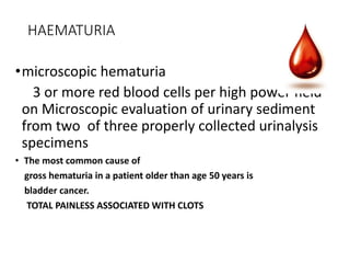
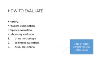
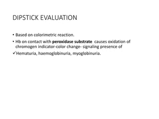
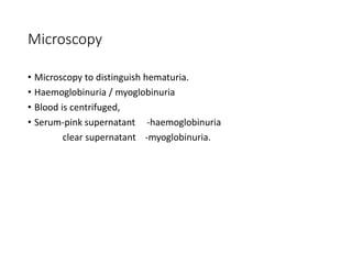
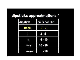
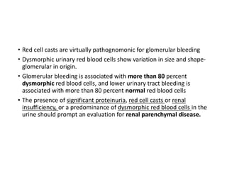
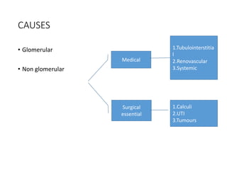
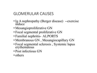
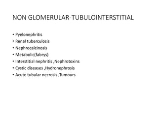
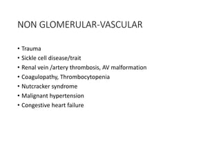
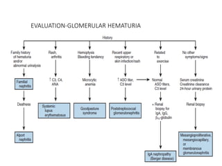
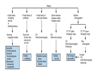
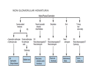
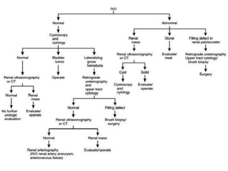
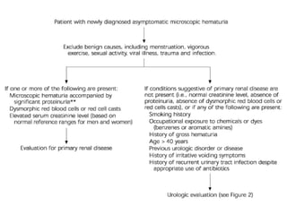
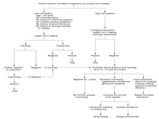
HEMATURIA CAUSES AND
EVALUATION
DR VIPIN SHARMA MCH 1 ST YEAR
CONTENT FOR ASSESSEMENT
• Define microscopic hematuria
• Presentation, Cause of hematuria
• Clots ---upper vs lower tract
• Diffentiation b/w myoglobimuria to hematuria
• Causes of heme negative red urine
• Evaluation-glomerular hematuria
• Excersice induced hematuria warns
• How many years to follow a pt of hematuria
• Basic treatment
HAEMATURIA
•microscopic hematuria
3 or more red blood cells per high power field
on Microscopic evaluation of urinary sediment
from two of three properly collected urinalysis
specimens
• The most common cause of
gross hematuria in a patient older than age 50 years is
bladder cancer.
TOTAL PAINLESS ASSOCIATED WITH CLOTS
WHAT TO ASK FOR ?
• Gross or microscopic.
• Initial or total or terminal.
• Associated loin pain.
• Presence or absence of clot
• Clot characteristics.
BASED ON TIMING
• Indicates the site of origin.
• Initial hematuria - arises from the urethra -least common-secondary
to inflammation.
• Total hematuria – most common -
from the bladder or upper urinary tracts.
• Terminal hematuria- end of micturition, secondary to inflammation in
the bladder neck or prostatic urethra
CLOTS. WHAT IT MEANS ?
amorphous
signifies bladder or prostatic urethral origin.
vermiform (wormlike) clots
associated with flank pain signifies origin from upper urinary tract
with formation of vermiform clots within the ureter.
HOW TO EVALUATE
• History.
• Physical examination.
• Dipstick evaluation.
• Laboratory evaluation
1. Urine microscopy.
2. Sediment evaluation.
3. Asso. proteinuria
1.NO OF RBCS.
2.MORPHOLOGY
3.RBC CASTS
DIPSTICK EVALUATION
• Based on colorimetric reaction.
• Hb on contact with peroxidase substrate causes oxidation of
chromogen indicator-color change- signaling presence of
Hematuria, haemoglobinuria, myoglobinuria.
Microscopy
• Microscopy to distinguish hematuria.
• Haemoglobinuria / myoglobinuria
• Blood is centrifuged,
• Serum-pink supernatant -haemoglobinuria
clear supernatant -myoglobinuria.
• Red cell casts are virtually pathognomonic for glomerular bleeding
• Dysmorphic urinary red blood cells show variation in size and shape-
glomerular in origin.
• Glomerular bleeding is associated with more than 80 percent
dysmorphic red blood cells, and lower urinary tract bleeding is
associated with more than 80 percent normal red blood cells
• The presence of significant proteinuria, red cell casts or renal
insufficiency, or a predominance of dysmorphic red blood cells in the
urine should prompt an evaluation for renal parenchymal disease.
CAUSES
• Glomerular
• Non glomerular
Medical
Surgical
essential
1.Tubulointerstitia
l
2.Renovascular
3.Systemic
1.Calculi
2.UTI
3.Tumours
GLOMERULAR CAUSES
• Ig A nephropathy (Berger disease) --exercise
induce
• Mesangioproliferative GN
• Focal segmental proliferative GN
• Familial nephritis- ALPORTS
• Membranous GN , Mesangiocapillary GN
• Focal segmental sclerosis , Systemic lupus
erythematous
• Post infectious GN
• others
NON GLOMERULAR-TUBULOINTERSTITIAL
• Pyelonephritis
• Renal tuberculosis
• Nephrocalcinosis
• Metabolic(fabrys)
• Interstitial nephritis ,Nephrotoxins
• Cystic diseases ,Hydronephrosis
• Acute tubular necrosis ,Tumours
NON GLOMERULAR-VASCULAR
• Trauma
• Sickle cell disease/trait
• Renal vein /artery thrombosis, AV malformation
• Coagulopathy, Thrombocytopenia
• Nutcracker syndrome
• Malignant hypertension
• Congestive heart failure
NON GLOMERULAR -ESSENTIAL
• Infection
• Hemorrhagic cystitis
• Urethritis
• Nephrolithiasis
• Hypercalciuria
• Obstruction
• Tumor.
DIFFERENCE
NON GLOMERULAR GLOMERULAR
COLOUR RED OR PINK RED,SMOKY BROWN OR COLA
COLOUR
CLOTS MAY BE PRESENT ABSENT
PROTEINURIA < 500 MGS/DAY > 500 MGS/DAY
RBC MORPHOLOGY NORMAL DYSMORPHIC
RBC CASTS ABSENT MAY BE PRESENT
CAUSES OF HEME NEGATIVE RED URINE
DRUGS FOOD DYES METABOLITES
CHLOROQUIN FOOD COLOURING BILE PIGMENTS
DOXORUBICIN BEETS HOMOGENTISIC ACID
DESFEROXAMIN BLACK BERRIES METHHAEMOGLOBIN
IBUPROFEN MELANIN
IRON SORBITOL PORPHYRIN
NITROFURANTOIN TYROSINOSIS
PHENOLPHTHALIN URATES
PHENOZOPYRIDIN
RIFAMPICIN
EVALUATION-GLOMERULAR HEMATURIA
NON GLOMERULAR HEMATURIA
Q
Risk factors for significant disease in
asymptomatic microhaematuria
• Smoking.
• Occupational exposure to chemicals/dyes.
• H/o gross hematuria.
• Age > 40 yrs.
• H/o irritative LUTS.
• H/o urologic disease.
• Analgesic abuse.
• Pelvic irradiation.
ALGORITHM
Isolated hematuria
• Patients with microscopic hematuria, a negative initial urologic
evaluation and no evidence of glomerular bleeding.
• May have structural glomerular abnormalities, they appear to have
low risk for progressive renal disease.
• should be followed for the development of hypertension, renal
insufficiency or proteinuria.
HOW TO PROCEED FURTHER
• Intravenous urography,
• ultrasonography and
• computed tomography
IMAGING APPROPRIATENESS SCALE COMMENTS
MULTI DETECTOR
CT UROGRAPHY
8 METHOD OF CHOICE
IVU 8 BEST INITIAL INV.
USG KUB,ABDOMINAL 6 MISS URETERAL UROTHELIAL
LESIONS
RETROGRADE UROGRAPHY 5
MRI UROGRAPHY 4
CT ABDOMEN PELVIS 4
RENAL ANGIO 4
RADIOGRAPHY KUB 2
MRI ABDOMEN PELVIS 2
SCINTIGRAPHY 2
VIRTUAL CYSTOSCOPY 2
FOLLOW UP
• Immediate urologic reevaluation, with consideration of cystoscopy,
cytology or repeat imaging, should be performed if any of the
following occur:
(1) Gross hematuria,
(2) Abnormal urinary cytology
(3) Irritative voiding symptoms in the absence of infection.
• If none of these occurs within three years, the patient does not
require further urologic monitoring.
INTRACTABLE HAEMATURIA
• Massive uncontrollable hematuria despite irrigation & fulguration.
• Causes
• Radiation cystitis.
• Bladder carcinoma.
• Cyclophosphamide induced cystitis.
• Severe infection
TREATMENT OPTIONS
• Intravesical alum irrigation.
• Intravesical formalin.
• Hydrostatic pressure.
• Embolization.
• Hyperbaric oxygen for radiation cystitis.
• Sodium pentosanpolysulphate for chronic gross hematuria.
• Intravesical PG for cyclophosphamide-induced hematuria.
• First step—clot evacuation.
MESSAGE
• Never ignore any degree of hematuria.
• Proper history taking and judicious use of investigations helps in
reaching a diagnosis.
• Never over react and over investigate.
• Follow standard protocols.
REFERENCE CAMPBELL AND WALSH UROLOGY
Management will be in next presentation

Haematuria causes and evaluation

  • 1.
    HEMATURIA CAUSES AND EVALUATION DRVIPIN SHARMA MCH 1 ST YEAR
  • 2.
    CONTENT FOR ASSESSEMENT •Define microscopic hematuria • Presentation, Cause of hematuria • Clots ---upper vs lower tract • Diffentiation b/w myoglobimuria to hematuria • Causes of heme negative red urine • Evaluation-glomerular hematuria • Excersice induced hematuria warns • How many years to follow a pt of hematuria • Basic treatment
  • 3.
    HAEMATURIA •microscopic hematuria 3 ormore red blood cells per high power field on Microscopic evaluation of urinary sediment from two of three properly collected urinalysis specimens • The most common cause of gross hematuria in a patient older than age 50 years is bladder cancer. TOTAL PAINLESS ASSOCIATED WITH CLOTS
  • 4.
    WHAT TO ASKFOR ? • Gross or microscopic. • Initial or total or terminal. • Associated loin pain. • Presence or absence of clot • Clot characteristics.
  • 5.
    BASED ON TIMING •Indicates the site of origin. • Initial hematuria - arises from the urethra -least common-secondary to inflammation. • Total hematuria – most common - from the bladder or upper urinary tracts. • Terminal hematuria- end of micturition, secondary to inflammation in the bladder neck or prostatic urethra
  • 6.
    CLOTS. WHAT ITMEANS ? amorphous signifies bladder or prostatic urethral origin. vermiform (wormlike) clots associated with flank pain signifies origin from upper urinary tract with formation of vermiform clots within the ureter.
  • 7.
    HOW TO EVALUATE •History. • Physical examination. • Dipstick evaluation. • Laboratory evaluation 1. Urine microscopy. 2. Sediment evaluation. 3. Asso. proteinuria 1.NO OF RBCS. 2.MORPHOLOGY 3.RBC CASTS
  • 8.
    DIPSTICK EVALUATION • Basedon colorimetric reaction. • Hb on contact with peroxidase substrate causes oxidation of chromogen indicator-color change- signaling presence of Hematuria, haemoglobinuria, myoglobinuria.
  • 9.
    Microscopy • Microscopy todistinguish hematuria. • Haemoglobinuria / myoglobinuria • Blood is centrifuged, • Serum-pink supernatant -haemoglobinuria clear supernatant -myoglobinuria.
  • 11.
    • Red cellcasts are virtually pathognomonic for glomerular bleeding • Dysmorphic urinary red blood cells show variation in size and shape- glomerular in origin. • Glomerular bleeding is associated with more than 80 percent dysmorphic red blood cells, and lower urinary tract bleeding is associated with more than 80 percent normal red blood cells • The presence of significant proteinuria, red cell casts or renal insufficiency, or a predominance of dysmorphic red blood cells in the urine should prompt an evaluation for renal parenchymal disease.
  • 12.
    CAUSES • Glomerular • Nonglomerular Medical Surgical essential 1.Tubulointerstitia l 2.Renovascular 3.Systemic 1.Calculi 2.UTI 3.Tumours
  • 13.
    GLOMERULAR CAUSES • IgA nephropathy (Berger disease) --exercise induce • Mesangioproliferative GN • Focal segmental proliferative GN • Familial nephritis- ALPORTS • Membranous GN , Mesangiocapillary GN • Focal segmental sclerosis , Systemic lupus erythematous • Post infectious GN • others
  • 14.
    NON GLOMERULAR-TUBULOINTERSTITIAL • Pyelonephritis •Renal tuberculosis • Nephrocalcinosis • Metabolic(fabrys) • Interstitial nephritis ,Nephrotoxins • Cystic diseases ,Hydronephrosis • Acute tubular necrosis ,Tumours
  • 15.
    NON GLOMERULAR-VASCULAR • Trauma •Sickle cell disease/trait • Renal vein /artery thrombosis, AV malformation • Coagulopathy, Thrombocytopenia • Nutcracker syndrome • Malignant hypertension • Congestive heart failure
  • 16.
    NON GLOMERULAR -ESSENTIAL •Infection • Hemorrhagic cystitis • Urethritis • Nephrolithiasis • Hypercalciuria • Obstruction • Tumor.
  • 17.
    DIFFERENCE NON GLOMERULAR GLOMERULAR COLOURRED OR PINK RED,SMOKY BROWN OR COLA COLOUR CLOTS MAY BE PRESENT ABSENT PROTEINURIA < 500 MGS/DAY > 500 MGS/DAY RBC MORPHOLOGY NORMAL DYSMORPHIC RBC CASTS ABSENT MAY BE PRESENT
  • 18.
    CAUSES OF HEMENEGATIVE RED URINE DRUGS FOOD DYES METABOLITES CHLOROQUIN FOOD COLOURING BILE PIGMENTS DOXORUBICIN BEETS HOMOGENTISIC ACID DESFEROXAMIN BLACK BERRIES METHHAEMOGLOBIN IBUPROFEN MELANIN IRON SORBITOL PORPHYRIN NITROFURANTOIN TYROSINOSIS PHENOLPHTHALIN URATES PHENOZOPYRIDIN RIFAMPICIN
  • 19.
  • 21.
  • 23.
    Risk factors forsignificant disease in asymptomatic microhaematuria • Smoking. • Occupational exposure to chemicals/dyes. • H/o gross hematuria. • Age > 40 yrs. • H/o irritative LUTS. • H/o urologic disease. • Analgesic abuse. • Pelvic irradiation.
  • 24.
  • 26.
    Isolated hematuria • Patientswith microscopic hematuria, a negative initial urologic evaluation and no evidence of glomerular bleeding. • May have structural glomerular abnormalities, they appear to have low risk for progressive renal disease. • should be followed for the development of hypertension, renal insufficiency or proteinuria.
  • 27.
    HOW TO PROCEEDFURTHER • Intravenous urography, • ultrasonography and • computed tomography
  • 28.
    IMAGING APPROPRIATENESS SCALECOMMENTS MULTI DETECTOR CT UROGRAPHY 8 METHOD OF CHOICE IVU 8 BEST INITIAL INV. USG KUB,ABDOMINAL 6 MISS URETERAL UROTHELIAL LESIONS RETROGRADE UROGRAPHY 5 MRI UROGRAPHY 4 CT ABDOMEN PELVIS 4 RENAL ANGIO 4 RADIOGRAPHY KUB 2 MRI ABDOMEN PELVIS 2 SCINTIGRAPHY 2 VIRTUAL CYSTOSCOPY 2
  • 29.
    FOLLOW UP • Immediateurologic reevaluation, with consideration of cystoscopy, cytology or repeat imaging, should be performed if any of the following occur: (1) Gross hematuria, (2) Abnormal urinary cytology (3) Irritative voiding symptoms in the absence of infection. • If none of these occurs within three years, the patient does not require further urologic monitoring.
  • 30.
    INTRACTABLE HAEMATURIA • Massiveuncontrollable hematuria despite irrigation & fulguration. • Causes • Radiation cystitis. • Bladder carcinoma. • Cyclophosphamide induced cystitis. • Severe infection
  • 31.
    TREATMENT OPTIONS • Intravesicalalum irrigation. • Intravesical formalin. • Hydrostatic pressure. • Embolization. • Hyperbaric oxygen for radiation cystitis. • Sodium pentosanpolysulphate for chronic gross hematuria. • Intravesical PG for cyclophosphamide-induced hematuria. • First step—clot evacuation.
  • 32.
    MESSAGE • Never ignoreany degree of hematuria. • Proper history taking and judicious use of investigations helps in reaching a diagnosis. • Never over react and over investigate. • Follow standard protocols.
  • 33.
    REFERENCE CAMPBELL ANDWALSH UROLOGY Management will be in next presentation