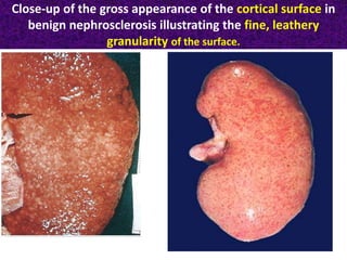
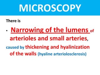
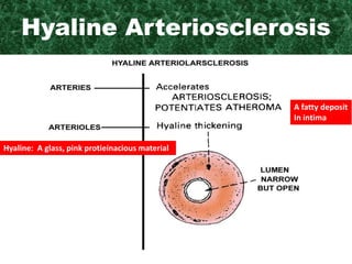
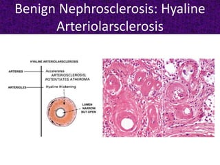
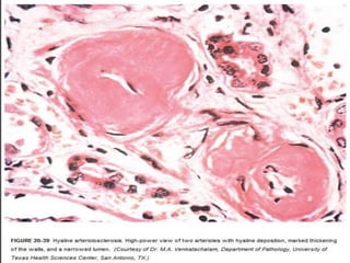
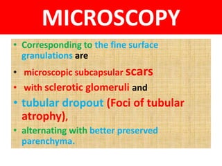
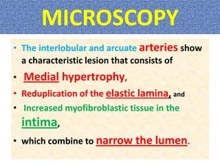
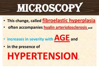
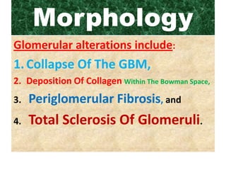
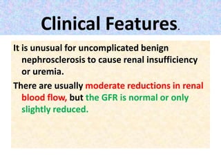
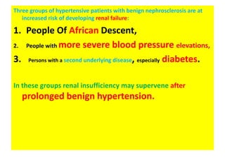
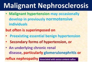
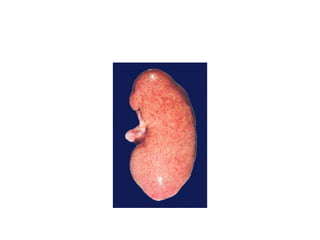
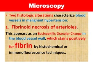
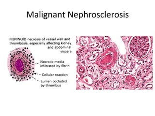
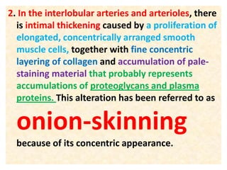
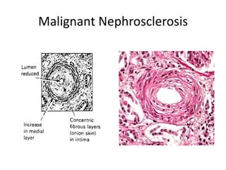
Nephrosclerosis
Lecture 49
Vascular Diseases
Types
• I. Benign Nephrosclerosis
• II. Malignant Nephrosclerosis
BENIGN NEPHROSCLEROSIS
• Benign nephrosclerosis is the term used for
the renal pathology associated with
sclerosis (Hardening) of renal arterioles
and small arteries.
• The resultant effect is focal ischemia
of parenchyma supplied by vessels with
thickened walls and consequent
narrowed lumens.
Benign
NephrosclerosisThe parenchymal effects include
• glomerulosclerosisand
• chronic tubulointersititial injury,
producing a reduction in functional renal mass.
Hypertension and diabetes
mellitus, increase the incidence and
severity of the lesions.
Pathogenesis
Two processes participate in the arterial
lesions:
• I. Medial and intimal thickening,
as a response to hemodynamic changes, aging,
genetic defects, or some combination of
these.
The wall of a blood vessel is composed of 3 layers:
Intima, media & adventitia.
Pathogenesis
II. Hyaline deposition in
arterioles, caused partly by extravasation
of plasma proteins through injured
endothelium and
partly by increased deposition
of basement membrane
matrix.
Gross- Morphology
• The kidneys are either normal or moderately
reduced in size, with average weights
between 110 and 130 gm.
• The cortical surfaces have a fine, even
granularity that resembles grain leather.
• The loss of mass is due mainly to cortical
scarring and shrinking.
The weight of one kidney averages about 120-150 g
Size: 12x 6x3 cm.
Close-up of the gross appearance of the cortical surface in
benign nephrosclerosis illustrating the fine, leathery
granularity of the surface.
MICROSCOPY
There is
• Narrowing of the lumens of
arterioles and small arteries,
caused by thickening and hyalinization
of the walls (hyaline arteriolosclerosis)
Hyaline Arteriosclerosis
A fatty deposit
In intima
Hyaline: A glass, pink protieinacious material
Benign Nephrosclerosis: Hyaline
Arteriolarsclerosis
MICROSCOPY
• Corresponding to the fine surface
granulations are
• microscopic subcapsular scars
• with sclerotic glomeruli and
• tubular dropout (Foci of tubular
atrophy),
• alternating with better preserved
parenchyma.
MICROSCOPY
• The interlobular and arcuate arteries show
a characteristic lesion that consists of
• Medial hypertrophy,
• Reduplication of the elastic lamina, and
• Increased myofibroblastic tissue in the
intima,
• which combine to narrow the lumen.
MICROSCOPY
• This change, called fibroelastic hyperplasia
• often accompanies hyalin arteriolosclerosis and
• increases in severity with AGEand
• in the presence of
HYPERTENSION.
Morphology cont….
There is Patchy Ischemic
Atrophy, which consists of
(1) Foci Of Tubular Atrophy and
Interstitial Fibrosis and
(2) A Variety Of Glomerular Alterations.
Morphology
Glomerular alterations include:
1.Collapse Of The GBM,
2. Deposition Of Collagen Within The Bowman Space,
3. Periglomerular Fibrosis, and
4. Total Sclerosis Of Glomeruli.
Morphology
• When the ischemic changes
are pronounced and affect large areas of
parenchyma, they can produce
regional scars.
Clinical Features.
It is unusual for uncomplicated benign
nephrosclerosis to cause renal insufficiency
or uremia.
There are usually moderate reductions in renal
blood flow, but the GFR is normal or only
slightly reduced.
Three groups of hypertensive patients with benign nephrosclerosis are at
increased risk of developing renal failure:
1. People Of African Descent,
2. People with more severe blood pressure elevations,
3. Persons with a second underlying disease, especially diabetes.
In these groups renal insufficiency may supervene after
prolonged benign hypertension.
Malignant Nephrosclerosis
• Malignant nephrosclerosis is the
form of Renal Disease associated
with The Malignant or Accelerated
Phase of
HYPERTENSION.
Malignant Nephrosclerosis
• Malignant hypertension may occasionally
develop in previously normotensive
individuals
but often is superimposed on
• Preexisting essential benign hypertension
• Secondary forms of hypertension, or
• An underlying chronic renal
disease, particularly glomerulonephritis or
reflux nephropathy Associated with vesico-ureteric reflux
Malignant Nephrosclerosis
• It is also a frequent cause of death from
uremia in individuals with scleroderma.
Malignant hypertension is relatively
uncommon, occurring in 1% to 5% of all
people with elevated blood pressure.
• In its pure form it usually affects younger
individuals, and occurs more often in men
and in blacks.
Pathogenesis- Unclear
• The basis for this turn for the worse (Zawaal)
in hypertensive subjects is
unclear, but the
following sequence of
events is suggested.
• 1.The initial insult seems to be some form of
vascular damage to the kidneys.
This might result
• from long-standing benign hypertension,
with eventual injury to the arteriolar walls, or
• 2. The initiating injury may spring de novo from
arteritis,
• a coagulopathy, or
• some injury causing acute exacerbation of the hypertension.
Pathogenesis
• In any case, the result is
• 1. increased permeability of the small
vessels to fibrinogen and other plasma
proteins,
• 2. endothelial injury,
• 3. focal death of cells of the vascular
wall, and
• 4. platelet deposition.
Pathogenesis
• This leads to the appearance of
1. Fibrinoid necrosis of arterioles and small
arteries,
2. Swelling of the vascular intima, and
3. Intravascular Thrombosis.
Pathogenesis
• Mitogenic factors from platelets (e.g., PDGF),
plasma, and other cells cause hyperplasia of
intimal smooth muscle of vessels, resulting in
the hyperplastic arteriolosclerosis that is
typical of malignant hypertension and further
narrowing of the lumens.
• The kidneys become
markedly ischemic.
Pathogenesis
• With severe involvement of the renal
afferent arterioles, the renin-angiotensin
system receives a powerful stimulus;
indeed, patients with malignant
hypertension have markedly elevated levels
of plasma renin.
Pathogenesis
• This sets up a self-perpetuating cycle in which
angiotensin II causes intrarenal
vasoconstriction, and the attendant renal
ischemia perpetuates renin secretion.
Pathogenesis
• Other vasoconstrictors
(e.g., endothelin) and loss of
vasodilators (nitric oxide) may
also contribute to
vasoconstriction.
Pathogenesis
• Aldosterone levels are also
elevated, and salt retention
undoubtedly contributes to the
elevation of blood
pressure.
Pathogenesis
• The consequences of the markedly elevated
blood pressure on the blood vessels
throughout the body are known as
malignant arteriosclerosis,and
the renal disorder is malignant
nephrosclerosis.
Gross Morphology.
• On gross inspection the kidney size
depends on the duration and severity of the
hypertensive disease.
• Small, pinpoint petechial HEMORRHAGES
may appear on the cortical surface from
RUPTURE of arterioles or glomerular
capillaries, giving the kidney a peculiar
“flea-bitten” appearance.
Microscopy
• Two histologic alterations characterize blood
vessels in malignant hypertension:
1. Fibrinoid necrosis of arterioles.
This appears as an Eosinophilic Granular Change in
the blood vessel wall, which stains positively
for fibrin by histochemical or
immunofluorescence techniques.
Malignant Nephrosclerosis
Microscopy
• This change represents an acute event; it may
be accompanied by limited inflammatory
infiltrate within the wall.
• Sometimes the glomeruli become necrotic
and infiltrated with neutrophils, and the
glomerular capillaries may thrombose.
2. In the interlobular arteries and arterioles, there
is intimal thickening caused by a proliferation of
elongated, concentrically arranged smooth
muscle cells, together with fine concentric
layering of collagen and accumulation of pale-
staining material that probably represents
accumulations of proteoglycans and plasma
proteins. This alteration has been referred to as
onion-skinning
because of its concentric appearance.
Malignant Nephrosclerosis
• The lesion, also called hyperplastic
arteriolitis, correlates well with renal failure
in malignant hypertension.
• There may be superimposed intraluminal
thrombosis.
• The arteriolar and arterial lesions result in
considerable narrowing of all vascular
lumens, ischemic atrophy and, at
times, infarction distal to the abnormal
vessels.
Clinical Features
• SP>200 mm Hg & DP>120 mm Hg,
• Papilledema,
• Retinal hemorrhages,
• Encephalopathy,
• Cardiovascular abnormalities, &
• Renal failure.
Clinical features
• Most often, the early symptoms are related to
increased intracranial pressure and include
• headaches,
• nausea,
• vomiting, and
• visual impairments,
particularly scotomas or spots before the eyes.
Clinical Features
•Hypertensive crises
are sometimes encountered, characterized by
• episodes of loss of consciousness or
even convulsions.
Clinical Features
• At the onset of rapidly mounting blood
pressure, there is marked proteinuria
and microscopic or sometimes macroscopic
hematuria but no significant alteration
in renal function.
• Soon, however, renal failure makes its
appearance.
Treatment
• The syndrome is
• a true medical emergency requiring
the institution of aggressive and prompt
antihypertensive therapy to
prevent the development of irreversible renal
lesions.
Prognosis
• Before the introduction of current
antihypertensive drugs, malignant
hypertension was associated with a
• 50% mortality rate within 3 months of
onset, progressing to 90%within a year.
Prognosis
• At present, however,
• about 75% of patients survive 5
years, and 50% survive with
restoration of pre-crisis renal
function.
Nephrosclerosis

Nephrosclerosis

  • 1.
  • 2.
    Types • I. BenignNephrosclerosis • II. Malignant Nephrosclerosis
  • 3.
    BENIGN NEPHROSCLEROSIS • Benignnephrosclerosis is the term used for the renal pathology associated with sclerosis (Hardening) of renal arterioles and small arteries. • The resultant effect is focal ischemia of parenchyma supplied by vessels with thickened walls and consequent narrowed lumens.
  • 4.
    Benign NephrosclerosisThe parenchymal effectsinclude • glomerulosclerosisand • chronic tubulointersititial injury, producing a reduction in functional renal mass. Hypertension and diabetes mellitus, increase the incidence and severity of the lesions.
  • 5.
    Pathogenesis Two processes participatein the arterial lesions: • I. Medial and intimal thickening, as a response to hemodynamic changes, aging, genetic defects, or some combination of these. The wall of a blood vessel is composed of 3 layers: Intima, media & adventitia.
  • 6.
    Pathogenesis II. Hyaline depositionin arterioles, caused partly by extravasation of plasma proteins through injured endothelium and partly by increased deposition of basement membrane matrix.
  • 7.
    Gross- Morphology • Thekidneys are either normal or moderately reduced in size, with average weights between 110 and 130 gm. • The cortical surfaces have a fine, even granularity that resembles grain leather. • The loss of mass is due mainly to cortical scarring and shrinking. The weight of one kidney averages about 120-150 g Size: 12x 6x3 cm.
  • 8.
    Close-up of thegross appearance of the cortical surface in benign nephrosclerosis illustrating the fine, leathery granularity of the surface.
  • 9.
    MICROSCOPY There is • Narrowingof the lumens of arterioles and small arteries, caused by thickening and hyalinization of the walls (hyaline arteriolosclerosis)
  • 10.
    Hyaline Arteriosclerosis A fattydeposit In intima Hyaline: A glass, pink protieinacious material
  • 11.
  • 13.
    MICROSCOPY • Corresponding tothe fine surface granulations are • microscopic subcapsular scars • with sclerotic glomeruli and • tubular dropout (Foci of tubular atrophy), • alternating with better preserved parenchyma.
  • 14.
    MICROSCOPY • The interlobularand arcuate arteries show a characteristic lesion that consists of • Medial hypertrophy, • Reduplication of the elastic lamina, and • Increased myofibroblastic tissue in the intima, • which combine to narrow the lumen.
  • 15.
    MICROSCOPY • This change,called fibroelastic hyperplasia • often accompanies hyalin arteriolosclerosis and • increases in severity with AGEand • in the presence of HYPERTENSION.
  • 16.
    Morphology cont…. There isPatchy Ischemic Atrophy, which consists of (1) Foci Of Tubular Atrophy and Interstitial Fibrosis and (2) A Variety Of Glomerular Alterations.
  • 17.
    Morphology Glomerular alterations include: 1.CollapseOf The GBM, 2. Deposition Of Collagen Within The Bowman Space, 3. Periglomerular Fibrosis, and 4. Total Sclerosis Of Glomeruli.
  • 18.
    Morphology • When theischemic changes are pronounced and affect large areas of parenchyma, they can produce regional scars.
  • 19.
    Clinical Features. It isunusual for uncomplicated benign nephrosclerosis to cause renal insufficiency or uremia. There are usually moderate reductions in renal blood flow, but the GFR is normal or only slightly reduced.
  • 20.
    Three groups ofhypertensive patients with benign nephrosclerosis are at increased risk of developing renal failure: 1. People Of African Descent, 2. People with more severe blood pressure elevations, 3. Persons with a second underlying disease, especially diabetes. In these groups renal insufficiency may supervene after prolonged benign hypertension.
  • 22.
    Malignant Nephrosclerosis • Malignantnephrosclerosis is the form of Renal Disease associated with The Malignant or Accelerated Phase of HYPERTENSION.
  • 23.
    Malignant Nephrosclerosis • Malignanthypertension may occasionally develop in previously normotensive individuals but often is superimposed on • Preexisting essential benign hypertension • Secondary forms of hypertension, or • An underlying chronic renal disease, particularly glomerulonephritis or reflux nephropathy Associated with vesico-ureteric reflux
  • 24.
    Malignant Nephrosclerosis • Itis also a frequent cause of death from uremia in individuals with scleroderma. Malignant hypertension is relatively uncommon, occurring in 1% to 5% of all people with elevated blood pressure. • In its pure form it usually affects younger individuals, and occurs more often in men and in blacks.
  • 25.
    Pathogenesis- Unclear • Thebasis for this turn for the worse (Zawaal) in hypertensive subjects is unclear, but the following sequence of events is suggested.
  • 26.
    • 1.The initialinsult seems to be some form of vascular damage to the kidneys. This might result • from long-standing benign hypertension, with eventual injury to the arteriolar walls, or • 2. The initiating injury may spring de novo from arteritis, • a coagulopathy, or • some injury causing acute exacerbation of the hypertension.
  • 27.
    Pathogenesis • In anycase, the result is • 1. increased permeability of the small vessels to fibrinogen and other plasma proteins, • 2. endothelial injury, • 3. focal death of cells of the vascular wall, and • 4. platelet deposition.
  • 28.
    Pathogenesis • This leadsto the appearance of 1. Fibrinoid necrosis of arterioles and small arteries, 2. Swelling of the vascular intima, and 3. Intravascular Thrombosis.
  • 29.
    Pathogenesis • Mitogenic factorsfrom platelets (e.g., PDGF), plasma, and other cells cause hyperplasia of intimal smooth muscle of vessels, resulting in the hyperplastic arteriolosclerosis that is typical of malignant hypertension and further narrowing of the lumens. • The kidneys become markedly ischemic.
  • 30.
    Pathogenesis • With severeinvolvement of the renal afferent arterioles, the renin-angiotensin system receives a powerful stimulus; indeed, patients with malignant hypertension have markedly elevated levels of plasma renin.
  • 31.
    Pathogenesis • This setsup a self-perpetuating cycle in which angiotensin II causes intrarenal vasoconstriction, and the attendant renal ischemia perpetuates renin secretion.
  • 32.
    Pathogenesis • Other vasoconstrictors (e.g.,endothelin) and loss of vasodilators (nitric oxide) may also contribute to vasoconstriction.
  • 33.
    Pathogenesis • Aldosterone levelsare also elevated, and salt retention undoubtedly contributes to the elevation of blood pressure.
  • 34.
    Pathogenesis • The consequencesof the markedly elevated blood pressure on the blood vessels throughout the body are known as malignant arteriosclerosis,and the renal disorder is malignant nephrosclerosis.
  • 35.
    Gross Morphology. • Ongross inspection the kidney size depends on the duration and severity of the hypertensive disease. • Small, pinpoint petechial HEMORRHAGES may appear on the cortical surface from RUPTURE of arterioles or glomerular capillaries, giving the kidney a peculiar “flea-bitten” appearance.
  • 37.
    Microscopy • Two histologicalterations characterize blood vessels in malignant hypertension: 1. Fibrinoid necrosis of arterioles. This appears as an Eosinophilic Granular Change in the blood vessel wall, which stains positively for fibrin by histochemical or immunofluorescence techniques.
  • 38.
  • 39.
    Microscopy • This changerepresents an acute event; it may be accompanied by limited inflammatory infiltrate within the wall. • Sometimes the glomeruli become necrotic and infiltrated with neutrophils, and the glomerular capillaries may thrombose.
  • 40.
    2. In theinterlobular arteries and arterioles, there is intimal thickening caused by a proliferation of elongated, concentrically arranged smooth muscle cells, together with fine concentric layering of collagen and accumulation of pale- staining material that probably represents accumulations of proteoglycans and plasma proteins. This alteration has been referred to as onion-skinning because of its concentric appearance.
  • 41.
  • 42.
    • The lesion,also called hyperplastic arteriolitis, correlates well with renal failure in malignant hypertension. • There may be superimposed intraluminal thrombosis. • The arteriolar and arterial lesions result in considerable narrowing of all vascular lumens, ischemic atrophy and, at times, infarction distal to the abnormal vessels.
  • 43.
    Clinical Features • SP>200mm Hg & DP>120 mm Hg, • Papilledema, • Retinal hemorrhages, • Encephalopathy, • Cardiovascular abnormalities, & • Renal failure.
  • 44.
    Clinical features • Mostoften, the early symptoms are related to increased intracranial pressure and include • headaches, • nausea, • vomiting, and • visual impairments, particularly scotomas or spots before the eyes.
  • 45.
    Clinical Features •Hypertensive crises aresometimes encountered, characterized by • episodes of loss of consciousness or even convulsions.
  • 46.
    Clinical Features • Atthe onset of rapidly mounting blood pressure, there is marked proteinuria and microscopic or sometimes macroscopic hematuria but no significant alteration in renal function. • Soon, however, renal failure makes its appearance.
  • 47.
    Treatment • The syndromeis • a true medical emergency requiring the institution of aggressive and prompt antihypertensive therapy to prevent the development of irreversible renal lesions.
  • 48.
    Prognosis • Before theintroduction of current antihypertensive drugs, malignant hypertension was associated with a • 50% mortality rate within 3 months of onset, progressing to 90%within a year.
  • 49.
    Prognosis • At present,however, • about 75% of patients survive 5 years, and 50% survive with restoration of pre-crisis renal function.