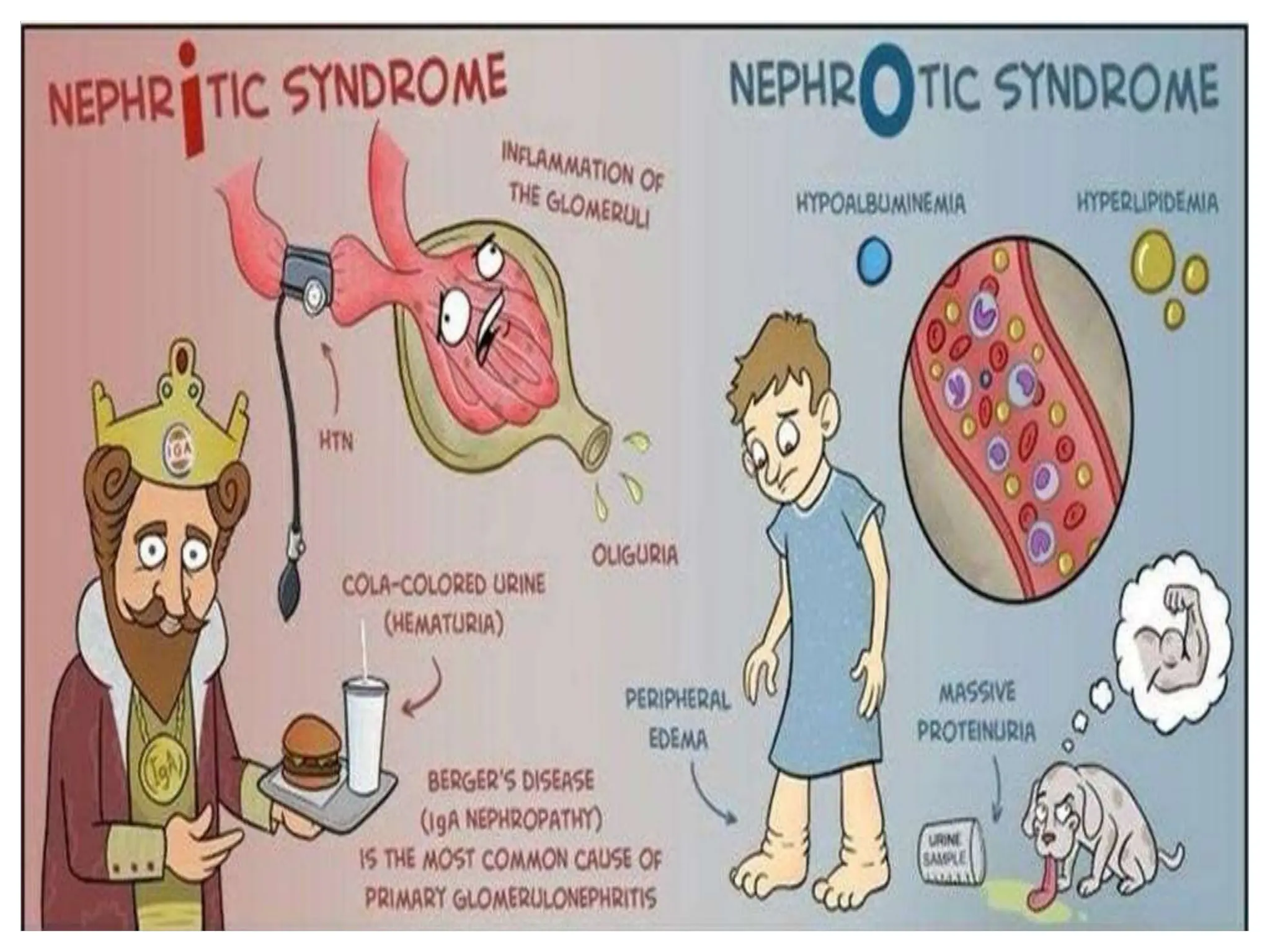
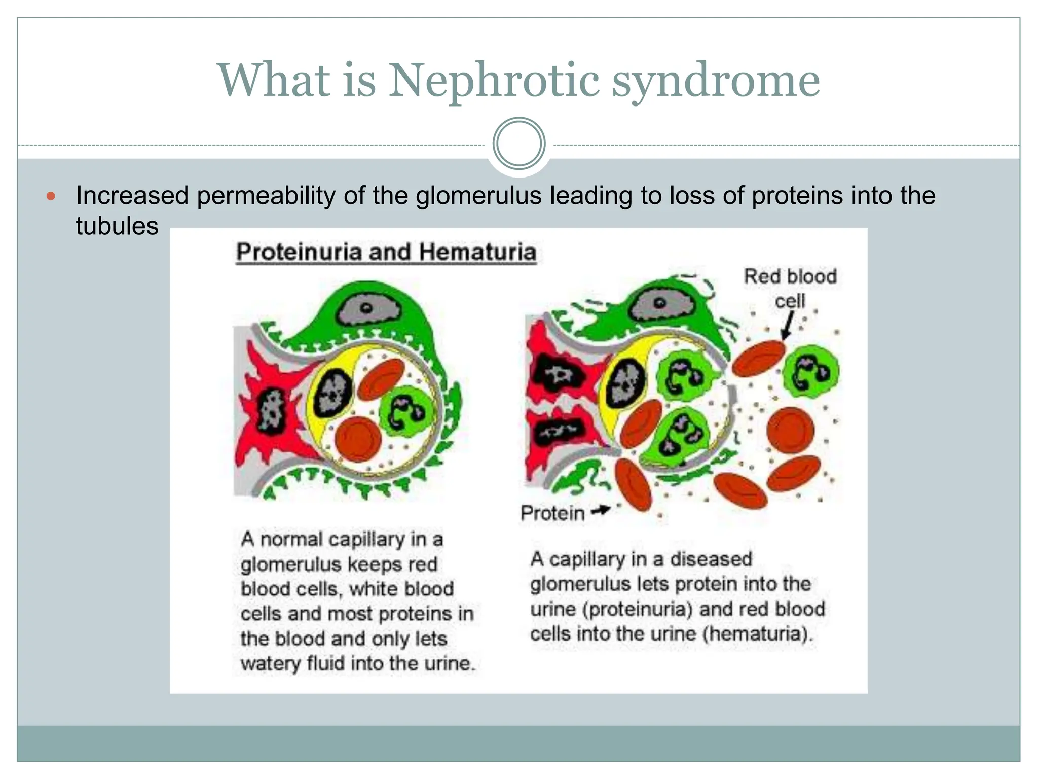
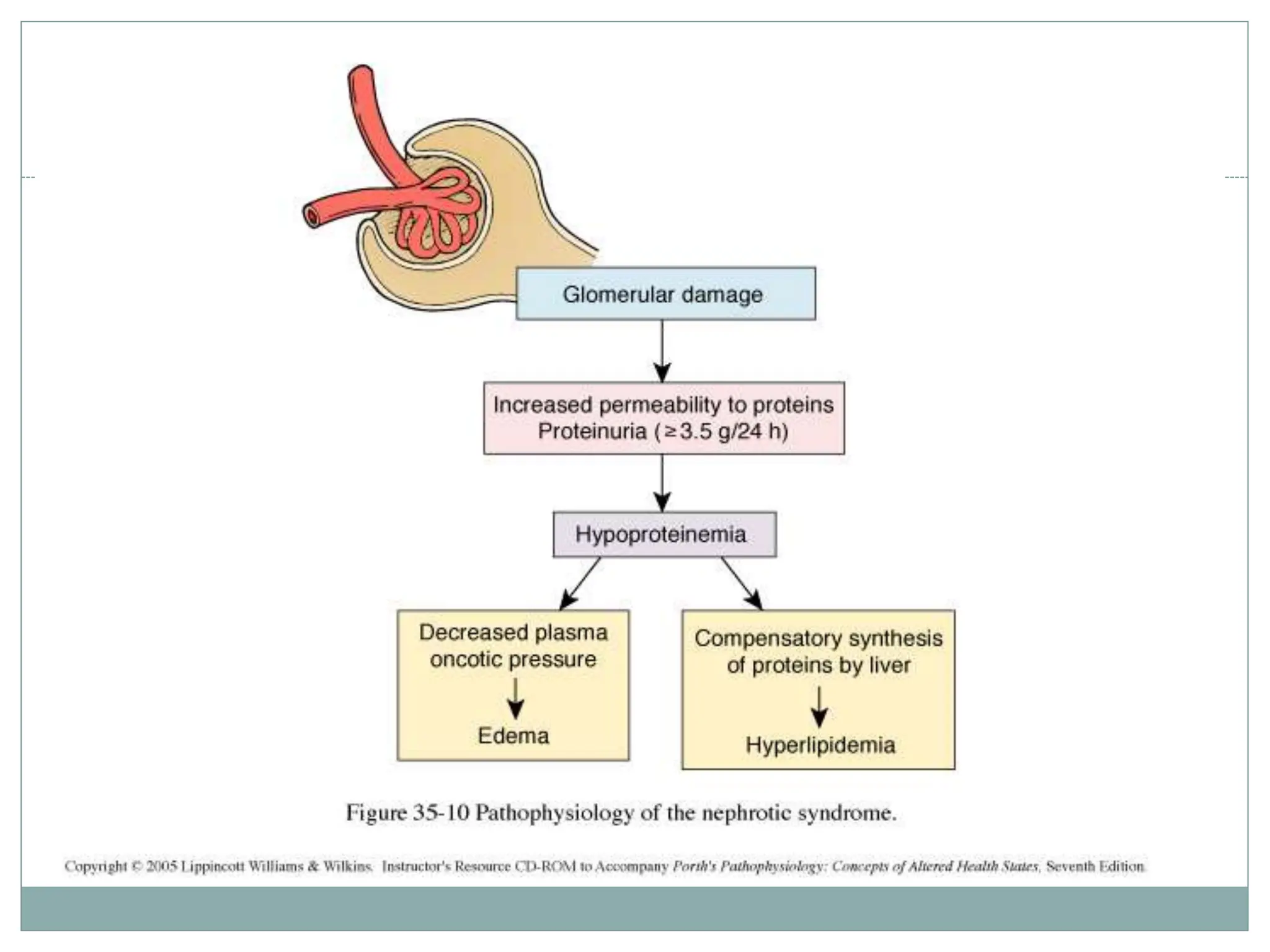
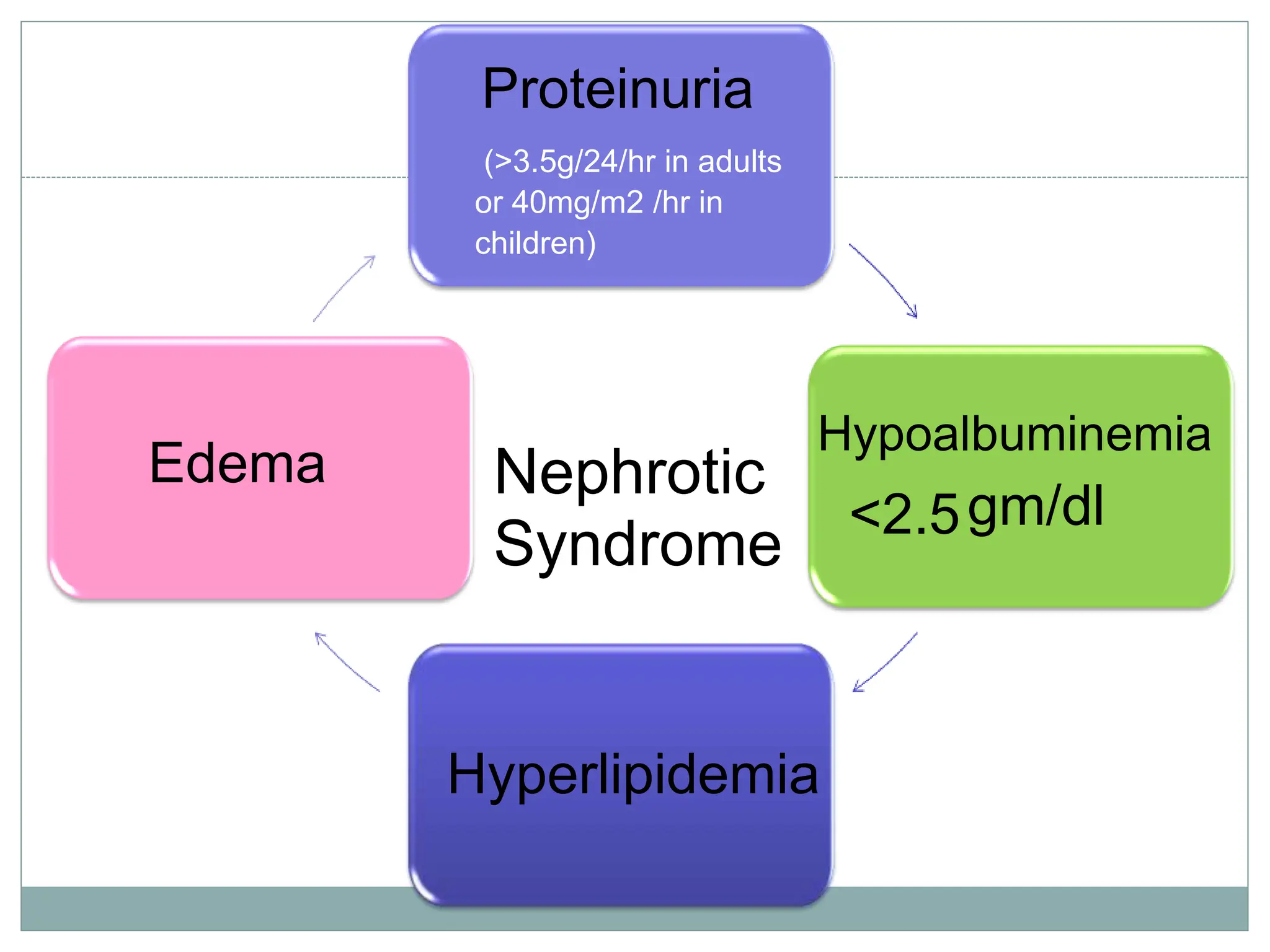
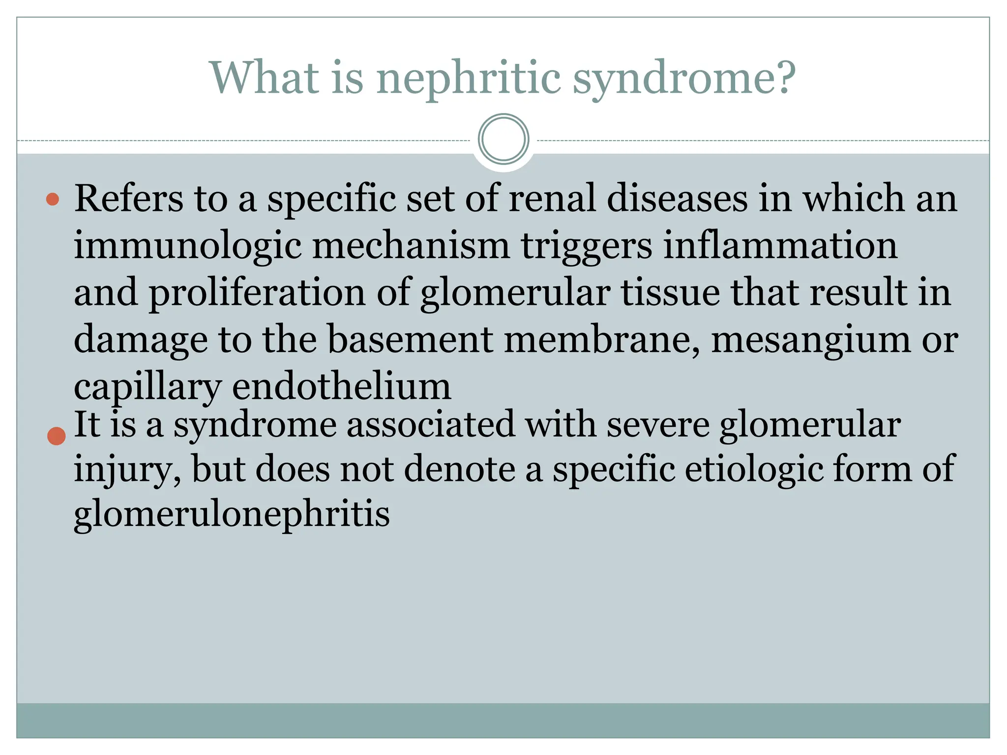
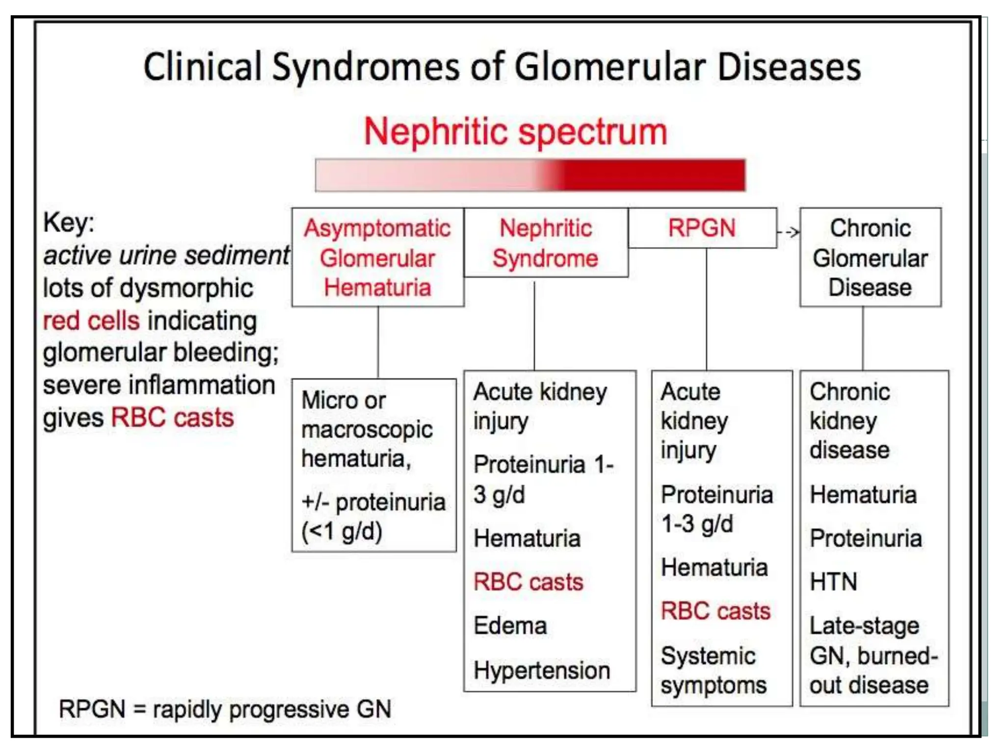
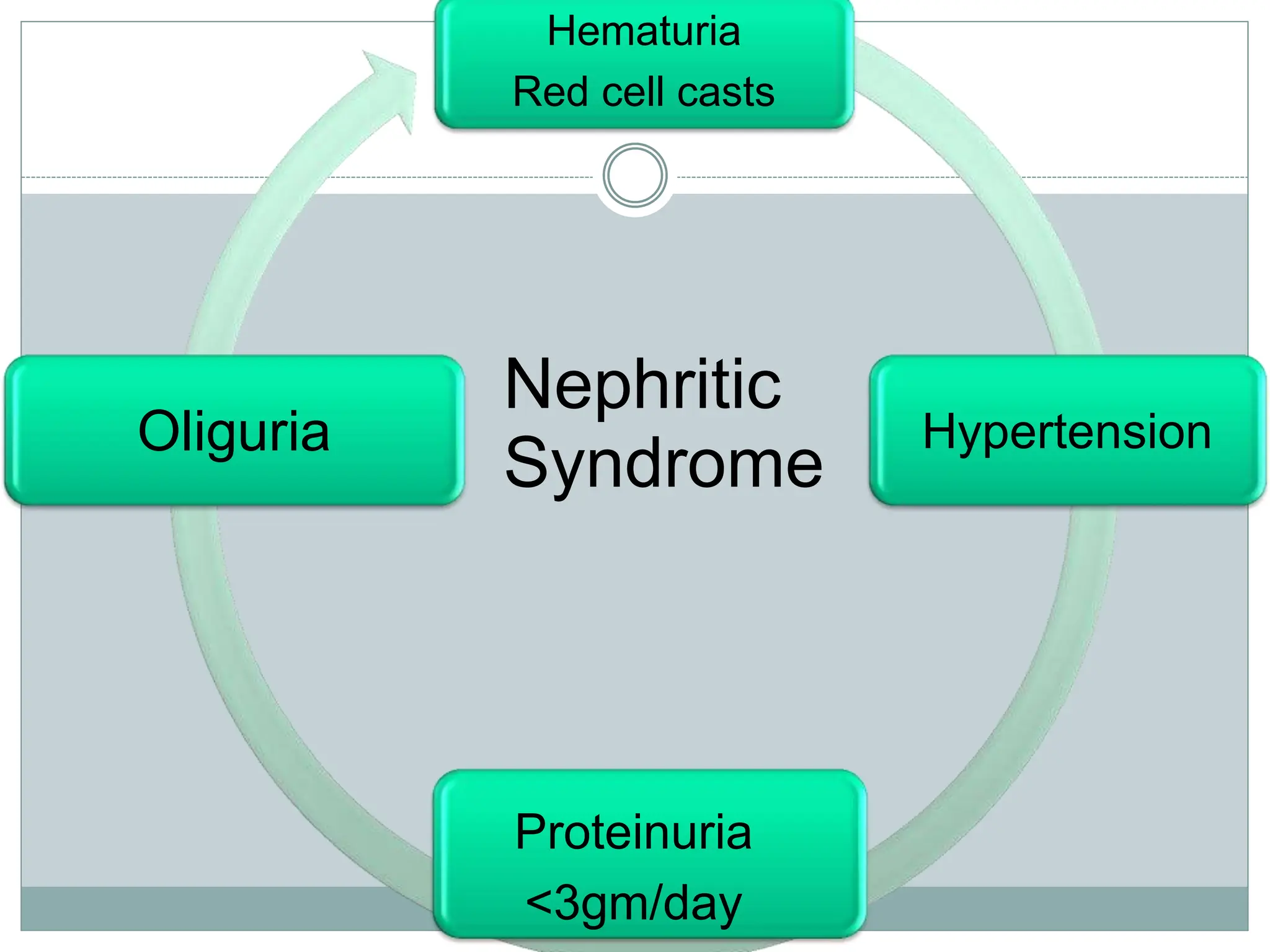
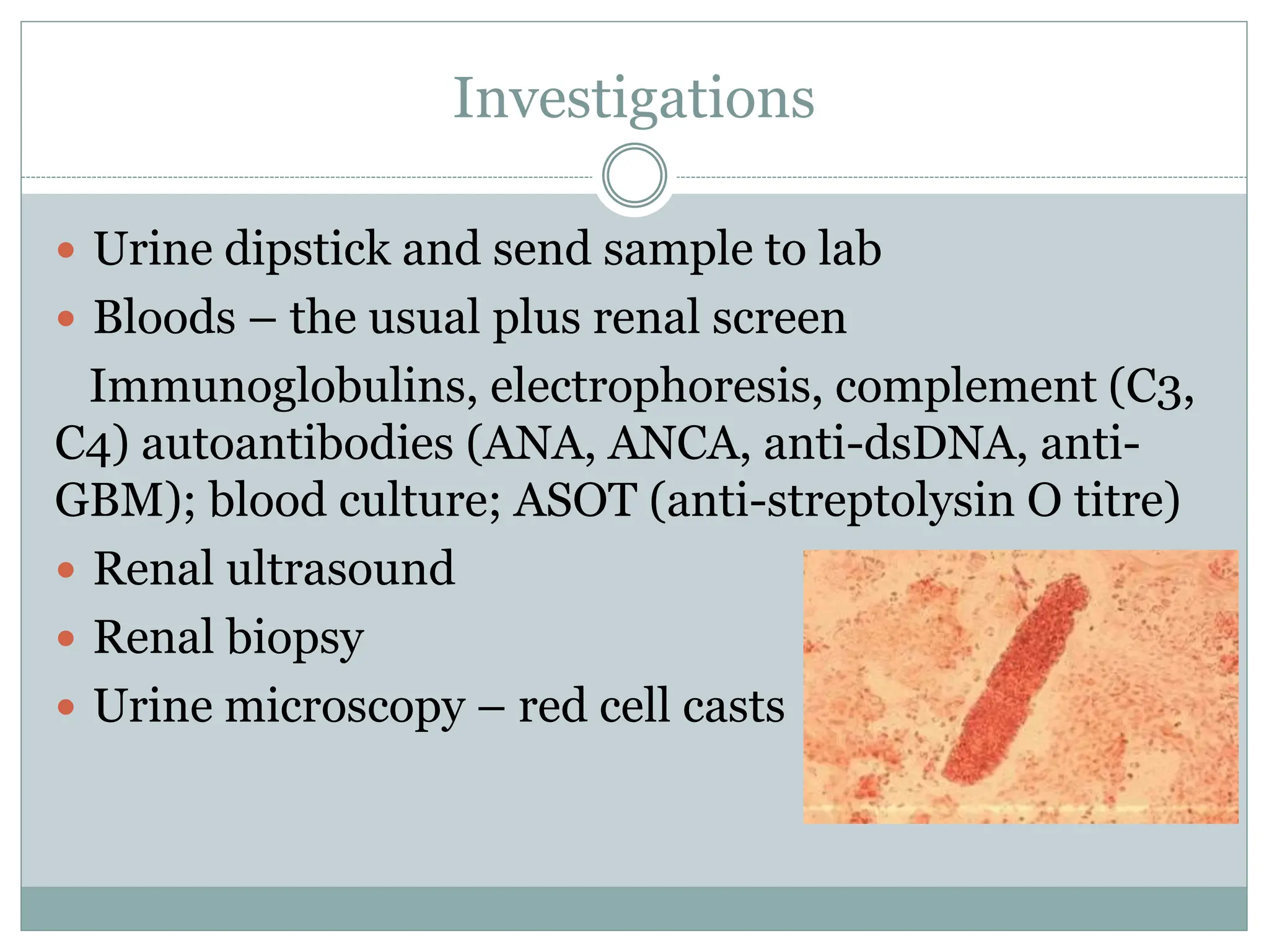
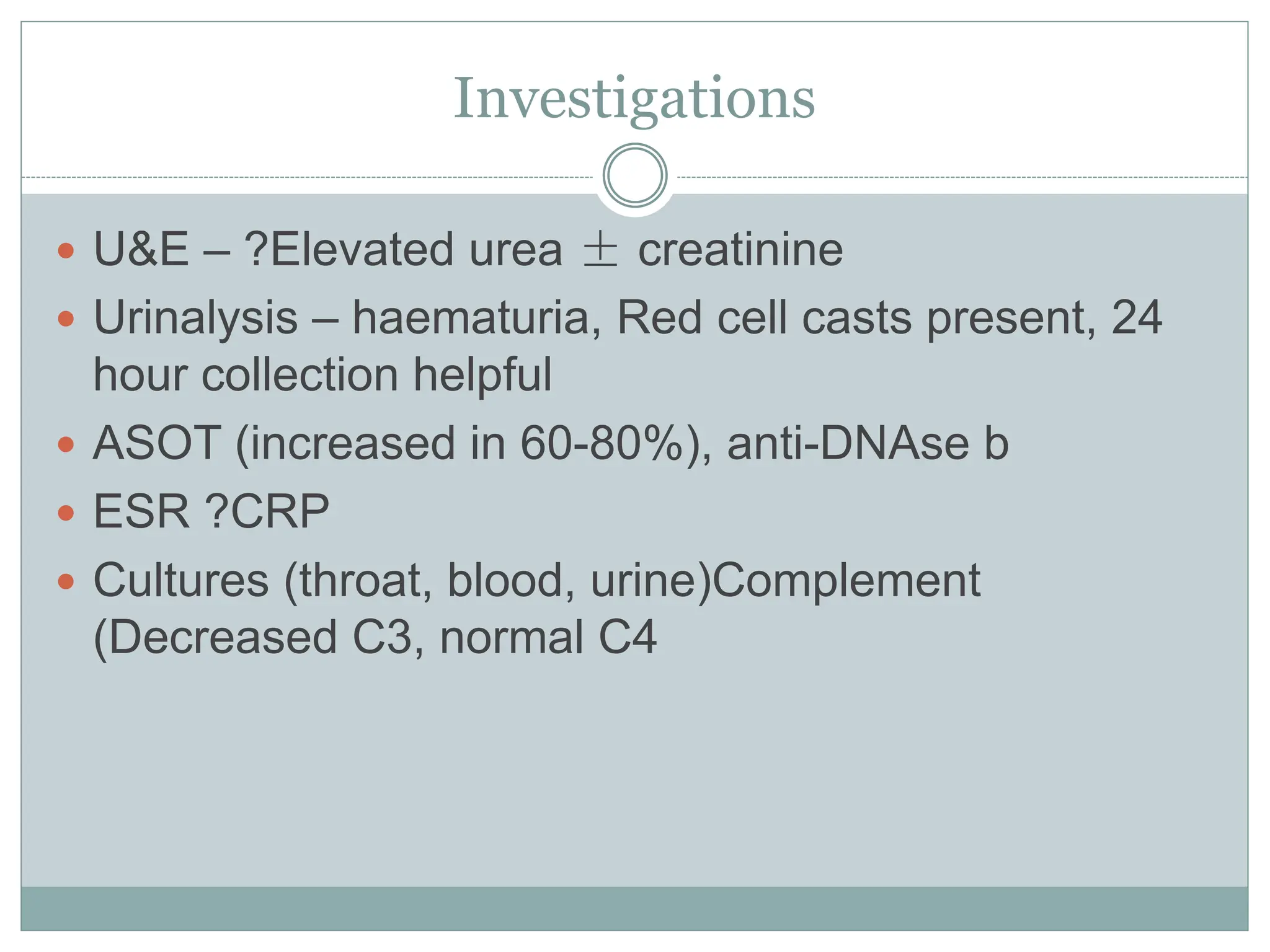
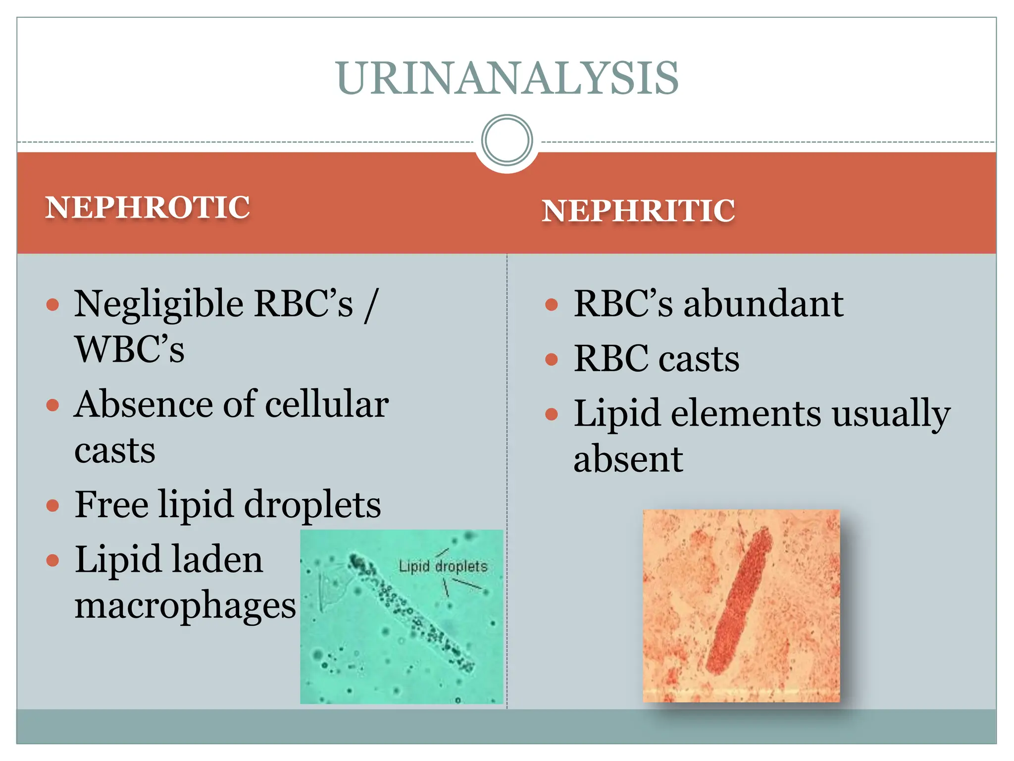
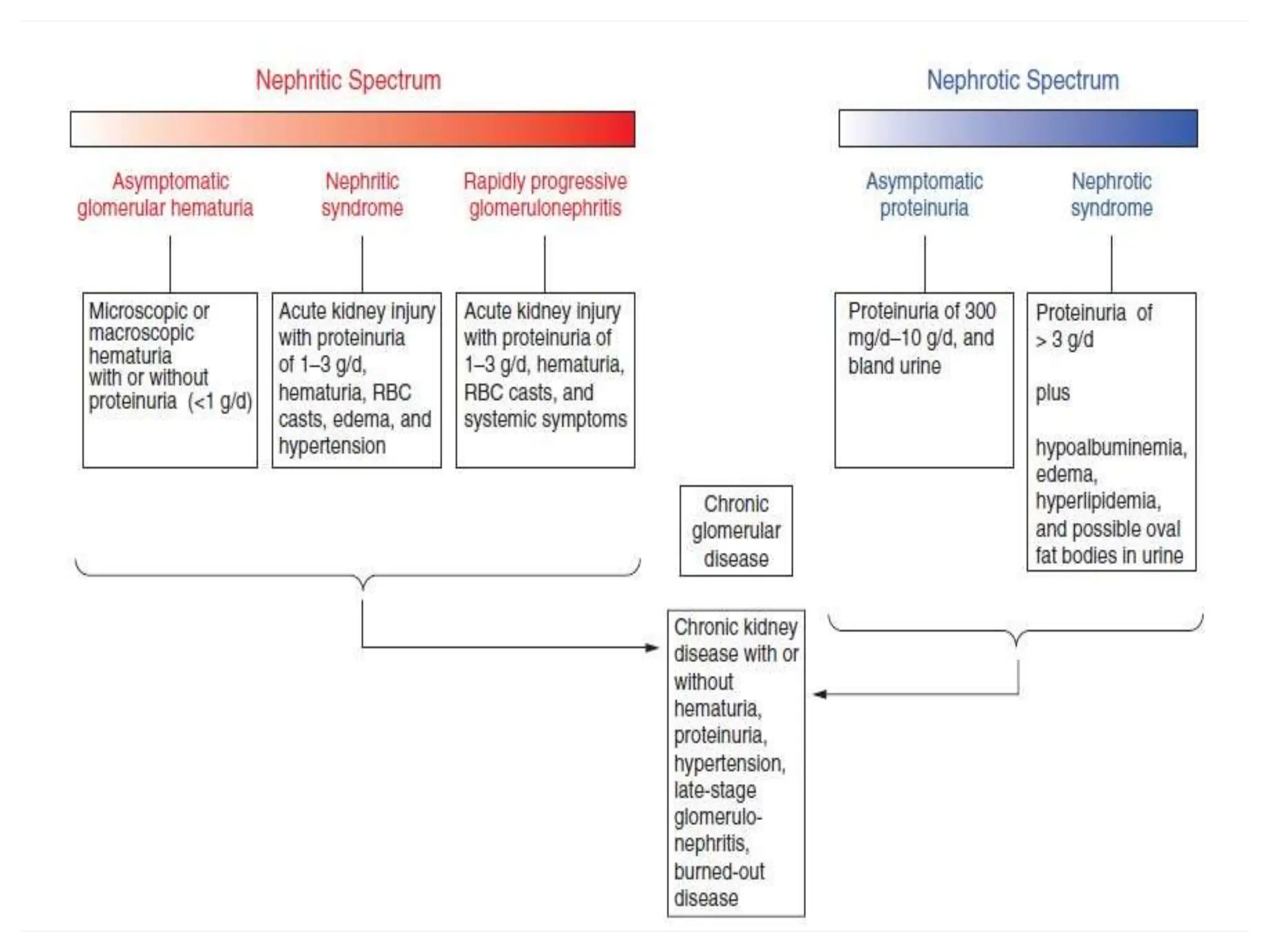
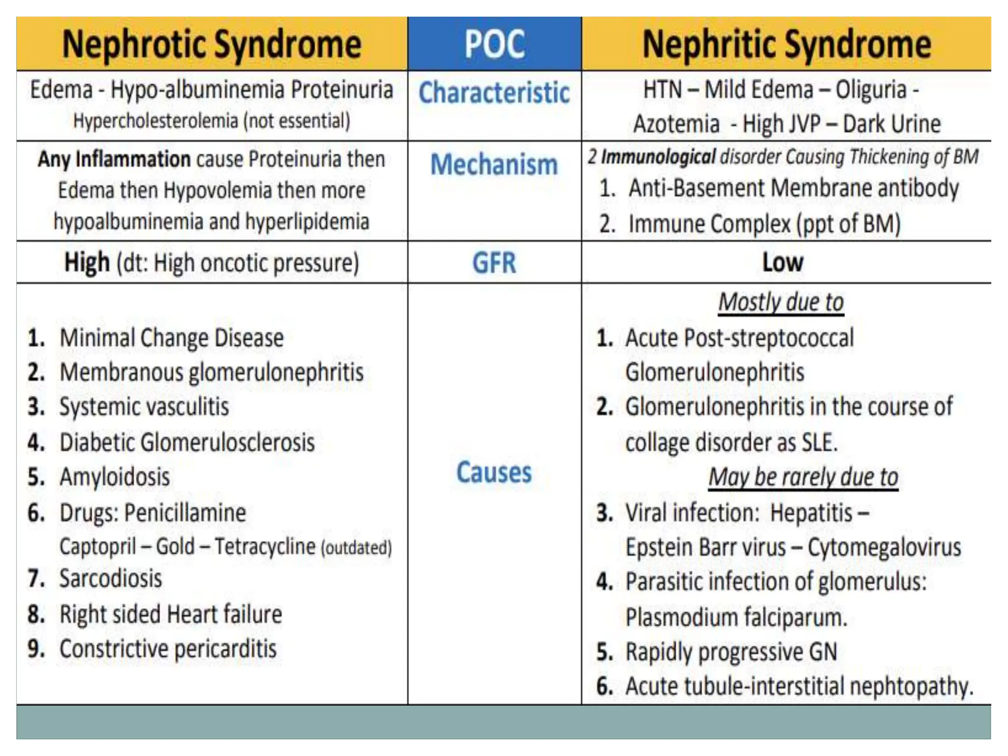
Nephrotic And Nephrotic
Syndrome
NEPHROTIC NEPHRITIC
• Loss of foot processes  Proliferative changes and
inflammation of the glomeruli
Bottom line- “increased permeability of the glomeruli”
Pathophysiology
What is Nephrotic syndrome
 Increased permeability of the glomerulus leading to loss of proteins into the
tubules
How many pathological types causes
nephrotic syndrome?
Proteinuria
(>3.5g/24/hr in adults
or 40mg/m2 /hr in
children)
Hypoalbuminemia
<2.5gm/dl
Hyperlipidemia
Edema Nephrotic
Syndrome
PRESENTATION
 New-onset oedema
Initially periorbital or peripheral
Later genitals, ascites, anasarca
 Frothy urine
 Generalised symptoms –
lethargy, fatigue, reduced
appetite
Further Possible Presentations
 Oedema
 BP normal/raised
 Leukonychia
 Breathlessness:
Pleural effusion, fluid overload,
AKI
 DVT/PE/MI
 Eruptive xanthomata/
xanthalosmata
Differential Diagnosis for Oedema
 Congestive Cardiac Failure
Raised JVP, pulmonary oedema, mild proteinuria
 Liver disease
Hypoalbuminaemia, ascites/oedema
 Most children (90%) with nephrotic syndrome have a form of
the idiopathic nephrotic syndrome.
Investigations
 Urine dipstick for protein
 Urine microscopy
 Bloods – the usual ones, plus renal screen
Immunoglobulins, electrophoresis (myeloma
screen), complement (C3, C4) autoantibodies (ANA,
ANCA, anti-dsDNA, anti-GBM)
 Renal ultrasound
 Renal biopsy (all adults)
Children generally trial of steroids first
Investigations:-
1-Urine analysis:-
 Proteinuria : 3-4 + SELECTIVE.
 Urine collection for protein
>40mg/m2/hr for children
 volume: oliguria (during stage of edema formation)
 Microscopically:-
microscopic hematuria 20%, large number of hyaline cast
Investigations:-
2-Blood:
 Serum protein: decrease >5.5gm/dL , Albumin levels are low
(<2.5gm/dL).
 Serum cholesterol and triglycerides:
Cholesterol >5.7mmol/L (220mg/dl).
 ESR↑>100mm/hr during activity phase
3.Serum complement: Vary with clinical type.
4.Renal function
Management
 Conservative
Monitor U&E, BP, fluid balance, weight
Salt and fluid restriction
Treat underlying cause
Management
 General (non-specific )
 *Corticosteroid therapy
General therapy:-
 Hospitalization:- for initial work-up and evaluation of
treatment.
 Activity: usually no restriction , except
massive edema,heavy hypertension and infection.
 Diet
Hypertension and edema: Low salt diet (<2gNa/ day) only
during period of edema or salt-free diet.
Severe edema: Restricting fluid intake
 Avoiding infection: very important.
 Diuresis: Hydrochlorothiazide (HCT) :2mg/kg.d
Antisterone : 2~4mg/kg.d
Dextran : 10~15ml/kg , after 30~60m,
followed by Furosemide (Lasix) at 2mg/kg .
Induction use of albumin:-
Albumin + Lasix (20 % salt poor)
 1-Severe edema
 2-Ascites
 3-Pleural effusion
 4-Genital edema
 5-Low serum albumin
Corticosteroid—prednisone therapy:-
 Prednisone tablets at a dose of 60 mg/m2/day
(maximum daily dose, 80 mg divided into 2-3 doses) for
at least 4 consecutive weeks.
 After complete absence of proteinuria, prednisone dose
should be tapered to 40 mg/m2/day given every other
day as a single morning dose.
 The alternate-day dose is then slowly tapered and
discontinued over the next 2-3 mo.
Treatment of relapse in NS:
 Many children with nephrotic syndrome will
experience at least 1 relapse (3-4+proteinuria plus
edema).
 daily divided-dose prednisone at the doses noted earlier
(where he has the relapse) until the child enters
remission (urine trace or negative for protein for 3
consecutive days).
 The pred-nisone dose is then changed to alternate-day
dosing and tapered over 1-2 mo.
According to response to prednisone
therapy:
 *Remission: no edema, urine is protein free for 5 consecutive
days.
 * Relapse: edema, or first morning urine sample contains > 2 +
protein for 7 consecutive days.
 *Frequent relapsing: > 2 relapses within 6 months (> 4/year).
 *Steroid resistant: failure to achieve remission with
prednisolone given daily for 28 days.
Side Effects With Long Term Use of
Steroids “Steroid toxicity
 hyperglycemia
 myopathy
 peptic ulcer
 poor healing of wound.
 Hirsutism
 Thromboembolism
-Stunted growth
Cataracts
- Pseudotumor cerebri
-Psycosis
-Osteoporosis
- Cushingoid features
-Adrenal gland suppression
Alternative agent:-
 When can be used:
 Steroid-dependent patients, frequent relapsers, and steroid-
resistant patients.
 Cyclophosphamide Pulse steroids
 Cyclosporin A
 Tacrolimus
 Microphenolate
Complications
Thromboembolism
Hyperlipidaemia
Increased
Susceptibility to
infection
What is nephritic syndrome?
 Refers to a specific set of renal diseases in which an
immunologic mechanism triggers inflammation
and proliferation of glomerular tissue that result in
damage to the basement membrane, mesangium or
capillary endothelium
It is a syndrome associated with severe glomerular
injury, but does not denote a specific etiologic form of
glomerulonephritis
Pathophysiology
 Thin glomerular basement membrane with pores that allow protein and blood
into the tubule.
Hematuria
Red cell casts
Hypertension
Proteinuria
<3gm/day
Oliguria
Nephritic
Syndrome
Signs and Symptoms
 Haematuria (E.g. cola coloured)
 Proteinuria
 Hypertension
 Oliguria
 Flank pain
 General systemic symptoms
 Post-infectious = 2-3 weeks
after strep-throat/URTI
Investigations
 Urine dipstick and send sample to lab
 Bloods – the usual plus renal screen
Immunoglobulins, electrophoresis, complement (C3,
C4) autoantibodies (ANA, ANCA, anti-dsDNA, anti-
GBM); blood culture; ASOT (anti-streptolysin O titre)
 Renal ultrasound
 Renal biopsy
 Urine microscopy – red cell casts
Investigations
 U&E – ?Elevated urea ± creatinine
 Urinalysis – haematuria, Red cell casts present, 24
hour collection helpful
 ASOT (increased in 60-80%), anti-DNAse b
 ESR ?CRP
 Cultures (throat, blood, urine)Complement
(Decreased C3, normal C4
Management
 Correct electrolyte abnormalities if present
 Post Streptococcal – penicillin therapy
 Admission if oliguria and renal failure
 Fluid restriction with significant oedema
Management
 Conservative
o Monitor U&E, BP, fluid balance, weight
o Salt and fluid restriction
o Treat underlying cause
 Medical
o Diuretics
o Treat hypertension
Corticosteroids/immunosuppression
o Dialysis
 Surgical
o Renal transplant
NEPHROTIC NEPHRITIC
 Negligible RBC’s /
WBC’s
 Absence of cellular
casts
 Free lipid droplets
 Lipid laden
macrophages
 RBC’s abundant
 RBC casts
 Lipid elements usually
absent
URINANALYSIS
Summary
 Nephrotic syndrome = MASSIVE proteinuria
 Nephritic syndrome = haematuria/red cell casts
 May be a mixed presentation
 New oedema? Dipstick that urine!
 Haematuria? Exclude malignancy!
 THE END….
THANK YOU….

Pediatrics Nephrotic and Nephritic Syndrome 7.ppt

  • 1.
  • 3.
    NEPHROTIC NEPHRITIC • Lossof foot processes  Proliferative changes and inflammation of the glomeruli Bottom line- “increased permeability of the glomeruli” Pathophysiology
  • 4.
    What is Nephroticsyndrome  Increased permeability of the glomerulus leading to loss of proteins into the tubules
  • 5.
    How many pathologicaltypes causes nephrotic syndrome?
  • 7.
    Proteinuria (>3.5g/24/hr in adults or40mg/m2 /hr in children) Hypoalbuminemia <2.5gm/dl Hyperlipidemia Edema Nephrotic Syndrome
  • 8.
    PRESENTATION  New-onset oedema Initiallyperiorbital or peripheral Later genitals, ascites, anasarca  Frothy urine  Generalised symptoms – lethargy, fatigue, reduced appetite
  • 10.
    Further Possible Presentations Oedema  BP normal/raised  Leukonychia  Breathlessness: Pleural effusion, fluid overload, AKI  DVT/PE/MI  Eruptive xanthomata/ xanthalosmata
  • 11.
    Differential Diagnosis forOedema  Congestive Cardiac Failure Raised JVP, pulmonary oedema, mild proteinuria  Liver disease Hypoalbuminaemia, ascites/oedema
  • 12.
     Most children(90%) with nephrotic syndrome have a form of the idiopathic nephrotic syndrome.
  • 13.
    Investigations  Urine dipstickfor protein  Urine microscopy  Bloods – the usual ones, plus renal screen Immunoglobulins, electrophoresis (myeloma screen), complement (C3, C4) autoantibodies (ANA, ANCA, anti-dsDNA, anti-GBM)  Renal ultrasound  Renal biopsy (all adults) Children generally trial of steroids first
  • 14.
    Investigations:- 1-Urine analysis:-  Proteinuria: 3-4 + SELECTIVE.  Urine collection for protein >40mg/m2/hr for children  volume: oliguria (during stage of edema formation)  Microscopically:- microscopic hematuria 20%, large number of hyaline cast
  • 15.
    Investigations:- 2-Blood:  Serum protein:decrease >5.5gm/dL , Albumin levels are low (<2.5gm/dL).  Serum cholesterol and triglycerides: Cholesterol >5.7mmol/L (220mg/dl).  ESR↑>100mm/hr during activity phase 3.Serum complement: Vary with clinical type. 4.Renal function
  • 16.
    Management  Conservative Monitor U&E,BP, fluid balance, weight Salt and fluid restriction Treat underlying cause
  • 17.
    Management  General (non-specific)  *Corticosteroid therapy
  • 18.
    General therapy:-  Hospitalization:-for initial work-up and evaluation of treatment.  Activity: usually no restriction , except massive edema,heavy hypertension and infection.  Diet Hypertension and edema: Low salt diet (<2gNa/ day) only during period of edema or salt-free diet. Severe edema: Restricting fluid intake  Avoiding infection: very important.  Diuresis: Hydrochlorothiazide (HCT) :2mg/kg.d Antisterone : 2~4mg/kg.d Dextran : 10~15ml/kg , after 30~60m, followed by Furosemide (Lasix) at 2mg/kg .
  • 19.
    Induction use ofalbumin:- Albumin + Lasix (20 % salt poor)  1-Severe edema  2-Ascites  3-Pleural effusion  4-Genital edema  5-Low serum albumin
  • 20.
    Corticosteroid—prednisone therapy:-  Prednisonetablets at a dose of 60 mg/m2/day (maximum daily dose, 80 mg divided into 2-3 doses) for at least 4 consecutive weeks.  After complete absence of proteinuria, prednisone dose should be tapered to 40 mg/m2/day given every other day as a single morning dose.  The alternate-day dose is then slowly tapered and discontinued over the next 2-3 mo.
  • 21.
    Treatment of relapsein NS:  Many children with nephrotic syndrome will experience at least 1 relapse (3-4+proteinuria plus edema).  daily divided-dose prednisone at the doses noted earlier (where he has the relapse) until the child enters remission (urine trace or negative for protein for 3 consecutive days).  The pred-nisone dose is then changed to alternate-day dosing and tapered over 1-2 mo.
  • 22.
    According to responseto prednisone therapy:  *Remission: no edema, urine is protein free for 5 consecutive days.  * Relapse: edema, or first morning urine sample contains > 2 + protein for 7 consecutive days.  *Frequent relapsing: > 2 relapses within 6 months (> 4/year).  *Steroid resistant: failure to achieve remission with prednisolone given daily for 28 days.
  • 23.
    Side Effects WithLong Term Use of Steroids “Steroid toxicity  hyperglycemia  myopathy  peptic ulcer  poor healing of wound.  Hirsutism  Thromboembolism -Stunted growth Cataracts - Pseudotumor cerebri -Psycosis -Osteoporosis - Cushingoid features -Adrenal gland suppression
  • 24.
    Alternative agent:-  Whencan be used:  Steroid-dependent patients, frequent relapsers, and steroid- resistant patients.  Cyclophosphamide Pulse steroids  Cyclosporin A  Tacrolimus  Microphenolate
  • 25.
  • 26.
    What is nephriticsyndrome?  Refers to a specific set of renal diseases in which an immunologic mechanism triggers inflammation and proliferation of glomerular tissue that result in damage to the basement membrane, mesangium or capillary endothelium It is a syndrome associated with severe glomerular injury, but does not denote a specific etiologic form of glomerulonephritis
  • 28.
    Pathophysiology  Thin glomerularbasement membrane with pores that allow protein and blood into the tubule.
  • 30.
  • 31.
    Signs and Symptoms Haematuria (E.g. cola coloured)  Proteinuria  Hypertension  Oliguria  Flank pain  General systemic symptoms  Post-infectious = 2-3 weeks after strep-throat/URTI
  • 32.
    Investigations  Urine dipstickand send sample to lab  Bloods – the usual plus renal screen Immunoglobulins, electrophoresis, complement (C3, C4) autoantibodies (ANA, ANCA, anti-dsDNA, anti- GBM); blood culture; ASOT (anti-streptolysin O titre)  Renal ultrasound  Renal biopsy  Urine microscopy – red cell casts
  • 33.
    Investigations  U&E –?Elevated urea ± creatinine  Urinalysis – haematuria, Red cell casts present, 24 hour collection helpful  ASOT (increased in 60-80%), anti-DNAse b  ESR ?CRP  Cultures (throat, blood, urine)Complement (Decreased C3, normal C4
  • 34.
    Management  Correct electrolyteabnormalities if present  Post Streptococcal – penicillin therapy  Admission if oliguria and renal failure  Fluid restriction with significant oedema
  • 35.
    Management  Conservative o MonitorU&E, BP, fluid balance, weight o Salt and fluid restriction o Treat underlying cause  Medical o Diuretics o Treat hypertension Corticosteroids/immunosuppression o Dialysis  Surgical o Renal transplant
  • 36.
    NEPHROTIC NEPHRITIC  NegligibleRBC’s / WBC’s  Absence of cellular casts  Free lipid droplets  Lipid laden macrophages  RBC’s abundant  RBC casts  Lipid elements usually absent URINANALYSIS
  • 37.
    Summary  Nephrotic syndrome= MASSIVE proteinuria  Nephritic syndrome = haematuria/red cell casts  May be a mixed presentation  New oedema? Dipstick that urine!  Haematuria? Exclude malignancy!
  • 42.