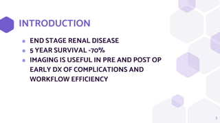
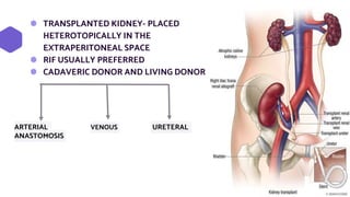
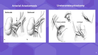
SlideShare a Scribd company logo
IMAGING IN RENAL
TRANSPLANT
PRESENTED BY : DR ALMA SWATHI DSOUZA
MODERATOR : DR. LENON J DSOUZA
Radiology- Nephrology Interdepartmental
meet
INTRODUCTION
INTRODUCTION
⬢ END STAGE RENAL DISEASE
⬢ 5 YEAR SURVIVAL -70%
⬢ IMAGING IS USEFUL IN PRE AND POST OP
EARLY DX OF COMPLICATIONS AND
WORKFLOW EFFICIENCY
3
RENAL TRANSPLANT
TECHNIQUES
⬢ TRANSPLANTED KIDNEY- PLACED
HETEROTOPICALLY IN THE
EXTRAPERITONEAL SPACE
⬢ RIF USUALLY PREFERRED
⬢ CADAVERIC DONOR AND LIVING DONOR
5
ARTERIAL VENOUS URETERAL
ANASTOMOSIS
6
Ureteroneocystostomy
Arterial Anastomosis
7
RADIOLOGICAL ANALYSIS OF
DONOR
⬢ kidney size
⬢ + focal cystic / solid lesions
⬢ condition of vascular structures and their anatomy
collecting system anomalies
⬢ nephrolithiasis.
8
CT ANGIOGRAPHY
Doppler ultrasound
RADIOLOGICAL ANALYSIS OF
RECIPIENTS
⬢ usual algorithm includes chest and abdominal x-ray and
ultrasound of the abdomen
⬢ presence and intensity of atherosclerotic changes in
the iliac vessels
9
pelvic x-ray CT
COMPLICATIONS
Timeline of post-transplant complications based on most likely occurrence periods
Perioperative or Iatrogenic
Complications
12
● Routine use of immediate postoperative US in the
postanesthesia care unit (PACU)
● rapid diagnosis of surgical and perioperative
complications
● renal allograft compartment syndrome (RACS)
● arteriovenous fistula (AVF)
● Pseudoaneurysm
● Hemorrhage.
13
14
Post Biopsy Renal AVF and Pseudoaneurysm
● AVF may form when an artery and vein are
lacerated, whereas PA results when only the
artery is lacerated
● AVF Pseudoaneurysm
AVFs have a feeding
artery with a high-
velocity low-resistance
waveform at spectral
analysis
● narrow neck
● to-and-fro pattern of
blood flow
● yin-yang sign
15
Bidirectional flow
Renal artery
Early draining
vein
pseudoaneurysm
16
pseudoaneurysm
early draining vein
from the AVF
17
PERINEPHRIC FLUID COLLECTIONS
18
Perinephric hematoma
Common
Resolve spontaneously
Some require surgery
Usg: peritransplant blood products can appear
anechoic, hypoechoic, or hyperechoic, depending
on the degree of blood product evolution.
19
20
⬢ Large clinically significant hematomas- 4-8%
⬢ C/F Red blood cell count
Peritransplant Subcapsular
● Double contour
● Compression of renal collecting
system, vascular pedicle
21
Small subcapsular hematoma.
22
Double contour
23
24
Urinoma
⬢ Fluid collection from renal collecting
system
⬢ 10 days after transplant
⬢ Inadequate blood supply to ureter
⬢ Usg appearance overlap --lymphocele
/seroma
25
26
27
28
Abscesses
● Not frequent
● Early post renal
● Perirenal collection + febrile
● Plain CT - fluid collections
containing dense fluid
content
● CECT or gadolinium
enhanced MRI - postcontrast
enhancement of capsule
Perinephric
collection
29
30
LYMPHOCELES SEROMAS
⬢ Sx disruption of lymphatics
⬢ Late post transplant(
months)
⬢ Tend to grow
⬢ Chylous & triglycerides
⬢ Clear fluid
● Usg : anechoic/ hypo fluid collections
● CT round, hypoattenuating collections of clear fluid
● MRI visualizes lymphoceles, seromas and urinomas very similarly
31
Large lymphocele
32
hilum
lymphocele
33
34
VASCULAR COMPLICATIONS
● Renal artery stenosis/thrombosis
● Renal vein stenosis/thrombosis
● Infarct
● Torsion of the graft
35
RTA is a rare but very serious complication, occurs in < 1% of cases
No signs of arterial blood flow
Renal artery is not opacified
36
Renal infarct
37
Renal vein filled with a large thrombus
38
Renal artery stenosis
⬢ 10% of renal transplant patients
⬢ first post transplant week
⬢ also several years after
⬢ M/C in cadaveric transplants, transplants with
multiple renal arteries, and patients with
complex vascular anastomosis
39
40
41
RENAL COLLECTING SYSTEM ABNORMALITIES
⬢ Obstructive hydronephrosis
⬢ flaccid or non-obstructive HN
⬢ formation of urinoma
⬢ Pyonephrosis
⬢ fungal infections
⬢ Renal stones
⬢ Transitional-cell carcinoma
(TCC).
42
● HN - 2-3%
● Causes : Edema
Blood clots
External compression
Stenosis d/t fibrosis
Dilatation of the collecting system of
the transplanted kidney.
43
● Thickening of the
urothelium - nonspecific
sign
● may suggest graft
rejection, pyelonephritis
● exclude tumor of the
transitional epithelium
Thickening of the urothelium
PARENCHYMAL ABNORMALITIES
FOCAL
44
DIFFUSE
RCC PTLD
RCC
same appearance in native and
in transplanted kidneys
PTLD
⬢ in transplanted kidneys
⬢ direct sequel of
immunosuppression
⬢ EBV infection
⬢ perihilar soft tissue thickening,
perinephric masses
lymphadenopathy
⬢ may regress after
immunosuppressive agents
⬢ untreated --progress to
aggressive lymphoma.
45
46
RCC
47
PARENCHYMAL ABNORMALITIES
FOCAL
48
DIFFUSE
● Acute tubular necrosis
● Hyperacute rejection
● Acute rejection
● Chronic rejection
● drug-induced nephrotoxicity.
Delayed graft function
⬢ need for dialysis in the 1st week after renal
transplantation
⬢ cold ischemia time
⬢ Doppler assessment may reveal absent or
diminished diastolic blood flow with
elevated RIs
49
50
● Hyperacute
rejection
● No signs of blood
flow
51
Afunctional and avascular kidney
52
53
54
⬢ diffuse parenchymal complications- CT demonstrates
decreased graft enhancement, with no contrast excretion in
excretory phase,
⬢ loss of corticomedullary differentiation on T1-weighted
images is nonspecific
⬢ but indicative of the presence of diffuse parenchymal
complications and impaired graft function
55
DRUG RELATED NEPHROTOXIC
EFFECTS
calcineurin inhibitors, while known to have
excellent short-term advantages, can result
in chronic nephrotoxic effects with
irreversible damage
56
57
58
59
Neoplastic complications
There is significantly increased incidence of some
cancers (nonmelanomatous skin cancer, lymphoma, and
colon cancer)
and no increased incidence of others (breast, prostate,
and brain cancers)
3rd M/C cause of death
60
New diagnostic methods
● blood flow in large intrarenal blood vessels does not correlate
with intrarenal parenchymal perfusion in transplanted kidney
● Kidney autonomously regulates blood flow at capillary level
● On Doppler ultrasound and nuclear isotopic methods, renal
perfusion is measured only in segmental and interlobar renal
arteries,
● contrast enhanced sonography and functional MRI techniques
provide measurement of cortical and medullary perfusion
61
Complications of renal transplantation continue to evolve alongside
the changing and improving surgical techniques,
immunosuppression regimens, surveillance imaging, and overall
understanding of rejection.
Duplex US is the primary tool for routine surveillance and initial
diagnostic imaging for allograft dysfunction.
Advanced imaging techniques including MRI and contrast-enhanced
US can be used as adjuncts to traditional duplex US and renal
allograft biopsy in workup of posttransplant complications.
“
THANK YOU
62

More Related Content

What's hot

Doppler ultrasound in peripheral arterial disease
Doppler ultrasound in peripheral arterial diseaseDoppler ultrasound in peripheral arterial disease
Doppler ultrasound in peripheral arterial disease
Dr. Naveed Quetta
 
Doppler ultrasound of the kidneys
Doppler ultrasound of the kidneysDoppler ultrasound of the kidneys
Doppler ultrasound of the kidneys
Samir Haffar
 
Presentation1.pptx, radiological imaging of prostatic diseases
Presentation1.pptx, radiological imaging of prostatic diseasesPresentation1.pptx, radiological imaging of prostatic diseases
Presentation1.pptx, radiological imaging of prostatic diseasesAbdellah Nazeer
 
Diagnostic Imaging of Cholangiocarcinoma
Diagnostic Imaging of CholangiocarcinomaDiagnostic Imaging of Cholangiocarcinoma
Diagnostic Imaging of Cholangiocarcinoma
Mohamed M.A. Zaitoun
 
Radiology in portal hypertension
Radiology in portal hypertensionRadiology in portal hypertension
Radiology in portal hypertension
Sunil Kumar
 
Venous Doppler Lower limb Dr Mukesh Tilgam
Venous Doppler Lower limb Dr Mukesh TilgamVenous Doppler Lower limb Dr Mukesh Tilgam
Venous Doppler Lower limb Dr Mukesh Tilgam
Drmukesh Tilgam
 
Presentation1, mri imaging of the prostate
Presentation1, mri imaging of the prostatePresentation1, mri imaging of the prostate
Presentation1, mri imaging of the prostate
Abdellah Nazeer
 
Radiology spotters
Radiology spottersRadiology spotters
Radiology spotters
priyanka rana
 
radiological anatomy of retroperitoneum powerpoint
radiological anatomy of  retroperitoneum powerpointradiological anatomy of  retroperitoneum powerpoint
radiological anatomy of retroperitoneum powerpoint
DactarAdhikari
 
Renal doppler
Renal dopplerRenal doppler
Renal doppler
Anish Choudhary
 
Presentation1.pptx, radiological imaging of cholangiocarcinoma.
Presentation1.pptx, radiological imaging of cholangiocarcinoma.Presentation1.pptx, radiological imaging of cholangiocarcinoma.
Presentation1.pptx, radiological imaging of cholangiocarcinoma.Abdellah Nazeer
 
Doppler of the portal system
Doppler of the portal systemDoppler of the portal system
Doppler of the portal system
Dr. Muhammad Bin Zulfiqar
 
Basics of Renal Doppler
Basics of Renal DopplerBasics of Renal Doppler
Basics of Renal Doppler
Dr. Yash Kumar Achantani
 
Perfusion MRI (DSC and DCE perfusion techniques) for radiology residents
Perfusion MRI (DSC and DCE perfusion techniques) for radiology residentsPerfusion MRI (DSC and DCE perfusion techniques) for radiology residents
Perfusion MRI (DSC and DCE perfusion techniques) for radiology residents
Riham Dessouky
 
Radiology Spots PPT- 2 by Dr Chandni Wadhwani
Radiology Spots PPT- 2 by Dr Chandni WadhwaniRadiology Spots PPT- 2 by Dr Chandni Wadhwani
Radiology Spots PPT- 2 by Dr Chandni Wadhwani
Chandni Wadhwani
 
Presentation1.pptx, radiological anatomy of the thigh and leg.
Presentation1.pptx, radiological anatomy of the thigh and leg.Presentation1.pptx, radiological anatomy of the thigh and leg.
Presentation1.pptx, radiological anatomy of the thigh and leg.Abdellah Nazeer
 
Retroperitoneal masses radiology
Retroperitoneal masses radiologyRetroperitoneal masses radiology
Retroperitoneal masses radiology
Dr. Mohit Goel
 
Doppler ultrasound of the portal system - Pathological findings
Doppler ultrasound of the portal system - Pathological findingsDoppler ultrasound of the portal system - Pathological findings
Doppler ultrasound of the portal system - Pathological findings
Samir Haffar
 
TRANSCRANIAL ULTRASOUND
TRANSCRANIAL ULTRASOUNDTRANSCRANIAL ULTRASOUND
TRANSCRANIAL ULTRASOUND
Ameen Rageh
 
Middle mediastinal masses
Middle mediastinal massesMiddle mediastinal masses
Middle mediastinal massesairwave12
 

What's hot (20)

Doppler ultrasound in peripheral arterial disease
Doppler ultrasound in peripheral arterial diseaseDoppler ultrasound in peripheral arterial disease
Doppler ultrasound in peripheral arterial disease
 
Doppler ultrasound of the kidneys
Doppler ultrasound of the kidneysDoppler ultrasound of the kidneys
Doppler ultrasound of the kidneys
 
Presentation1.pptx, radiological imaging of prostatic diseases
Presentation1.pptx, radiological imaging of prostatic diseasesPresentation1.pptx, radiological imaging of prostatic diseases
Presentation1.pptx, radiological imaging of prostatic diseases
 
Diagnostic Imaging of Cholangiocarcinoma
Diagnostic Imaging of CholangiocarcinomaDiagnostic Imaging of Cholangiocarcinoma
Diagnostic Imaging of Cholangiocarcinoma
 
Radiology in portal hypertension
Radiology in portal hypertensionRadiology in portal hypertension
Radiology in portal hypertension
 
Venous Doppler Lower limb Dr Mukesh Tilgam
Venous Doppler Lower limb Dr Mukesh TilgamVenous Doppler Lower limb Dr Mukesh Tilgam
Venous Doppler Lower limb Dr Mukesh Tilgam
 
Presentation1, mri imaging of the prostate
Presentation1, mri imaging of the prostatePresentation1, mri imaging of the prostate
Presentation1, mri imaging of the prostate
 
Radiology spotters
Radiology spottersRadiology spotters
Radiology spotters
 
radiological anatomy of retroperitoneum powerpoint
radiological anatomy of  retroperitoneum powerpointradiological anatomy of  retroperitoneum powerpoint
radiological anatomy of retroperitoneum powerpoint
 
Renal doppler
Renal dopplerRenal doppler
Renal doppler
 
Presentation1.pptx, radiological imaging of cholangiocarcinoma.
Presentation1.pptx, radiological imaging of cholangiocarcinoma.Presentation1.pptx, radiological imaging of cholangiocarcinoma.
Presentation1.pptx, radiological imaging of cholangiocarcinoma.
 
Doppler of the portal system
Doppler of the portal systemDoppler of the portal system
Doppler of the portal system
 
Basics of Renal Doppler
Basics of Renal DopplerBasics of Renal Doppler
Basics of Renal Doppler
 
Perfusion MRI (DSC and DCE perfusion techniques) for radiology residents
Perfusion MRI (DSC and DCE perfusion techniques) for radiology residentsPerfusion MRI (DSC and DCE perfusion techniques) for radiology residents
Perfusion MRI (DSC and DCE perfusion techniques) for radiology residents
 
Radiology Spots PPT- 2 by Dr Chandni Wadhwani
Radiology Spots PPT- 2 by Dr Chandni WadhwaniRadiology Spots PPT- 2 by Dr Chandni Wadhwani
Radiology Spots PPT- 2 by Dr Chandni Wadhwani
 
Presentation1.pptx, radiological anatomy of the thigh and leg.
Presentation1.pptx, radiological anatomy of the thigh and leg.Presentation1.pptx, radiological anatomy of the thigh and leg.
Presentation1.pptx, radiological anatomy of the thigh and leg.
 
Retroperitoneal masses radiology
Retroperitoneal masses radiologyRetroperitoneal masses radiology
Retroperitoneal masses radiology
 
Doppler ultrasound of the portal system - Pathological findings
Doppler ultrasound of the portal system - Pathological findingsDoppler ultrasound of the portal system - Pathological findings
Doppler ultrasound of the portal system - Pathological findings
 
TRANSCRANIAL ULTRASOUND
TRANSCRANIAL ULTRASOUNDTRANSCRANIAL ULTRASOUND
TRANSCRANIAL ULTRASOUND
 
Middle mediastinal masses
Middle mediastinal massesMiddle mediastinal masses
Middle mediastinal masses
 

Similar to Imaging in renal transplant

medicine.Kidney 2.(dr.ala)
medicine.Kidney 2.(dr.ala)medicine.Kidney 2.(dr.ala)
medicine.Kidney 2.(dr.ala)student
 
Budd chiary syndrome presentation latest.pptx
Budd chiary syndrome presentation latest.pptxBudd chiary syndrome presentation latest.pptx
Budd chiary syndrome presentation latest.pptx
katanchhabra
 
Budd chiari syndrome
Budd chiari syndromeBudd chiari syndrome
Budd chiari syndrome
Dr. Thakur Prashant Singh
 
RVD
RVDRVD
Bilateral Renal Lymphangiectasia: Case Presentation
Bilateral Renal Lymphangiectasia: Case PresentationBilateral Renal Lymphangiectasia: Case Presentation
Bilateral Renal Lymphangiectasia: Case Presentation
Arushi Bhartiya
 
Acute pancreatitis
Acute pancreatitisAcute pancreatitis
Acute pancreatitis
Nandinii Ramasenderan
 
Renal biopsy fadl
Renal biopsy fadlRenal biopsy fadl
Renal biopsy fadl
Ahmed Fadl
 
endoscopy learn about our in simple way
endoscopy  learn about our in simple wayendoscopy  learn about our in simple way
endoscopy learn about our in simple way
Manoj847525
 
Oncological emergencies
Oncological emergenciesOncological emergencies
Oncological emergencies
Chanchal Sharma
 
CLINICAL FEATURES & PROGNOSTIC FACTORS OF RCC
CLINICAL FEATURES & PROGNOSTIC FACTORS OF RCCCLINICAL FEATURES & PROGNOSTIC FACTORS OF RCC
CLINICAL FEATURES & PROGNOSTIC FACTORS OF RCC
GovtRoyapettahHospit
 
Liver resection and Metastasectomy.pptx
Liver resection and Metastasectomy.pptxLiver resection and Metastasectomy.pptx
Liver resection and Metastasectomy.pptx
Pushpa Lal Bhadel
 
cystickidneydiseases-161210202710 (1).pdf
cystickidneydiseases-161210202710 (1).pdfcystickidneydiseases-161210202710 (1).pdf
cystickidneydiseases-161210202710 (1).pdf
HARYANVITRAVLLER
 
Cystic kidney diseases
Cystic kidney diseasesCystic kidney diseases
Cystic kidney diseases
sahar Hamdy
 
Renal malignancy
Renal malignancyRenal malignancy
Renal malignancy
suriyaprakash nagarajan
 
2. LIVER CIRRHOSIS-1-1.pptx
2. LIVER CIRRHOSIS-1-1.pptx2. LIVER CIRRHOSIS-1-1.pptx
2. LIVER CIRRHOSIS-1-1.pptx
LameckNyaisa
 
Carcinoma Rectum
Carcinoma RectumCarcinoma Rectum
Carcinoma Rectum
Ankita Singh
 
Prostate diseases for General practitioners
Prostate diseases for General practitionersProstate diseases for General practitioners
Prostate diseases for General practitioners
Priyatham Kasaraneni
 
Early graft dysfunction
Early graft dysfunctionEarly graft dysfunction
Early graft dysfunction
Kushal Dp
 

Similar to Imaging in renal transplant (20)

medicine.Kidney 2.(dr.ala)
medicine.Kidney 2.(dr.ala)medicine.Kidney 2.(dr.ala)
medicine.Kidney 2.(dr.ala)
 
Budd chiary syndrome presentation latest.pptx
Budd chiary syndrome presentation latest.pptxBudd chiary syndrome presentation latest.pptx
Budd chiary syndrome presentation latest.pptx
 
Budd chiari syndrome
Budd chiari syndromeBudd chiari syndrome
Budd chiari syndrome
 
RVD
RVDRVD
RVD
 
Bilateral Renal Lymphangiectasia: Case Presentation
Bilateral Renal Lymphangiectasia: Case PresentationBilateral Renal Lymphangiectasia: Case Presentation
Bilateral Renal Lymphangiectasia: Case Presentation
 
Acute pancreatitis
Acute pancreatitisAcute pancreatitis
Acute pancreatitis
 
Renal biopsy fadl
Renal biopsy fadlRenal biopsy fadl
Renal biopsy fadl
 
endoscopy learn about our in simple way
endoscopy  learn about our in simple wayendoscopy  learn about our in simple way
endoscopy learn about our in simple way
 
Oncological emergencies
Oncological emergenciesOncological emergencies
Oncological emergencies
 
CLINICAL FEATURES & PROGNOSTIC FACTORS OF RCC
CLINICAL FEATURES & PROGNOSTIC FACTORS OF RCCCLINICAL FEATURES & PROGNOSTIC FACTORS OF RCC
CLINICAL FEATURES & PROGNOSTIC FACTORS OF RCC
 
Liver resection and Metastasectomy.pptx
Liver resection and Metastasectomy.pptxLiver resection and Metastasectomy.pptx
Liver resection and Metastasectomy.pptx
 
cystickidneydiseases-161210202710 (1).pdf
cystickidneydiseases-161210202710 (1).pdfcystickidneydiseases-161210202710 (1).pdf
cystickidneydiseases-161210202710 (1).pdf
 
Cystic kidney diseases
Cystic kidney diseasesCystic kidney diseases
Cystic kidney diseases
 
d.pptx
d.pptxd.pptx
d.pptx
 
Renal malignancy
Renal malignancyRenal malignancy
Renal malignancy
 
2. LIVER CIRRHOSIS-1-1.pptx
2. LIVER CIRRHOSIS-1-1.pptx2. LIVER CIRRHOSIS-1-1.pptx
2. LIVER CIRRHOSIS-1-1.pptx
 
Kidney
KidneyKidney
Kidney
 
Carcinoma Rectum
Carcinoma RectumCarcinoma Rectum
Carcinoma Rectum
 
Prostate diseases for General practitioners
Prostate diseases for General practitionersProstate diseases for General practitioners
Prostate diseases for General practitioners
 
Early graft dysfunction
Early graft dysfunctionEarly graft dysfunction
Early graft dysfunction
 

More from alma dsouza

doppler physics.pptx
doppler physics.pptxdoppler physics.pptx
doppler physics.pptx
alma dsouza
 
X-ray generators..pptx
X-ray generators..pptxX-ray generators..pptx
X-ray generators..pptx
alma dsouza
 
Endometrium and its pathologies
Endometrium and its pathologiesEndometrium and its pathologies
Endometrium and its pathologies
alma dsouza
 
Pulmonary tb presented by dr alma swathi dsouza moderator dr krishna kiran
Pulmonary tb presented by   dr alma swathi dsouza moderator  dr krishna kiranPulmonary tb presented by   dr alma swathi dsouza moderator  dr krishna kiran
Pulmonary tb presented by dr alma swathi dsouza moderator dr krishna kiran
alma dsouza
 
Mri prostate
Mri prostateMri prostate
Mri prostate
alma dsouza
 
Xeroradiography
XeroradiographyXeroradiography
Xeroradiography
alma dsouza
 

More from alma dsouza (6)

doppler physics.pptx
doppler physics.pptxdoppler physics.pptx
doppler physics.pptx
 
X-ray generators..pptx
X-ray generators..pptxX-ray generators..pptx
X-ray generators..pptx
 
Endometrium and its pathologies
Endometrium and its pathologiesEndometrium and its pathologies
Endometrium and its pathologies
 
Pulmonary tb presented by dr alma swathi dsouza moderator dr krishna kiran
Pulmonary tb presented by   dr alma swathi dsouza moderator  dr krishna kiranPulmonary tb presented by   dr alma swathi dsouza moderator  dr krishna kiran
Pulmonary tb presented by dr alma swathi dsouza moderator dr krishna kiran
 
Mri prostate
Mri prostateMri prostate
Mri prostate
 
Xeroradiography
XeroradiographyXeroradiography
Xeroradiography
 

Recently uploaded

Ocular injury ppt Upendra pal optometrist upums saifai etawah
Ocular injury  ppt  Upendra pal  optometrist upums saifai etawahOcular injury  ppt  Upendra pal  optometrist upums saifai etawah
Ocular injury ppt Upendra pal optometrist upums saifai etawah
pal078100
 
heat stroke and heat exhaustion in children
heat stroke and heat exhaustion in childrenheat stroke and heat exhaustion in children
heat stroke and heat exhaustion in children
SumeraAhmad5
 
Cervical & Brachial Plexus By Dr. RIG.pptx
Cervical & Brachial Plexus By Dr. RIG.pptxCervical & Brachial Plexus By Dr. RIG.pptx
Cervical & Brachial Plexus By Dr. RIG.pptx
Dr. Rabia Inam Gandapore
 
BRACHYTHERAPY OVERVIEW AND APPLICATORS
BRACHYTHERAPY OVERVIEW  AND  APPLICATORSBRACHYTHERAPY OVERVIEW  AND  APPLICATORS
BRACHYTHERAPY OVERVIEW AND APPLICATORS
Krishan Murari
 
ARTHROLOGY PPT NCISM SYLLABUS AYURVEDA STUDENTS
ARTHROLOGY PPT NCISM SYLLABUS AYURVEDA STUDENTSARTHROLOGY PPT NCISM SYLLABUS AYURVEDA STUDENTS
ARTHROLOGY PPT NCISM SYLLABUS AYURVEDA STUDENTS
Dr. Vinay Pareek
 
TEST BANK for Operations Management, 14th Edition by William J. Stevenson, Ve...
TEST BANK for Operations Management, 14th Edition by William J. Stevenson, Ve...TEST BANK for Operations Management, 14th Edition by William J. Stevenson, Ve...
TEST BANK for Operations Management, 14th Edition by William J. Stevenson, Ve...
kevinkariuki227
 
Maxilla, Mandible & Hyoid Bone & Clinical Correlations by Dr. RIG.pptx
Maxilla, Mandible & Hyoid Bone & Clinical Correlations by Dr. RIG.pptxMaxilla, Mandible & Hyoid Bone & Clinical Correlations by Dr. RIG.pptx
Maxilla, Mandible & Hyoid Bone & Clinical Correlations by Dr. RIG.pptx
Dr. Rabia Inam Gandapore
 
micro teaching on communication m.sc nursing.pdf
micro teaching on communication m.sc nursing.pdfmicro teaching on communication m.sc nursing.pdf
micro teaching on communication m.sc nursing.pdf
Anurag Sharma
 
Tom Selleck Health: A Comprehensive Look at the Iconic Actor’s Wellness Journey
Tom Selleck Health: A Comprehensive Look at the Iconic Actor’s Wellness JourneyTom Selleck Health: A Comprehensive Look at the Iconic Actor’s Wellness Journey
Tom Selleck Health: A Comprehensive Look at the Iconic Actor’s Wellness Journey
greendigital
 
Triangles of Neck and Clinical Correlation by Dr. RIG.pptx
Triangles of Neck and Clinical Correlation by Dr. RIG.pptxTriangles of Neck and Clinical Correlation by Dr. RIG.pptx
Triangles of Neck and Clinical Correlation by Dr. RIG.pptx
Dr. Rabia Inam Gandapore
 
263778731218 Abortion Clinic /Pills In Harare ,
263778731218 Abortion Clinic /Pills In Harare ,263778731218 Abortion Clinic /Pills In Harare ,
263778731218 Abortion Clinic /Pills In Harare ,
sisternakatoto
 
Alcohol_Dr. Jeenal Mistry MD Pharmacology.pdf
Alcohol_Dr. Jeenal Mistry MD Pharmacology.pdfAlcohol_Dr. Jeenal Mistry MD Pharmacology.pdf
Alcohol_Dr. Jeenal Mistry MD Pharmacology.pdf
Dr Jeenal Mistry
 
Knee anatomy and clinical tests 2024.pdf
Knee anatomy and clinical tests 2024.pdfKnee anatomy and clinical tests 2024.pdf
Knee anatomy and clinical tests 2024.pdf
vimalpl1234
 
New Drug Discovery and Development .....
New Drug Discovery and Development .....New Drug Discovery and Development .....
New Drug Discovery and Development .....
NEHA GUPTA
 
How STIs Influence the Development of Pelvic Inflammatory Disease.pptx
How STIs Influence the Development of Pelvic Inflammatory Disease.pptxHow STIs Influence the Development of Pelvic Inflammatory Disease.pptx
How STIs Influence the Development of Pelvic Inflammatory Disease.pptx
FFragrant
 
BENIGN PROSTATIC HYPERPLASIA.BPH. BPHpdf
BENIGN PROSTATIC HYPERPLASIA.BPH. BPHpdfBENIGN PROSTATIC HYPERPLASIA.BPH. BPHpdf
BENIGN PROSTATIC HYPERPLASIA.BPH. BPHpdf
DR SETH JOTHAM
 
Couples presenting to the infertility clinic- Do they really have infertility...
Couples presenting to the infertility clinic- Do they really have infertility...Couples presenting to the infertility clinic- Do they really have infertility...
Couples presenting to the infertility clinic- Do they really have infertility...
Sujoy Dasgupta
 
Ophthalmology Clinical Tests for OSCE exam
Ophthalmology Clinical Tests for OSCE examOphthalmology Clinical Tests for OSCE exam
Ophthalmology Clinical Tests for OSCE exam
KafrELShiekh University
 
Pulmonary Thromboembolism - etilogy, types, medical- Surgical and nursing man...
Pulmonary Thromboembolism - etilogy, types, medical- Surgical and nursing man...Pulmonary Thromboembolism - etilogy, types, medical- Surgical and nursing man...
Pulmonary Thromboembolism - etilogy, types, medical- Surgical and nursing man...
VarunMahajani
 
ANATOMY AND PHYSIOLOGY OF URINARY SYSTEM.pptx
ANATOMY AND PHYSIOLOGY OF URINARY SYSTEM.pptxANATOMY AND PHYSIOLOGY OF URINARY SYSTEM.pptx
ANATOMY AND PHYSIOLOGY OF URINARY SYSTEM.pptx
Swetaba Besh
 

Recently uploaded (20)

Ocular injury ppt Upendra pal optometrist upums saifai etawah
Ocular injury  ppt  Upendra pal  optometrist upums saifai etawahOcular injury  ppt  Upendra pal  optometrist upums saifai etawah
Ocular injury ppt Upendra pal optometrist upums saifai etawah
 
heat stroke and heat exhaustion in children
heat stroke and heat exhaustion in childrenheat stroke and heat exhaustion in children
heat stroke and heat exhaustion in children
 
Cervical & Brachial Plexus By Dr. RIG.pptx
Cervical & Brachial Plexus By Dr. RIG.pptxCervical & Brachial Plexus By Dr. RIG.pptx
Cervical & Brachial Plexus By Dr. RIG.pptx
 
BRACHYTHERAPY OVERVIEW AND APPLICATORS
BRACHYTHERAPY OVERVIEW  AND  APPLICATORSBRACHYTHERAPY OVERVIEW  AND  APPLICATORS
BRACHYTHERAPY OVERVIEW AND APPLICATORS
 
ARTHROLOGY PPT NCISM SYLLABUS AYURVEDA STUDENTS
ARTHROLOGY PPT NCISM SYLLABUS AYURVEDA STUDENTSARTHROLOGY PPT NCISM SYLLABUS AYURVEDA STUDENTS
ARTHROLOGY PPT NCISM SYLLABUS AYURVEDA STUDENTS
 
TEST BANK for Operations Management, 14th Edition by William J. Stevenson, Ve...
TEST BANK for Operations Management, 14th Edition by William J. Stevenson, Ve...TEST BANK for Operations Management, 14th Edition by William J. Stevenson, Ve...
TEST BANK for Operations Management, 14th Edition by William J. Stevenson, Ve...
 
Maxilla, Mandible & Hyoid Bone & Clinical Correlations by Dr. RIG.pptx
Maxilla, Mandible & Hyoid Bone & Clinical Correlations by Dr. RIG.pptxMaxilla, Mandible & Hyoid Bone & Clinical Correlations by Dr. RIG.pptx
Maxilla, Mandible & Hyoid Bone & Clinical Correlations by Dr. RIG.pptx
 
micro teaching on communication m.sc nursing.pdf
micro teaching on communication m.sc nursing.pdfmicro teaching on communication m.sc nursing.pdf
micro teaching on communication m.sc nursing.pdf
 
Tom Selleck Health: A Comprehensive Look at the Iconic Actor’s Wellness Journey
Tom Selleck Health: A Comprehensive Look at the Iconic Actor’s Wellness JourneyTom Selleck Health: A Comprehensive Look at the Iconic Actor’s Wellness Journey
Tom Selleck Health: A Comprehensive Look at the Iconic Actor’s Wellness Journey
 
Triangles of Neck and Clinical Correlation by Dr. RIG.pptx
Triangles of Neck and Clinical Correlation by Dr. RIG.pptxTriangles of Neck and Clinical Correlation by Dr. RIG.pptx
Triangles of Neck and Clinical Correlation by Dr. RIG.pptx
 
263778731218 Abortion Clinic /Pills In Harare ,
263778731218 Abortion Clinic /Pills In Harare ,263778731218 Abortion Clinic /Pills In Harare ,
263778731218 Abortion Clinic /Pills In Harare ,
 
Alcohol_Dr. Jeenal Mistry MD Pharmacology.pdf
Alcohol_Dr. Jeenal Mistry MD Pharmacology.pdfAlcohol_Dr. Jeenal Mistry MD Pharmacology.pdf
Alcohol_Dr. Jeenal Mistry MD Pharmacology.pdf
 
Knee anatomy and clinical tests 2024.pdf
Knee anatomy and clinical tests 2024.pdfKnee anatomy and clinical tests 2024.pdf
Knee anatomy and clinical tests 2024.pdf
 
New Drug Discovery and Development .....
New Drug Discovery and Development .....New Drug Discovery and Development .....
New Drug Discovery and Development .....
 
How STIs Influence the Development of Pelvic Inflammatory Disease.pptx
How STIs Influence the Development of Pelvic Inflammatory Disease.pptxHow STIs Influence the Development of Pelvic Inflammatory Disease.pptx
How STIs Influence the Development of Pelvic Inflammatory Disease.pptx
 
BENIGN PROSTATIC HYPERPLASIA.BPH. BPHpdf
BENIGN PROSTATIC HYPERPLASIA.BPH. BPHpdfBENIGN PROSTATIC HYPERPLASIA.BPH. BPHpdf
BENIGN PROSTATIC HYPERPLASIA.BPH. BPHpdf
 
Couples presenting to the infertility clinic- Do they really have infertility...
Couples presenting to the infertility clinic- Do they really have infertility...Couples presenting to the infertility clinic- Do they really have infertility...
Couples presenting to the infertility clinic- Do they really have infertility...
 
Ophthalmology Clinical Tests for OSCE exam
Ophthalmology Clinical Tests for OSCE examOphthalmology Clinical Tests for OSCE exam
Ophthalmology Clinical Tests for OSCE exam
 
Pulmonary Thromboembolism - etilogy, types, medical- Surgical and nursing man...
Pulmonary Thromboembolism - etilogy, types, medical- Surgical and nursing man...Pulmonary Thromboembolism - etilogy, types, medical- Surgical and nursing man...
Pulmonary Thromboembolism - etilogy, types, medical- Surgical and nursing man...
 
ANATOMY AND PHYSIOLOGY OF URINARY SYSTEM.pptx
ANATOMY AND PHYSIOLOGY OF URINARY SYSTEM.pptxANATOMY AND PHYSIOLOGY OF URINARY SYSTEM.pptx
ANATOMY AND PHYSIOLOGY OF URINARY SYSTEM.pptx
 

Imaging in renal transplant

  • 1. IMAGING IN RENAL TRANSPLANT PRESENTED BY : DR ALMA SWATHI DSOUZA MODERATOR : DR. LENON J DSOUZA Radiology- Nephrology Interdepartmental meet
  • 3. INTRODUCTION ⬢ END STAGE RENAL DISEASE ⬢ 5 YEAR SURVIVAL -70% ⬢ IMAGING IS USEFUL IN PRE AND POST OP EARLY DX OF COMPLICATIONS AND WORKFLOW EFFICIENCY 3
  • 5. ⬢ TRANSPLANTED KIDNEY- PLACED HETEROTOPICALLY IN THE EXTRAPERITONEAL SPACE ⬢ RIF USUALLY PREFERRED ⬢ CADAVERIC DONOR AND LIVING DONOR 5 ARTERIAL VENOUS URETERAL ANASTOMOSIS
  • 7. 7
  • 8. RADIOLOGICAL ANALYSIS OF DONOR ⬢ kidney size ⬢ + focal cystic / solid lesions ⬢ condition of vascular structures and their anatomy collecting system anomalies ⬢ nephrolithiasis. 8 CT ANGIOGRAPHY Doppler ultrasound
  • 9. RADIOLOGICAL ANALYSIS OF RECIPIENTS ⬢ usual algorithm includes chest and abdominal x-ray and ultrasound of the abdomen ⬢ presence and intensity of atherosclerotic changes in the iliac vessels 9 pelvic x-ray CT
  • 11. Timeline of post-transplant complications based on most likely occurrence periods
  • 12. Perioperative or Iatrogenic Complications 12 ● Routine use of immediate postoperative US in the postanesthesia care unit (PACU) ● rapid diagnosis of surgical and perioperative complications ● renal allograft compartment syndrome (RACS) ● arteriovenous fistula (AVF) ● Pseudoaneurysm ● Hemorrhage.
  • 13. 13
  • 14. 14 Post Biopsy Renal AVF and Pseudoaneurysm ● AVF may form when an artery and vein are lacerated, whereas PA results when only the artery is lacerated ● AVF Pseudoaneurysm AVFs have a feeding artery with a high- velocity low-resistance waveform at spectral analysis ● narrow neck ● to-and-fro pattern of blood flow ● yin-yang sign
  • 15. 15 Bidirectional flow Renal artery Early draining vein pseudoaneurysm
  • 18. 18 Perinephric hematoma Common Resolve spontaneously Some require surgery Usg: peritransplant blood products can appear anechoic, hypoechoic, or hyperechoic, depending on the degree of blood product evolution.
  • 19. 19
  • 20. 20 ⬢ Large clinically significant hematomas- 4-8% ⬢ C/F Red blood cell count Peritransplant Subcapsular ● Double contour ● Compression of renal collecting system, vascular pedicle
  • 23. 23
  • 24. 24 Urinoma ⬢ Fluid collection from renal collecting system ⬢ 10 days after transplant ⬢ Inadequate blood supply to ureter ⬢ Usg appearance overlap --lymphocele /seroma
  • 25. 25
  • 26. 26
  • 27. 27
  • 28. 28 Abscesses ● Not frequent ● Early post renal ● Perirenal collection + febrile ● Plain CT - fluid collections containing dense fluid content ● CECT or gadolinium enhanced MRI - postcontrast enhancement of capsule Perinephric collection
  • 29. 29
  • 30. 30 LYMPHOCELES SEROMAS ⬢ Sx disruption of lymphatics ⬢ Late post transplant( months) ⬢ Tend to grow ⬢ Chylous & triglycerides ⬢ Clear fluid ● Usg : anechoic/ hypo fluid collections ● CT round, hypoattenuating collections of clear fluid ● MRI visualizes lymphoceles, seromas and urinomas very similarly
  • 33. 33
  • 34. 34 VASCULAR COMPLICATIONS ● Renal artery stenosis/thrombosis ● Renal vein stenosis/thrombosis ● Infarct ● Torsion of the graft
  • 35. 35 RTA is a rare but very serious complication, occurs in < 1% of cases No signs of arterial blood flow Renal artery is not opacified
  • 37. 37 Renal vein filled with a large thrombus
  • 38. 38 Renal artery stenosis ⬢ 10% of renal transplant patients ⬢ first post transplant week ⬢ also several years after ⬢ M/C in cadaveric transplants, transplants with multiple renal arteries, and patients with complex vascular anastomosis
  • 39. 39
  • 40. 40
  • 41. 41 RENAL COLLECTING SYSTEM ABNORMALITIES ⬢ Obstructive hydronephrosis ⬢ flaccid or non-obstructive HN ⬢ formation of urinoma ⬢ Pyonephrosis ⬢ fungal infections ⬢ Renal stones ⬢ Transitional-cell carcinoma (TCC).
  • 42. 42 ● HN - 2-3% ● Causes : Edema Blood clots External compression Stenosis d/t fibrosis Dilatation of the collecting system of the transplanted kidney.
  • 43. 43 ● Thickening of the urothelium - nonspecific sign ● may suggest graft rejection, pyelonephritis ● exclude tumor of the transitional epithelium Thickening of the urothelium
  • 45. RCC same appearance in native and in transplanted kidneys PTLD ⬢ in transplanted kidneys ⬢ direct sequel of immunosuppression ⬢ EBV infection ⬢ perihilar soft tissue thickening, perinephric masses lymphadenopathy ⬢ may regress after immunosuppressive agents ⬢ untreated --progress to aggressive lymphoma. 45
  • 47. 47
  • 48. PARENCHYMAL ABNORMALITIES FOCAL 48 DIFFUSE ● Acute tubular necrosis ● Hyperacute rejection ● Acute rejection ● Chronic rejection ● drug-induced nephrotoxicity.
  • 49. Delayed graft function ⬢ need for dialysis in the 1st week after renal transplantation ⬢ cold ischemia time ⬢ Doppler assessment may reveal absent or diminished diastolic blood flow with elevated RIs 49
  • 50. 50 ● Hyperacute rejection ● No signs of blood flow
  • 52. 52
  • 53. 53
  • 54. 54 ⬢ diffuse parenchymal complications- CT demonstrates decreased graft enhancement, with no contrast excretion in excretory phase, ⬢ loss of corticomedullary differentiation on T1-weighted images is nonspecific ⬢ but indicative of the presence of diffuse parenchymal complications and impaired graft function
  • 55. 55 DRUG RELATED NEPHROTOXIC EFFECTS calcineurin inhibitors, while known to have excellent short-term advantages, can result in chronic nephrotoxic effects with irreversible damage
  • 56. 56
  • 57. 57
  • 58. 58
  • 59. 59 Neoplastic complications There is significantly increased incidence of some cancers (nonmelanomatous skin cancer, lymphoma, and colon cancer) and no increased incidence of others (breast, prostate, and brain cancers) 3rd M/C cause of death
  • 60. 60 New diagnostic methods ● blood flow in large intrarenal blood vessels does not correlate with intrarenal parenchymal perfusion in transplanted kidney ● Kidney autonomously regulates blood flow at capillary level ● On Doppler ultrasound and nuclear isotopic methods, renal perfusion is measured only in segmental and interlobar renal arteries, ● contrast enhanced sonography and functional MRI techniques provide measurement of cortical and medullary perfusion
  • 61. 61 Complications of renal transplantation continue to evolve alongside the changing and improving surgical techniques, immunosuppression regimens, surveillance imaging, and overall understanding of rejection. Duplex US is the primary tool for routine surveillance and initial diagnostic imaging for allograft dysfunction. Advanced imaging techniques including MRI and contrast-enhanced US can be used as adjuncts to traditional duplex US and renal allograft biopsy in workup of posttransplant complications.

Editor's Notes

  1. Renal transplantation is the most effective treatment option in patients with end-stage renal disease. Studies have shown that the 5-year survival after renal transplantation is 70%, as compared to 30% survival in patients receiving dialysis. The use of appropriate diagnostic method in preoperative analysis and also in postoperative follow up protocol is necessary for accurate preparation and early diagnosis of complications and workflow efficiency . The most important role of diagnostic radiological methods is to identify multiple complications in the posttransplant period
  2. Generally, the transplanted kidney is placed heterotopically in an extraperitoneal space in the pelvis; that is, a right kidney is placed in the left iliac fossa and vice versa The right iliac fossa is usually preferred, since the right iliac vein runs a more superficial and horizontal course on this side of the pelvis, making the creation of vascular anastomoses easier.
  3. Venous Anastomosis are almost always placed end-to-side to the external iliac vein Arterial Anastomosis In patients who receive cadaveric transplants, the donor renal artery along with a portion of the aorta (Carrel patch) is anastomosed end-to-side to the external iliac artery. In patients who receive living donor kidneys that were harvested with only the main renal artery, the donor renal artery is anastomosed either end-to-end to the internal iliac artery or end-to-side to the recipient external iliac artery (Fig 1) ureter can be anastomosed to the recipient urinary bladder, often at the dome (ureteroneocystostomy), or alternatively to the recipient’s native ureter after removal of the diseased native kidney using an end-to-side anastomosis (ureteroureterostomy)
  4. Coronal contrast-enhanced CT image in the corticomedullary phase shows a single adult renal allograft in the right iliac fossa Coronal contrast-enhanced MIP (maximum intensity projection) MR image shows a renal transplant in the left iliac fossa
  5. It is necessary to evaluate certain important features in a donor kidney to establish if it is appropriate for transplantation, i.e. kidney size, presence of focal cystic or solid lesions, condition of vascular structures and their anatomy (presence of accessory arteries or early bifurcations), collecting system anomalies, or problems or presence of nephrolithiasis. Most of these issues can be visualized with Doppler ultrasound; however, CT angiography is usually necessary for more detailed evaluation of vascular anatomy
  6. It is important to assess the recipient before transplantation to establish any possible conditions that could present a threat to the recipient or functioning of the received kidney. The usual algorithm includes chest and abdominal x-ray and ultrasound of the abdomen. A very important feature is to analyze the presence and intensity of atherosclerotic changes in the iliac vessels because these patients tend to develop prominent arterial calcifications due to dialysis. This can sometimes be sufficiently analyzed with pelvic x-ray but sometimes a CT is needed. CT will also be needed to assess and plan nephrectomy that is occasionally performed at the same time in patients with large polycystic kidneys in order to make space for the transplanted kidney
  7. Renal transplant complications can generally categorized as early (which includes hyperacute and acute complications), intermediate, and late, depending on the time frame in which they occur after renal transplantation Major categories of complications include perioperative or iatrogenic complications, perinephric fluid collections, vascular complications, urinary complications, generalized abdominopelvic complications, allograft rejection, infections, and malignancies. Each of these major categories generally predictable time course after transplantation. For example, type of infection can vary on the basis of time from transplantation, with nosocomial and procedure-related infections occurring predominantly in the early period, latent and opportunistic infections occurring in the intermediate period, and community-acquired infections occurring in the late period.
  8. Routine use of immediate postoperative US in the postanesthesia care unit (PACU) allows rapid diagnosis of surgical and perioperative complications after transplantation. Perioperative and iatrogenic complications include those related to surgical technique, anatomic constraints in the case of renal allograft compartment syndrome (RACS), and procedure-related complications such as arteriovenous fistula (AVF), pseudoaneurysm, and hemorrhage. RACS and vascular thrombosis typically occur in the immediate postoperative to several days after surgery.
  9. Immediate postoperative power Doppler image of the left iliac fossa transplant kidney shows complete absence of cortical flow (arrows). Immediate The patient was returned to the operating room and the allograft was intraperitonealized, CASE OF compartment syndrome due to fascial compression. (b) Follow-up color Doppler image of the kidney shows good cortical flow.
  10. AVF may form when an artery and vein are lacerated, whereas PA results when only the artery is lacerated. It is important to distinguish between a small hemodynamically insignificant AVF and a pseudoaneurysm color Doppler both AVFs and pseudoaneurysms can appear as focal areas with disorganized blood flow extending beyond the margins of the normal vessel intrarenal pseudoaneurysm may appear as a mildly complex cystic structure but can also mimic a simple renal cyst. Classic color Doppler findings of postbiopsy pseudoaneurysm, particularly those with a narrow neck, include a to-and-fro pattern of blood flow within the neck and the yin-yang sign of swirling blood within the sac
  11. Postbiopsy pseudoaneurysm and arteriovenous fistula (AVF) Color Doppler image shows a round area of bidirectional flow (arrow) in the upper transplant kidney, consistent with a pseudoaneurysm. Spectral analysis at a site of focal aliasing near the pseudoaneurysm shows high-velocity systolic flow and low-resistance diastolic flow, characteristic features of an AVF Three-dimensional volume-rendered image shows the renal artery (white arrow) with an early draining vein (black arrow) as well as the pseudoaneurysm (arrowhead).
  12. Image from subtraction angiography shows the pseudoaneurysm (arrowhead) and the early draining vein from the AVF (arrow) Same AVF treated with coil embolization. postprocedure AVFs have been reported in up to 10% of renal allograft biopsies, most of which are asymptomatic with no clinically significant hemodynamic consequences. These cases can be treated conservatively and can be followed up with US as needed, with 70% regressing or resolving spontaneously. Large or symptomatic AVFs resulting in abnormal large or persistent gross hematuria or significant hypertension occur in only 1%–2% of cases and can be treated with catheter embolization
  13. There are four main types of perinephric fluid collections commonly encountered: hematoma, urinoma, abscess, and lymphocele, Imaging features at gray-scale US may overlap; Time from surgery is a key consideration in discriminating between these types of fluid collections in conjunction with clinical manifestations and imaging findings
  14. In the immediate postoperative period, perinephric hematomas are common and should be documented during postoperative baseline US. While hematomas resolve spontaneously, a small minority may require surgical evacuation, usually if it causes significant mass effect on the transplant kidney or continuous hemorrhage requiring transfusion At US, peritransplant blood products can appear anechoic, hypoechoic, or hyperechoic, depending on the degree of blood product evolution. With US alone, clinically significant perinephric hematomas may be underdiagnosed and their volume may be underestimated; need to correlate with hemoglobin levels At nonenhanced CT, acute hematomas are hyperattenuating collections (>30 HU), and at MRI, they typically have increased signal intensity on precontrast T1-weighted images
  15. Coronal nonenhanced CT image shows a hyperattenuating perinephric hematoma (arrows).
  16. Large and clinically significant hematomas occur in 4%-8% of cases and together with dramatic clinical manifestation and drop of red blood cell count Subcapsular hematomas can be more difficult to visualize by ultrasound (sign of ‘double contour’ of the kidney) (Fig. 8), but can also lead to compression of the renal collecting system, of the vascular pedicle, or to graft dysfunction.
  17. Subcapsular hematomas sign of ‘double contour’ of the kidney
  18. Unenhanced CT A large perirenal hematoma (arrow), showing increased absorption coefficients of 85 H.U., indicating the presence of fresh blood immediately after transplantation.
  19. A urinoma is a fluid collection that has leaked from the renal collecting system usually found in the first 10 days after transplantation, most commonly interposed between the allograft and the urinary bladder Urinomas usually occur inadequate blood supply to the ureter or elevated pressures from obstruction The US appearance of urinoma may overlap with that of lymphocele or seroma, appearing as a simple hypoechoic fluid collection. Further diagnostic workup may include delayed contrast-enhanced CT or MRI to assess the collecting system and ureteral anastomoses and detect leakage of excreted contrast material Retrograde urography or renal scintigraphy can also be performed.
  20. (a) US image shows a nonspecific perinephric fluid collection (arrows). (b)99mTc MAG3 renal scintigram shows prompt radiopharmaceutical uptake in the allograft (*) and subsequent excretion into the urinary bladder (arrowhead).
  21. delayed static images show ill-defined radiopharmaceutical accumulation anterior and lateral to the transplant kidney, compatible with a urinoma, which was confirmed at surgery Fluid aspirated from a urinoma will have creatinine and potassium concentrations greater than blood serum, whereas lymphocele aspirate will creatinine and potassium concentrations same has serologic concentrations
  22. Coronal contrast-enhanced delayed phase MIP CT image shows focal urinary contrast material leak from the collecting system (arrow) Note the contrast material–filled transplant ureter (*)
  23. Infected perinephric fluid collections usually develop within the first weeks to months after transplantation not as frequent as other fluid collections in the early posttransplant period. If perirenal collections are seen in febrile patients, they should be considered potentially infected. At US, these collections typically are heterogeneously hypoechoic with internal debris and septa, with blood flow present in the thickened wall and septa Unenhanced CT displays abscesses as fluid collections containing dense fluid content, sometimes with visible gas inside the collection Contrast enhanced CT or gadolinium enhanced MRI display postcontrast ienchancement of hypervascular capsule around the collection
  24. Coronal gadolinium-enhanced T1-weighted MR image shows a peripherally enhancing thick-walled abscess (arrow).
  25. Lymphoceles result from surgical disruption of lymphatics, they usually occur in the late posttransplant period, a month or several months after the surgery. lymphocele is the most commonly encountered perinephric fluid collection, typically occurring 2 weeks to 6 months after surgery, The key to differentiation of lymphoceles from seromas is that lymphoceles occur later and tend to grow. Seromas are composed of clear liquid, while lymphoceles have chylous content and contain triglycerides. In both cases, ultrasound shows an anechogenic cystic structure (Fig. 10), rarely containing internal debris or septations (15). CT displays round, hypoattenuating collections of clear liquid, without postcontrast imbibition. MRI visualizes lymphoceles, seromas and urinomas very similarly, on T1 low signal intensity collections and on T2 measured time as high signal intensity collections.
  26. Large lymphoceles located dorsally of the renal transplant, 45 days after transplantation.
  27. . Lymphocele extending into the hilum of the transplant, imitating dilated renal pelvis.
  28. . Dilatation of the collecting system due to compression of the ureter by lymphocele.
  29. Thrombosis of the transplant renal artery or vein may occur immediately after surgery or as late as 5 days postoperatively. Renal artery stenosis is usually seen 3 months or later after surgery, as its causes tend to be multifactorial.
  30. Renal artery thrombosis is a rare but serious complication, in early postoperative period (within minutes to hours) and can occur as a result of hyperacute rejection, anastomotic occlusion, kinking of the renal artery Clinical signs of renal artery thrombosis include abrupt cessation of urine output and worsening hypertension Contrast enhanced CT, arterial phase. Kidney transplant without signs of arterial blood flow. The contrast is in the iliac artery, renal artery is not opacified with contrast (marked with arrow). Occlusion of the renal artery.
  31. The most common cause of thrombosis is hyperacute rejection or damage to the intima of the artery during transplantation. Color Doppler shows the absence of flow in both the main transplant artery and the intra-renal vessels. Renal infarction can be segmental or global. At US, a segmental infarct appears as a hypoechoic masslike region, which can be ill defined or have a well-defined, with a corresponding wedge-shaped region of avascularity at color or power Doppler assessment Power Doppler. Segmental area without vascular flow in the renal parenchyma corresponding to renal infarct.
  32. Renal vein thrombosis complicates less than 3% of transplants, typically early complication within the first postoperative week. 5 days - peak at 48hrs Clinical manifestations include acute pain and swelling of the graft. Early findings US include edematous engorgement of the kidney, loss of corticomedullary differentiation, and perinephric fluid The causes are external compression by fluid collections or extension of iliac vein thrombosis, hypovolemia, and surgical complications. Age <6 yrs and > 60 yrs Reversed diastolic flow in the transplant renal artery at spectral Doppler US is highly suggestive of renal vein thrombosis (Fig 9b) but may also be seen less frequently with disorders such as allograft torsion, severe allograft rejection, or acute tubular necrosis (ATN) shows heterogeneity of the lateral renal moiety (arrows). (b) Color Doppler image shows classic reversed diastolic flow (arrows) with preserved systolic arterial upstroke
  33. Renal artery stenosis occurs in about 10% of renal transplant patients. It can occur in the first posttransplant week, but also several years after the kidney transplantation. 3Months to 2 years It is more common in cadaveric transplants, transplants with multiple renal arteries, and patients with complex vascular anastomosis
  34. The presenting symptom is hypertension. The main location of the stenosis is at the anastomotic site Doppler ultrasound is the best method for detection Direct signs of transplant renal artery stenosis are seen at the site of narrowing and include elevated peak systolic velocity (PSV), abnormal ratio of PSV in the main renal artery with respect to the upstream iliac artery, and presence of aliasing due to turbulence. Historically, PSV exceeding 250 cm/sec in the transplant main renal artery or ratio greater than 1.8 has been used to suggest significant transplant renal artery stenosis. absolute PSV of 340–400 cm/ sec at the anastomosis has been suggested as a more reliable cutoff (a) Color Doppler proximal renal artery shows elevated peak systolic velocity of 575 cm/sec. (b) Intrarenal arterial shows a classic tardus-parvus waveform distal to the arterial stenosis.
  35. When transplant renal artery stenosis is hemodynamically significant, endovascular techniques including percutaneous transluminal angioplasty and stent placement are first-line treatments, followed by surgery in refractory cases Coronal gadolinium-enhanced MIP T1-weighted MR image shows high-grade stenosis of the transplant renal artery at the anastomosis (arrow). (d) Angiogram after balloon angioplasty shows the artery as widely patent (arrow).
  36. Renal collecting system abnormalities include obstructive hydronephrosis, flaccid or non-obstructive hydronephrosis due to loss of tonicity from denervation urinary leak with formation of urinoma, pyonephros, fungal infections, renal stones, and tumors such as transitional-cell carcinoma (TCC). manifests as diminished renal function with elevated serum creatinine level and presence of hydronephrosis at imaging, usually at US
  37. Hydronephrosis in transplanted kidney is encountered in 2%-5% of renal transplant recipients, with obstruction at the level of ureter. The causes of obstruction can be temporary, such as edema at the ureteral anastomosis or blood clots after surgery. The cause can also be external compression by collections such as lymphoceles (Fig. 17). Late-onset obstruction --ureteral stenosis due to fibrosis or ischemia (Fig. 18).
  38. Kidney stones are rarely the cause of hydronephrosis in transplanted kidney, and either exist initially in the donor kidney or can occur over time as a late complication. Echogenicity in the collecting system can be a sign of pyonephros or fungal infection. Thickening of the urothelium is a nonspecific sign (Fig. 22), which may suggest graft rejection, pyelonephritis, and it is necessary to exclude tumor of the transitional epithelium (Fig. 23). B-mode ultrasound image. Thickening of the urothelium, a nonspecific sign that may indicate pyelonephritis but also renal rejection.
  39. Parenchymal abnormalities are grouped into focal and diffuse. The most important focal abnormality is a tumor mass such as renal cell carcinoma and post-transplant lymphoproliferative disorder (PTLD) Renal cell carcinomas have the same appearance in native and in transplanted kidneys. On the other hand, PTLD occurs only in transplanted kidneys and is a direct sequel of immunosuppression.
  40. It is connected to Epstein-Barr virus infection following transplantation, and occurs in less than 1% of patients. PTLD may present as perihilar soft tissue thickening, perinephric masses and lymphadenopathy (Fig. 39). When diagnosed early, PTLD may regress after reduction of immunosuppressive agents, but if untreated it can progress to aggressive lymphoma.
  41. Contrast enhanced CT, axial view. Hypervascularized tumor in the transplanted kidney. CT urography shows a solid tissue mass in the upper pole of the transplanted kidney. Histologically proven hypernephroma.
  42. Large solid mass located in the hilus of the transplanted kidney. The differential diagnosis includes hypernephroma, PTLD, and abscess.
  43. acute tubular necrosis, hyperacute rejection, acute rejection, chronic rejection, and drug-induced nephrotoxicity.
  44. Delayed graft function is defined as the need for dialysis in the 1st week after renal transplantation The greatest risk factor is cold ischemia time. Other factors include infarction, ATN, rejection, and presence of a peritransplant fluid collection At gray-scale US, the allograft parenchyma can appear normal. Doppler absent or diminished diastolic blood flow with elevated RIs, which are nonspecific findings may also be present in the setting of allograft rejection
  45. Hyperacute rejection can occur within minutes to hours but is usually identified in the operating room at the time of transplantation. This is a rare entity that involves abrupt global allograft nonperfusion and ischemia immediately after vascular anastomosis, secondary to small vessel thrombosis Power Doppler. Kidney transplant immediately after transplantation with no signs of blood flow due to hyperacute rejection. Acute rejection occurs approximately 5–7 days after transplantation and is the result of T-cell activation. Accelerated acute rejection, which can occur in the first 5 postoperative days, is an antibody-mediated aggressive form of rejection that occurs in patients with a history of blood transfusion, prior transplantation, or other causes of presensitization. Chronic rejection involves gradual progressive deterioration in allograft function, which can begin months to years after transplantation Elevated intrarenal RI (>0.74),
  46. Color Doppler. Afunctional and avascular transplanted kidney located in the right iliac fossa.
  47. Doppler exam can show reduction of intrarenal arborization, lower flow rate values intrarenally with elevated RIs Fig. 43a B-mode ultrasound image. Kidney with moderately reduced and hyperechogenic parenchyma in long-term graft. Fig. 43b Color Doppler. Substantial reduction in intrarenal arborization, the same patient. Fig. 43cPulsed Doppler. Intrarenal atypical spectrum, extremely low flow rates of about 10 cm/s, the same patient. Fig. 43d Pulsed Doppler. Renal artery shows still normal flow rates of about 1 m/s, but with high-resistance waveform and almost no diastolic flow. All the above indicates chronic graft dysfunction
  48. Acute tubular necrosis (ATN) is a common cause of anuria in the postoperative period Histopathologic finding is necrosis of tubular cells that relates to ischemic time and reperfusion injury. For all three diffuse parenchymal complications, i.e. acute rejection, acute tubular necrosis and nephrotoxicity, ultrasound presentation can be identical . It shows an enlarged kidney in both diameters, with thickened and hyperechogenic parenchyma, with prominent pyramids and effacement of the renal sinus. B-mode ultrasound image. Enlarged kidney, with thickened and hyperechogenic parenchyma, with prominent pyramids and narrowed renal sinus.
  49. In these diffuse parenchymal complications, CT demonstrates decreased graft enhancement, with no contrast excretion in excretory phase Loss of corticomedullary differentiation on T1-weighted images on MRI finding in acute rejection.
  50. Renal transplant immunosuppression regimens can be categorized into three phases or categories: induction therapy (intense immunosuppressive therapy immediately after transplantation), maintenance therapy, and treatment of rejection . Maintenance therapy can be comprised of antimetabolite agents, calcineurin inhibitors (eg, cyclosporine, tacrolimus), and corticosteroids (eg, oral prednisone) (111). In particular, calcineurin inhibitors, while known to have excellent short-term advantages, can result in chronic nephrotoxic effects with irreversible damage
  51. In the early phase (postoperative period to 1 month), most infections are related to surgery or are nosocomial and include antimicrobial resistant bacteria such as methicillin-resistant Staphylococcus aureus (MRSA) and vancomycin resistant enterococci (VRE) (Donor-derived infections are rare during this period, and when they occur, are often viral (eg, herpes simplex virus, West Nile virus, rhabdovirus, and human immunodeficiency virus). Recipient-derived infections can occur as a result of colonized organisms (eg, Aspergillus or Pseudomonas) In the 2nd to 6th months after surgery, the transplant recipient is more susceptible to opportunistic infections, most commonly cytomegalovirus (CMV), polyomavirus and Epstein-Barr virus (EBV) Another important infectious cause of renal allograft dysfunction or premature graft loss is polyomavirus nephropathy. BK virus is the predominant polyomavirus affecting renal transplant allografts, present in 5% of biopsy specimens BK virus is an otherwise innocuous latent in 75% of the adult population The virus is more often donor derived and usually reactivates in the allograft within the first 3 months after transplantation, inducing tubulointerstitial inflammatory changes that may mimic acute cellular rejection After 6 months, as the risk of early viral and latent infections diminishes, routine infections including community-acquired pneumonia and urinary tract infections are more commonly seen. However, as a result of continued immunosuppression, these patients remain at increased risk for opportunistic pathogens such as Nocardia and fungal organisms such as Aspergillus and Mucor species.
  52. US image shows a large complex perinephric collection (arrows) at the superior aspect of the transplant kidney. (b) Color Doppler image shows robust flow in the renal allograft without flow in the collection. ( c) Axial contrast-enhanced CT image shows a multiseptated low-attenuation collection (arrows) deep to the superior aspect of the renal allograft, Culture of a specimen from CT-guided aspiration demonstrated Nocardia species.
  53. Hrct shows soft tissue density in right upper lobe with central cavity and air lucencies and surrounding ggo
  54. Malignancy is the third most common cause of death in the renal transplant recipient, with three to five times the risk of malignancy as compared with the general population, There is significantly increased incidence of some cancers (nonmelanomatous skin cancer, lymphoma, and colon cancer) annual US or CT of the native kidneys have been suggested as recommended surveillance for skin cancers and native renal cell carcinomas (RCCs), respectively allograft is susceptible to the same forms of malignancy known to occur in the native kidney, but also additional risk for development of malignancies associated with chronic immunosuppression of the host PTLD is a common entity affecting posttransplant patients (
  55. New diagnostic methods (contrast enhanced sonography, functional MRI techniques) indicate a potential future It is an important physiological fact that the blood flow in large intrarenal blood vessels does not correlate with intrarenal parenchymal perfusion in transplanted kidney On Doppler ultrasound and nuclear isotopic methods, renal perfusion is measured only in segmental and interlobar renal arteries, whereas contrast enhanced sonography and functional MRI techniques provide measurement of cortical and medullary perfusion, after the blood has passed the glomerular unit. C ontrast enhanced sonography provides information on microvascular parenchymal perfusion, and also provides quantitative measurements, which can be useful in chronic graft dysfunction