Introduction to renal ct scan
•
15 likes•3,714 views

This is about Introduction To renal CT scan Protocol what are the indication and tailoring how to optimize the the right protocol for the patient according to the indication . Hopping you like it and helping you in daily practice . Dr Hisham AlKhatib Consultant Radiologist

Recommended
More Related Content
What's hot
What's hot (20)
Similar to Introduction to renal ct scan
Similar to Introduction to renal ct scan (20)
More from Hisham Khatib
More from Hisham Khatib (20)
Recently uploaded
Recently uploaded (20)
Introduction to renal ct scan
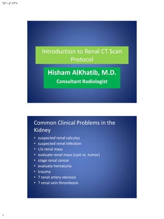
- 1. 9/10/1438 1 Introduction to Renal CT Scan Protocol Hisham AlKhatib, M.D. Consultant Radiologist Common Clinical Problems in the Kidney • suspected renal calculus • suspected renal infection • r/o renal mass • evaluate renal mass (cyst vs. tumor) • stage renal cancer • evaluate hematuria • trauma • ? renal artery stenosis • ? renal vein thrombosis
- 2. 9/10/1438 2 Patient preparation and technique Before study • Fasting 4 hours • Creatinine level • Pertinent history to decide the protocol needed • Protocol design
- 3. 9/10/1438 3 What are the risk factors for contrast induced acute renal failure? • preexisting renal failure • diabetes mellitus • dehydration • cardiovascular disease and diuretics • age over 75 years • multiple myeloma (in dehydrated person) • hypertension • uricosuria Scanning phases • precontrast phase • arterial • corticomedullary phase • nephrographic phase • excretory phase
- 4. 9/10/1438 4 Noncontrast scan • baseline density measurements for evaluating renal masses or renal cysts • urolithiasis, nephrolithiasis, renal calcifications • in patients unable to receive intravenous contrast (i.e., contrast allergy, poor renal function, etc.). Unenhanced CT of the Kidney • Optimal Phase For Detection of- - Calculus - Cyst versus mass (HDRC versus solid tumor) - High density renal cyst - Identify location of the kidneys to define coverage
- 5. 9/10/1438 5
- 6. 9/10/1438 6 arterial phase • is a short phase that occurs about 15–25 seconds after the start of intravenous contrast medium injection and is marked by maximum opacification of the renal arteries. • The renal veins also usually opacify in the late arterial phase.
- 7. 9/10/1438 7 corticomedullary (angionephrographic) phase • The starts at about 30–40 seconds after the start of contrast medium injection. • There is intense enhancement of the renal cortex due to preferential arterial flow to the cortex and glomerular filtration of the contrast material, while the medulla remains relatively less enhanced. • This is also the best phase for maximum opacification of the renal veins.
- 8. 9/10/1438 8 nephrographic phase • begins at 80–120 seconds after the start of contrast medium injection. • Tubular filtration of contrast material produces homogeneous enhancement of the renal parenchyma. • the best phase for detection of subtle parenchymal lesions. Nephrographic Phase • (60-140 sec): • Optimal Phase For Detection of- - Renal lesion detection - Pyelonephritis - Tumor invasion (renal vein/IVC) - Characterize lesion density - Perfusion changes - Renal vein or IVC thrombus
- 9. 9/10/1438 9
- 10. 9/10/1438 10 excretory or urographic phase • starts at 180 seconds (3 minutes) after the start of contrast medium injection. • Excretion of the contrast material allows opacification of the calyces, renal pelvises, and ureters, while the intensity of the nephrogram progressively declines. • routinely acquire excretory phase images at 4– 5 minutes to ensure opacification of the ureters.
- 11. 9/10/1438 11 Excretory (pyelogram)Phase • delayed scans ( 3-5 minutes) • Contrast in calyces ,pelvis and ureters • Excretory Phase Imaging is optimal for detecting; – pathology in the renal pelvis or collecting system – visualization of the renal parenchyma – pathology in the ureter CT Urography: Indications per Society of Uroradiology • - Painless gross and microscopic hematuria - Suspected transitional cell carcinoma - Follow up of transitional cell carcinoma - Recurrent UTI’s - Congenital anomalies - Renal trauma
- 12. 9/10/1438 12 Ct urogram Split Bolus Technique for CT Urography - Scan without contrast from top of kidneys thru the base of the bladder - Inject 50 ml of iodixanol at 3 cc/sec - Wait 5 minutes - Inject 80 ml of iodixanol at 3 cc/sec - Wait 100 seconds and then scan the patient from the top of the kidneys thru the pelvis (combined nephrographic and excretory phase)
- 13. 9/10/1438 13