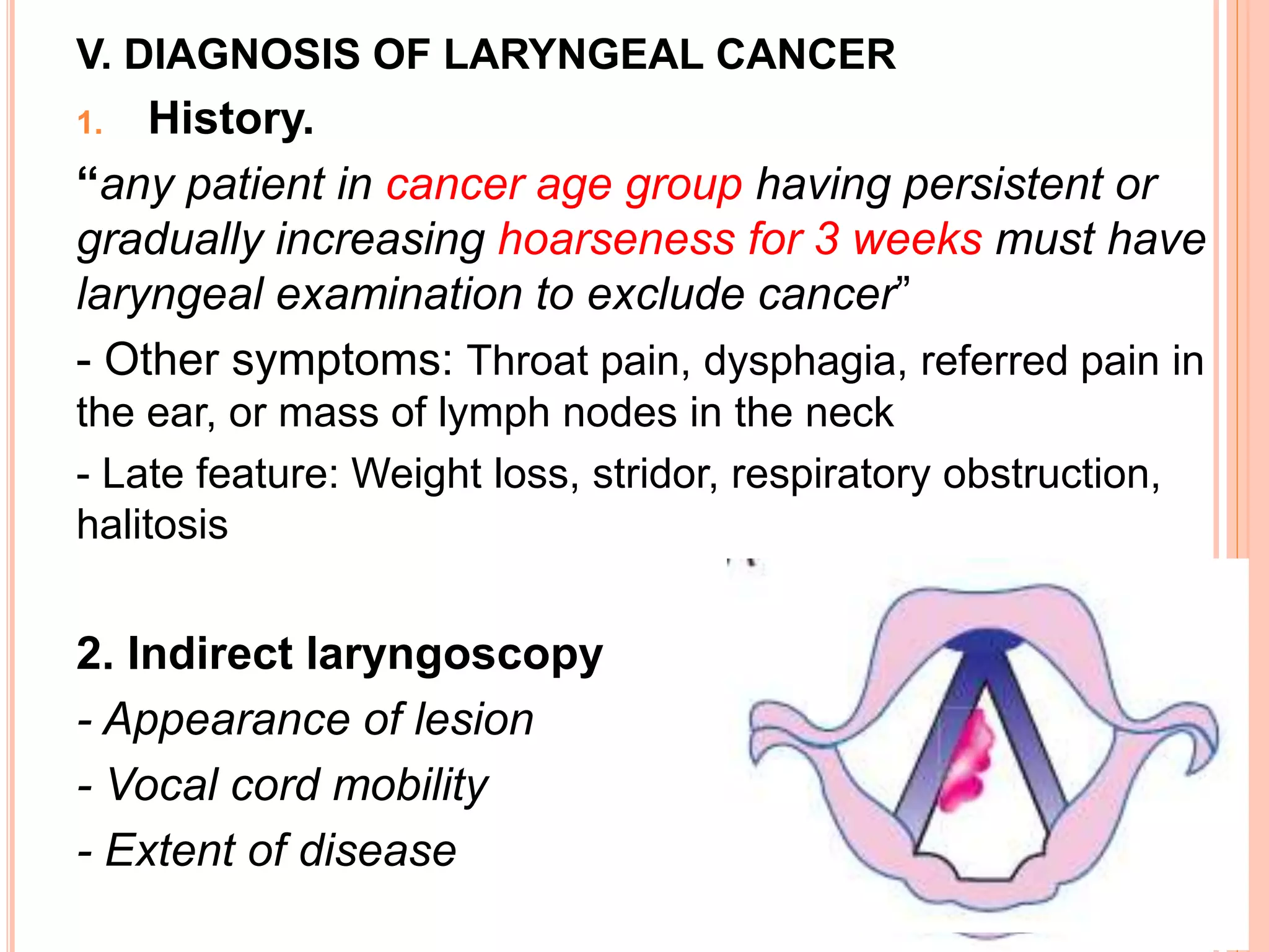
LARYNGEAL
CANCER
Duong Thi My
Cancer larynx, Disease of ear, nose, throat, head and neck
surgery
I. EPIDEMIOLOGY AND HISTOPATHOLOGY
II. AETIOLOGY
III. ANATOMY - SUBDIVISION
IV. TNM CLASSIFICATION AND STAGING
V. DIAGNOSIS OF LARYNGEAL CANCER
VI. TREATMENT OF LARYNGEAL CANCER
I. EPIDEMIOLOGY AND HISTOPATHOLOGY
- 2,63% of all body cancer in India
- M : F = 10:1
- Age group: 40-70 years
- 90–95% : squamous cell carcinoma
II. AETIOLOGY
American Cancer Society: Cancer Facts and Figures 2008. Atlanta, Ga: American
Cancer Society, 2008
III. ANATOMY - SUBDIVISION
IV. TNM CLASSIFICATION AND STAGING
Source: AJCC Cancer Staging Manual, 6th Ed (2002)
Fixed
Source: Greene FL, Page DL, Fleming ID, et al. (editors). American Joint Committee on
Cancer Staging Manual, 6th edition, New York: Springer-Verlag, 2002
IV. TNM CLASSIFICATION AND STAGING
V. DIAGNOSIS OF LARYNGEAL CANCER
1. History.
“any patient in cancer age group having persistent or
gradually increasing hoarseness for 3 weeks must have
laryngeal examination to exclude cancer”
- Other symptoms: Throat pain, dysphagia, referred pain in
the ear, or mass of lymph nodes in the neck
- Late feature: Weight loss, stridor, respiratory obstruction,
halitosis
2. Indirect laryngoscopy
- Appearance of lesion
- Vocal cord mobility
- Extent of disease
3. Examination of neck
- Extralaryngeal spread of disease
- Nodal metastasis
4. Radiography: CT scan or MRI:
- Evaluate pre-epiglottic or paraglottic space
- Laryngeal cartilage erosion
- Cervical node metastasis
5. Direct laryngoscopy
- The hidden areas of larynx
- Extent of disease
6. Supravital staining and biopsy
Certain
diagnostic test
V. DIAGNOSIS OF LARYNGEAL CANCER
VI. TREATMENT OF LARYNGEAL CANCER
Site and
extent of
lesion (T)
Presence or
absence of
Nodes (N)
Distant
Metastases (M)
Treatment
Radiotherapy Surgery Combined
therapy
Organ
preservation
Conservation
laryngeal surgery
Total
laryngectomy Surgery with
pre- or
postoperative
radiotherapy
Endoscopic
CO2 laser excision
(early stage)
GLOTTIC CARCINOMA
 1. Carcinoma in situ:
Transoral endoscopic CO2 laser
stripping of vocal cord biopsy
invasive carcinoma
radiotherapy
carcinoma in situ
Regular follow-up.
Invasive carcinoma
T1
carcinoma
Radiotherapy
Excision of cord
carcinoma with
extension
to anterior commissure
carcinoma with extension
to arytenoid
Radiotherapy
Frontolateral
partial laryngectomy
total laryngectomy
The
best
absence
Fails
Refused or not avalable
- Mobility of vocal cord?
- Involvement of anterior commissure
and/or arytenoid? T2N0
Cord mobile Cord mobility impaired
or
Involvement of anterior
commissure or arytenoid
Conservation
laryngectomy
Total laryngectomy
± neck dissection
Failure
Radiotherapy
Conservation
laryngectomy
Total laryngectomy
± neck dissection
Failure
Failure
Failure
Vertical
hemilaryngectomy
Frontolateral
laryngectomy
T3, T4 total laryngectomy + neck dissection
More advanced T4 : combined therapy: surgery + P.R
If nodes are palpable
Or only Palliative treatment
P.R: postoperative radiotherapy
T1,T2 T3,T4
Radiotherapy
Total laryngectomy
and P.R
Subglottic
cancer
Age of patient
Subglottic
cancer
Supraglottic cancer
T1 T2 T3 , T4
Radiation or
excised with CO2 laser
Lung
function
good poor
supraglottic laryngectomy
With or without
neck dissection radiotherapy
total laryngectomy
with neck
dissection
and P.R to neck
Radiotherapy
- Cord mobility: normal
- Invade cartilage or cervical nodes: no
Total laryngectomy:
- T3 lesions (i.e. with cord fixed)
- All T4 lesions
- Invasion of thyroid or cricoid cartilage
- Bilateral arytenoid cartilage involvement
- Lesions of posterior commissure
- Failure after radiotherapy or conservation surgery
- Transglottic cancers, i.e. tumours involving supraglottis
and glottis across the ventricle, causing fixation of the
vocal cord
Laryngeal cancer

Laryngeal cancer

  • 1.
    LARYNGEAL CANCER Duong Thi My Cancerlarynx, Disease of ear, nose, throat, head and neck surgery
  • 2.
    I. EPIDEMIOLOGY ANDHISTOPATHOLOGY II. AETIOLOGY III. ANATOMY - SUBDIVISION IV. TNM CLASSIFICATION AND STAGING V. DIAGNOSIS OF LARYNGEAL CANCER VI. TREATMENT OF LARYNGEAL CANCER
  • 3.
    I. EPIDEMIOLOGY ANDHISTOPATHOLOGY - 2,63% of all body cancer in India - M : F = 10:1 - Age group: 40-70 years - 90–95% : squamous cell carcinoma II. AETIOLOGY American Cancer Society: Cancer Facts and Figures 2008. Atlanta, Ga: American Cancer Society, 2008
  • 4.
    III. ANATOMY -SUBDIVISION
  • 5.
    IV. TNM CLASSIFICATIONAND STAGING Source: AJCC Cancer Staging Manual, 6th Ed (2002) Fixed
  • 6.
    Source: Greene FL,Page DL, Fleming ID, et al. (editors). American Joint Committee on Cancer Staging Manual, 6th edition, New York: Springer-Verlag, 2002 IV. TNM CLASSIFICATION AND STAGING
  • 7.
  • 8.
  • 9.
    V. DIAGNOSIS OFLARYNGEAL CANCER 1. History. “any patient in cancer age group having persistent or gradually increasing hoarseness for 3 weeks must have laryngeal examination to exclude cancer” - Other symptoms: Throat pain, dysphagia, referred pain in the ear, or mass of lymph nodes in the neck - Late feature: Weight loss, stridor, respiratory obstruction, halitosis 2. Indirect laryngoscopy - Appearance of lesion - Vocal cord mobility - Extent of disease
  • 10.
    3. Examination ofneck - Extralaryngeal spread of disease - Nodal metastasis 4. Radiography: CT scan or MRI: - Evaluate pre-epiglottic or paraglottic space - Laryngeal cartilage erosion - Cervical node metastasis 5. Direct laryngoscopy - The hidden areas of larynx - Extent of disease 6. Supravital staining and biopsy Certain diagnostic test V. DIAGNOSIS OF LARYNGEAL CANCER
  • 11.
    VI. TREATMENT OFLARYNGEAL CANCER Site and extent of lesion (T) Presence or absence of Nodes (N) Distant Metastases (M) Treatment Radiotherapy Surgery Combined therapy Organ preservation Conservation laryngeal surgery Total laryngectomy Surgery with pre- or postoperative radiotherapy Endoscopic CO2 laser excision (early stage)
  • 12.
    GLOTTIC CARCINOMA  1.Carcinoma in situ: Transoral endoscopic CO2 laser stripping of vocal cord biopsy invasive carcinoma radiotherapy carcinoma in situ Regular follow-up.
  • 13.
    Invasive carcinoma T1 carcinoma Radiotherapy Excision ofcord carcinoma with extension to anterior commissure carcinoma with extension to arytenoid Radiotherapy Frontolateral partial laryngectomy total laryngectomy The best absence Fails Refused or not avalable
  • 14.
    - Mobility ofvocal cord? - Involvement of anterior commissure and/or arytenoid? T2N0 Cord mobile Cord mobility impaired or Involvement of anterior commissure or arytenoid Conservation laryngectomy Total laryngectomy ± neck dissection Failure Radiotherapy Conservation laryngectomy Total laryngectomy ± neck dissection Failure Failure Failure Vertical hemilaryngectomy Frontolateral laryngectomy
  • 15.
    T3, T4 totallaryngectomy + neck dissection More advanced T4 : combined therapy: surgery + P.R If nodes are palpable Or only Palliative treatment P.R: postoperative radiotherapy T1,T2 T3,T4 Radiotherapy Total laryngectomy and P.R Subglottic cancer
  • 16.
  • 17.
    Supraglottic cancer T1 T2T3 , T4 Radiation or excised with CO2 laser Lung function good poor supraglottic laryngectomy With or without neck dissection radiotherapy total laryngectomy with neck dissection and P.R to neck
  • 18.
    Radiotherapy - Cord mobility:normal - Invade cartilage or cervical nodes: no Total laryngectomy: - T3 lesions (i.e. with cord fixed) - All T4 lesions - Invasion of thyroid or cricoid cartilage - Bilateral arytenoid cartilage involvement - Lesions of posterior commissure - Failure after radiotherapy or conservation surgery - Transglottic cancers, i.e. tumours involving supraglottis and glottis across the ventricle, causing fixation of the vocal cord