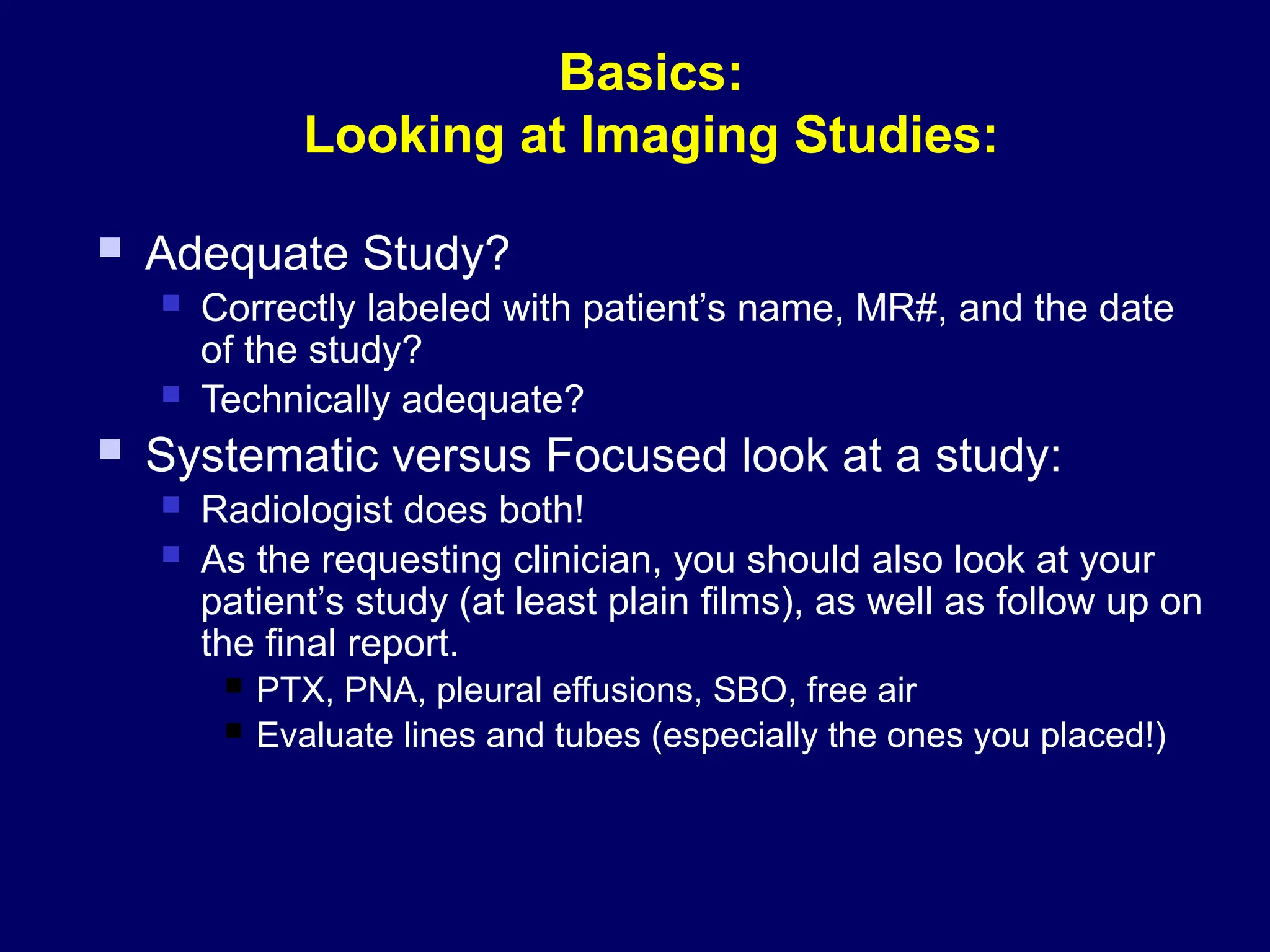
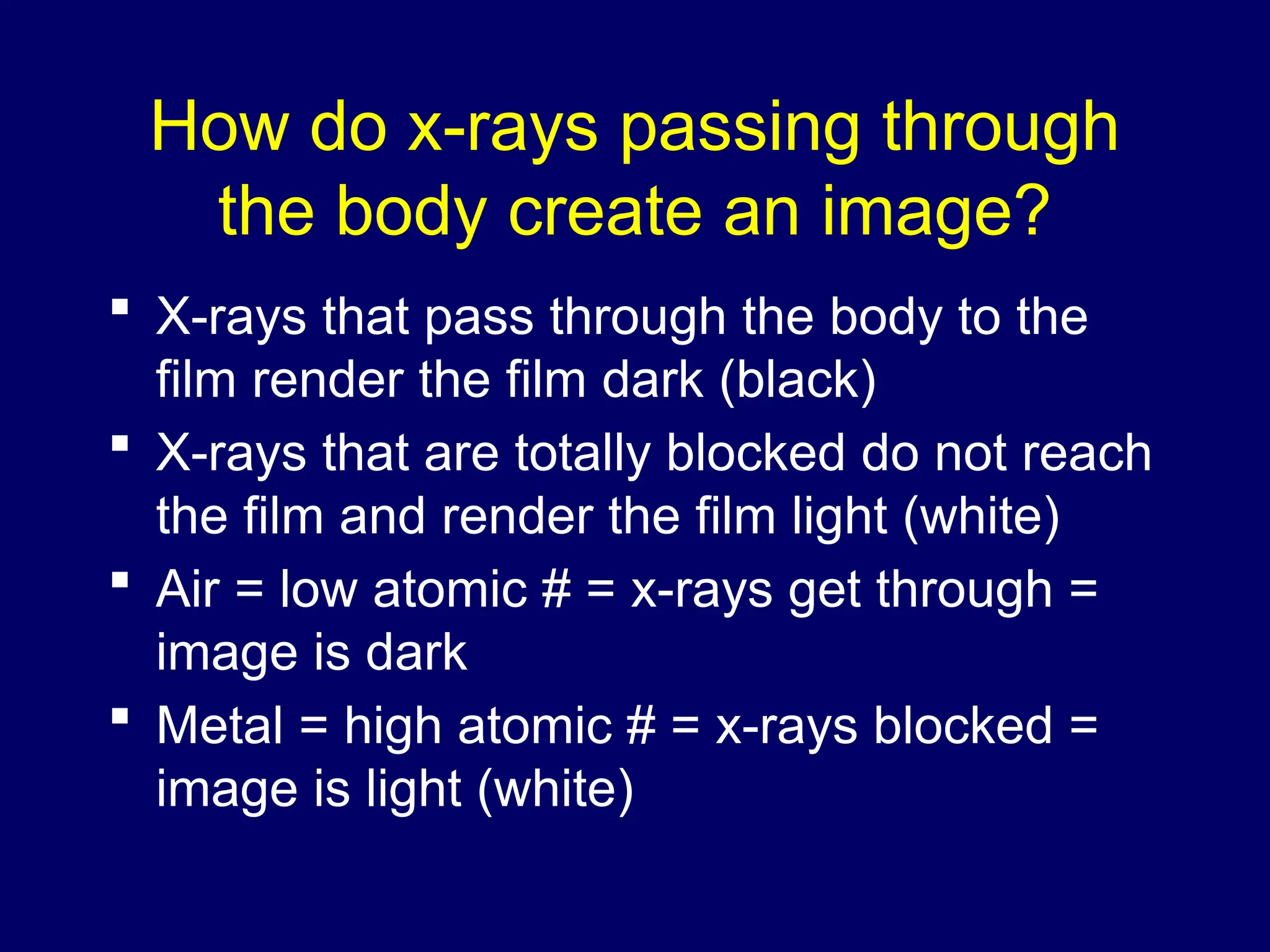
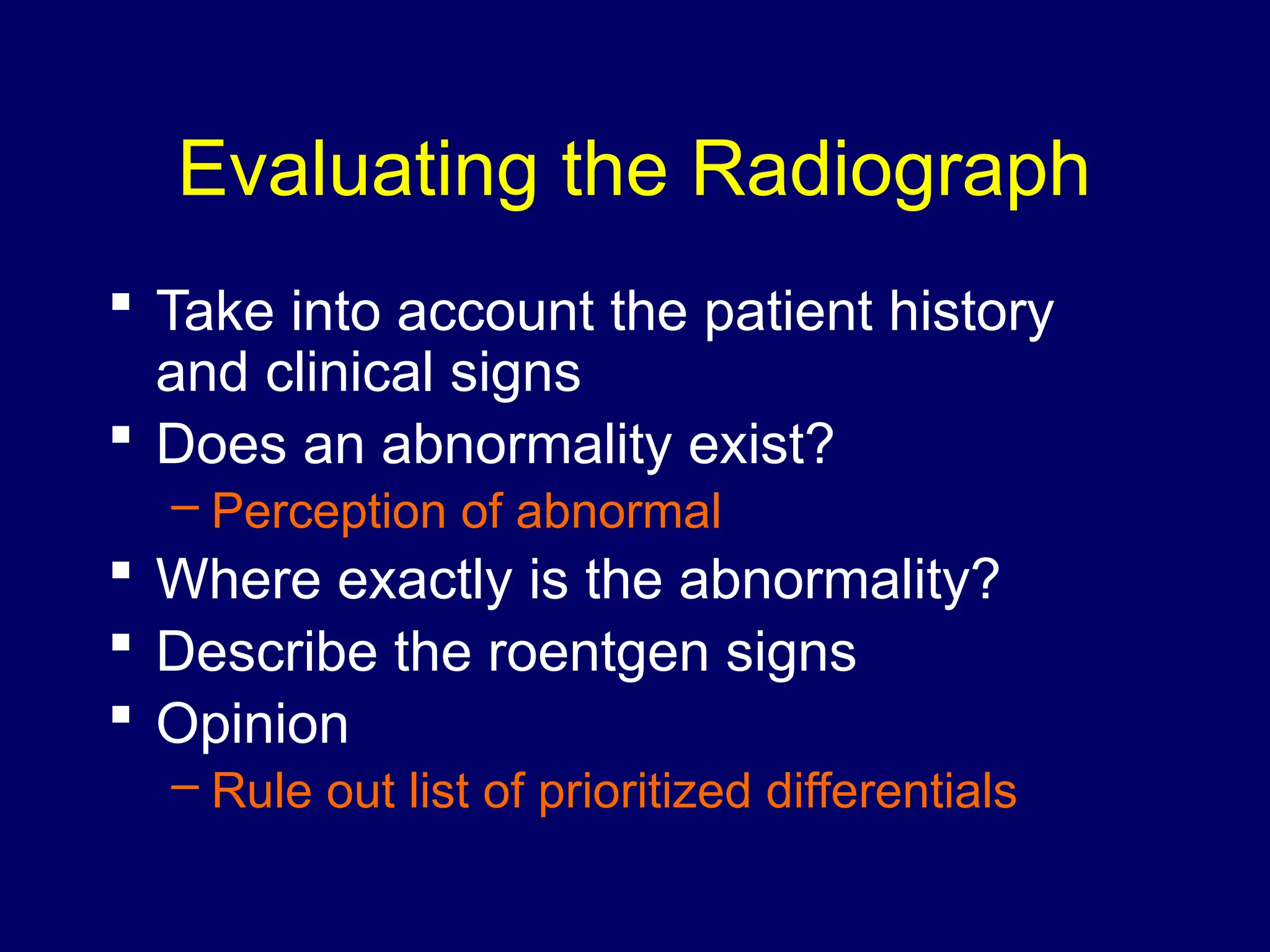
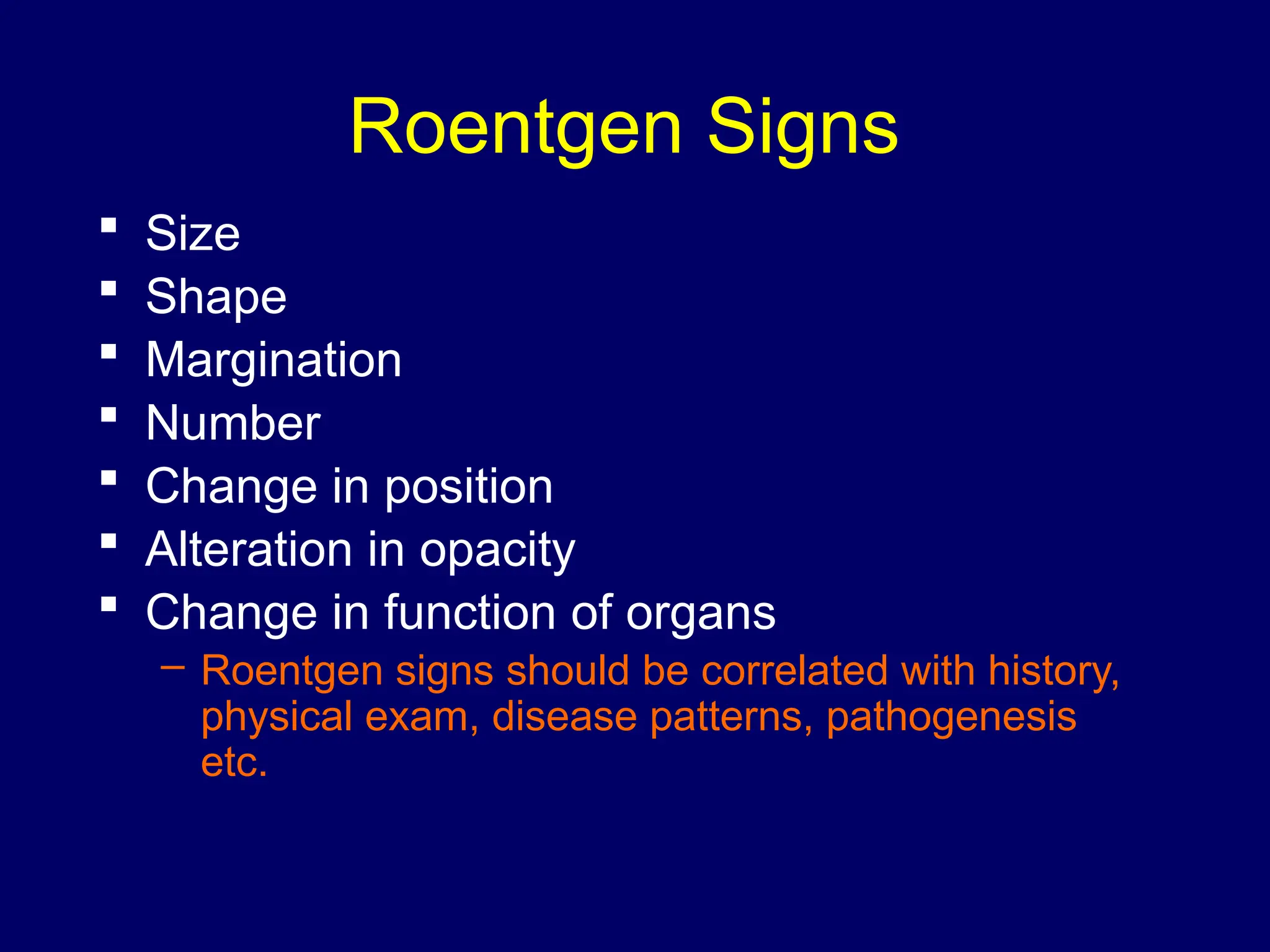
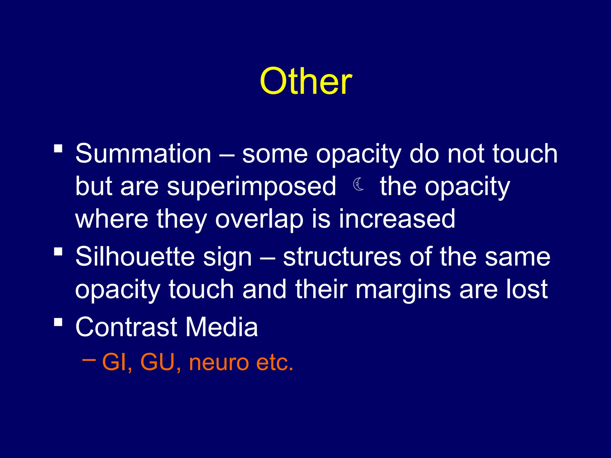
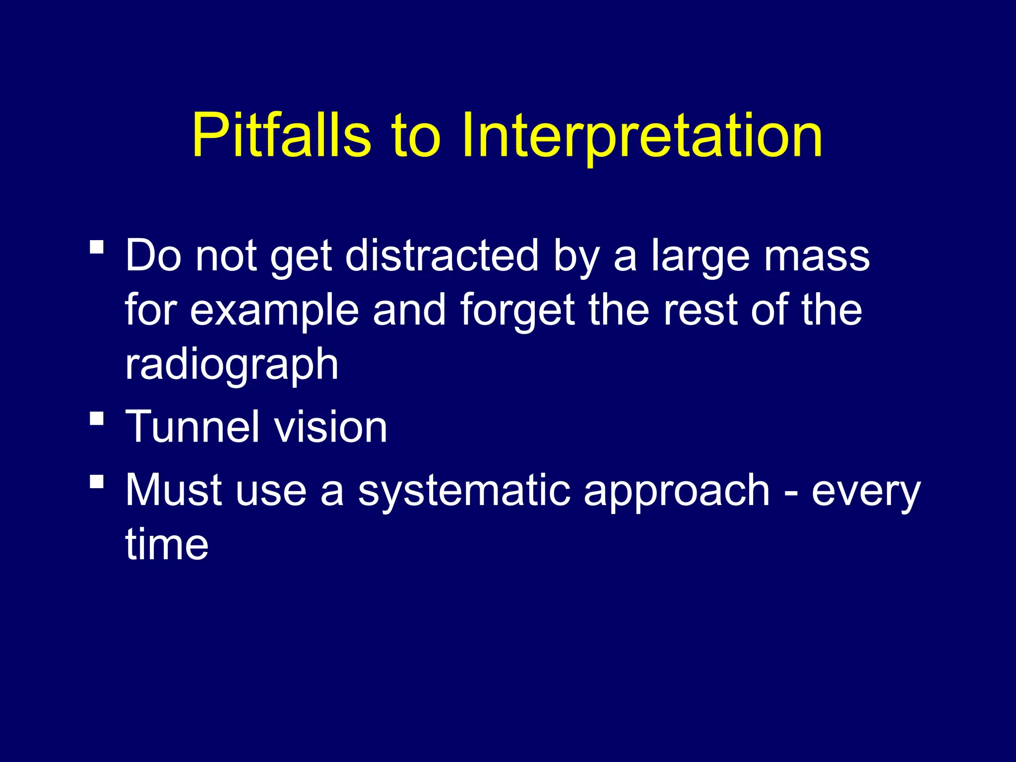
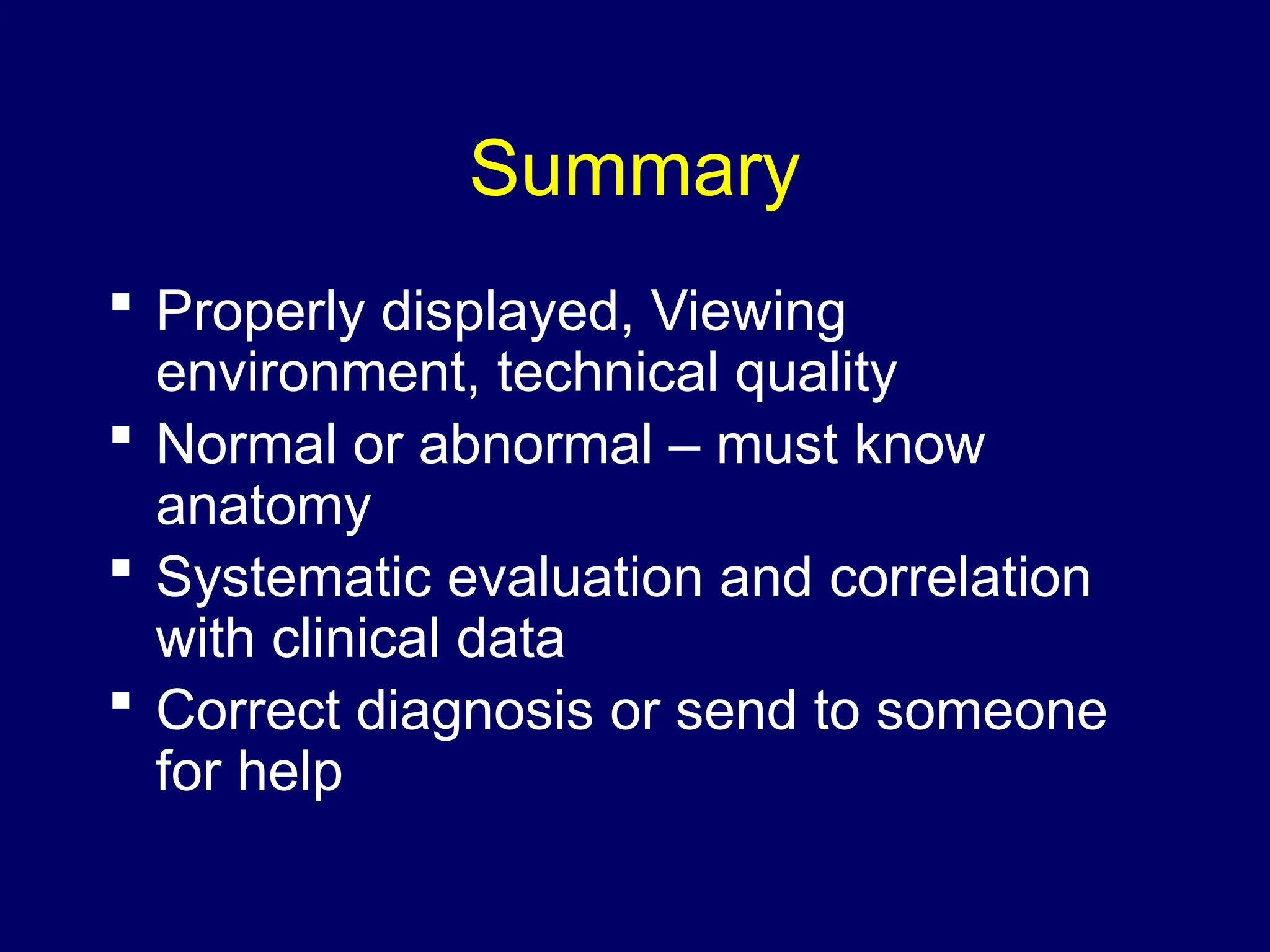
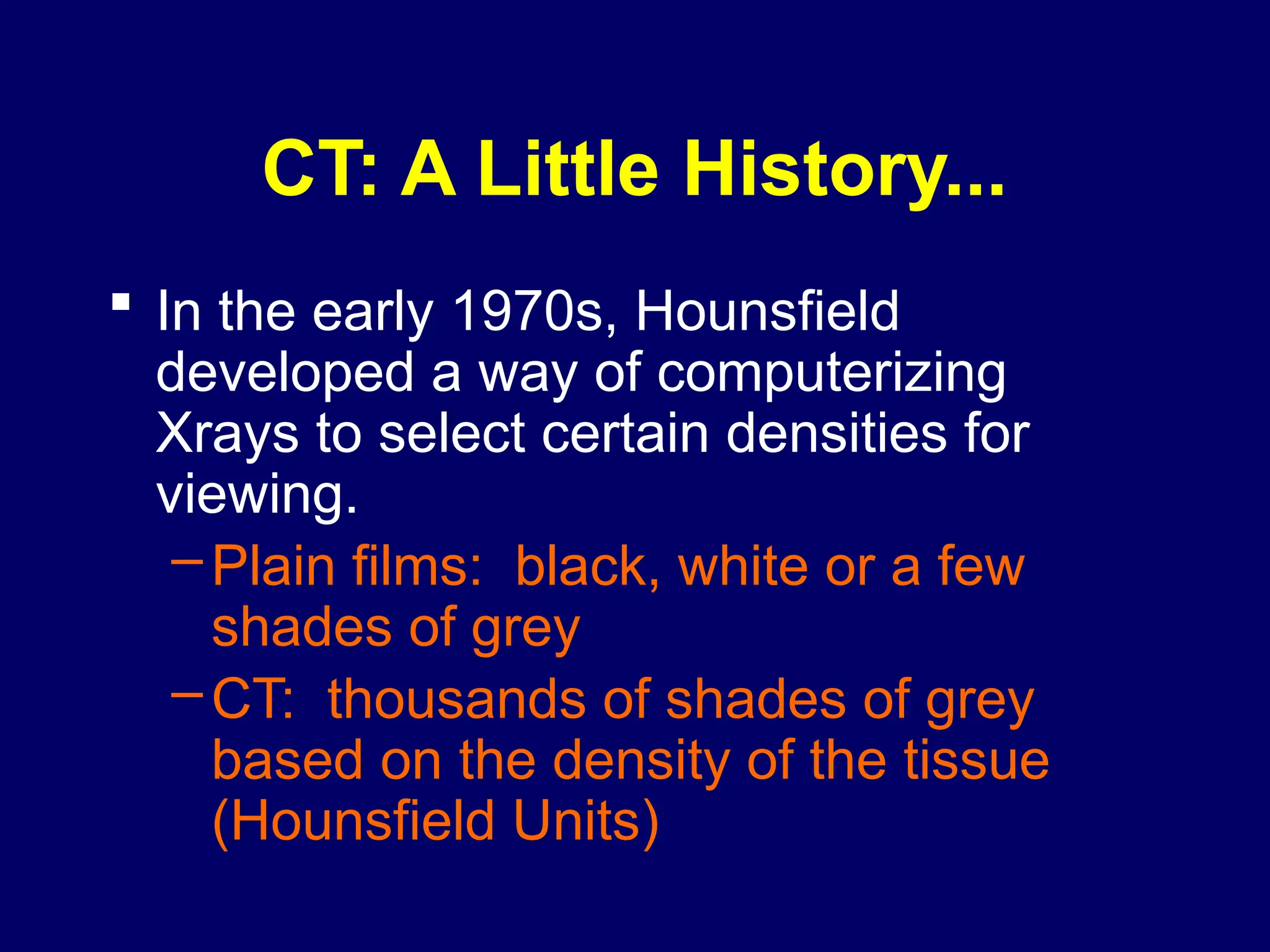
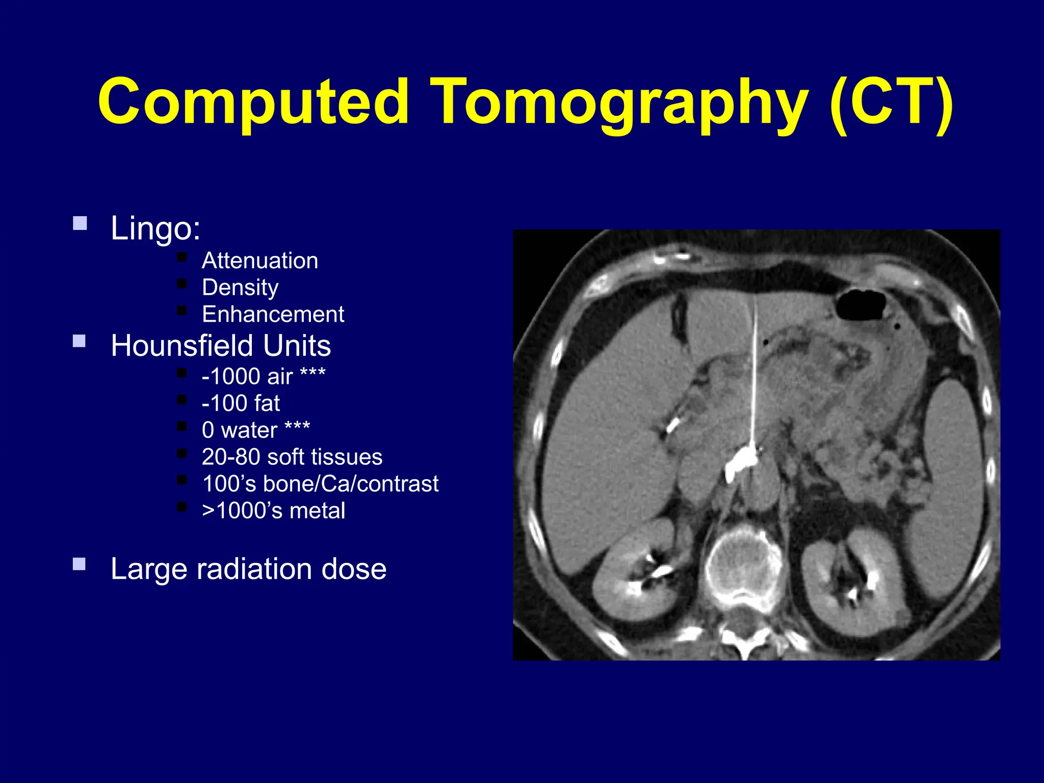
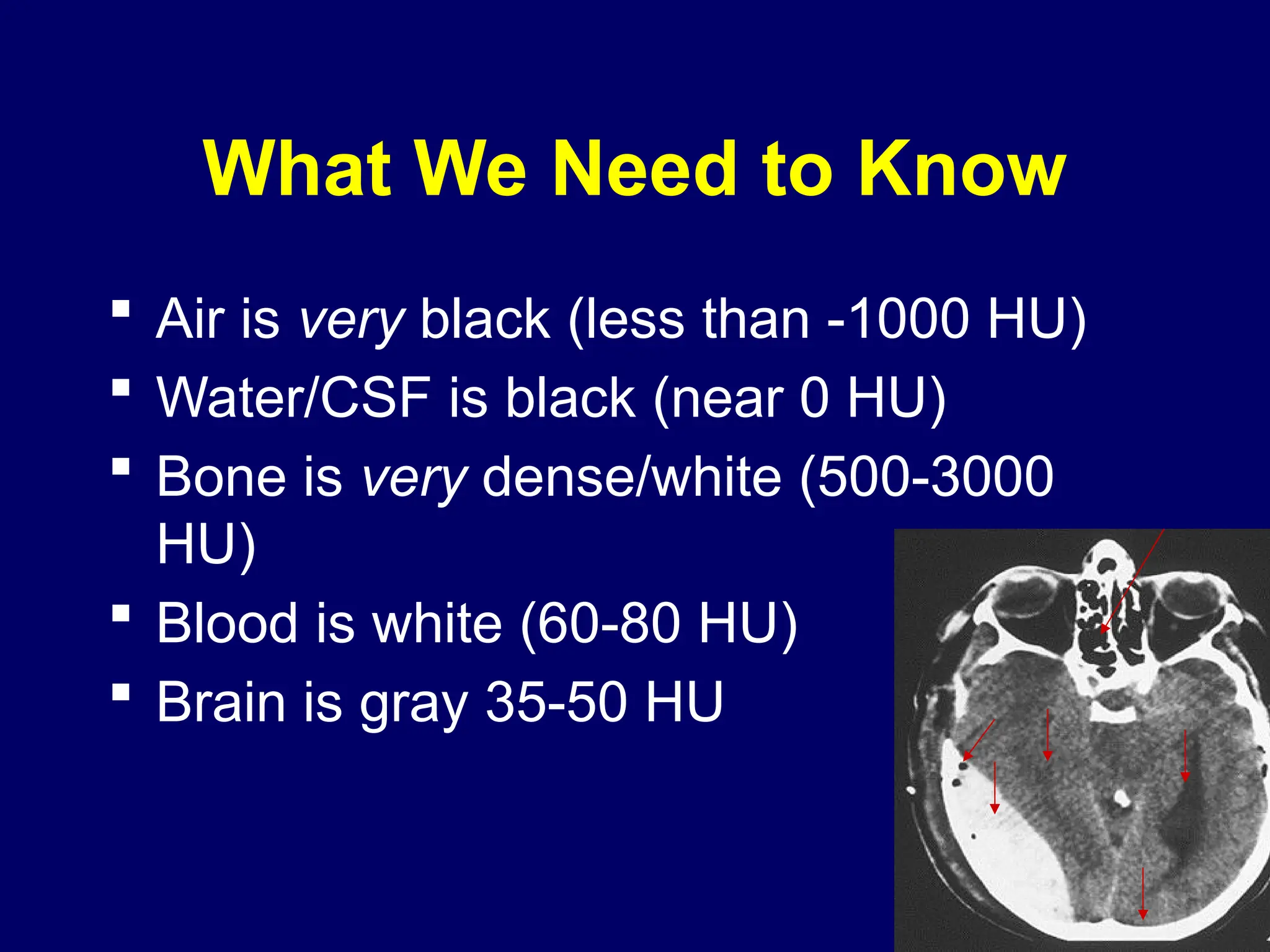
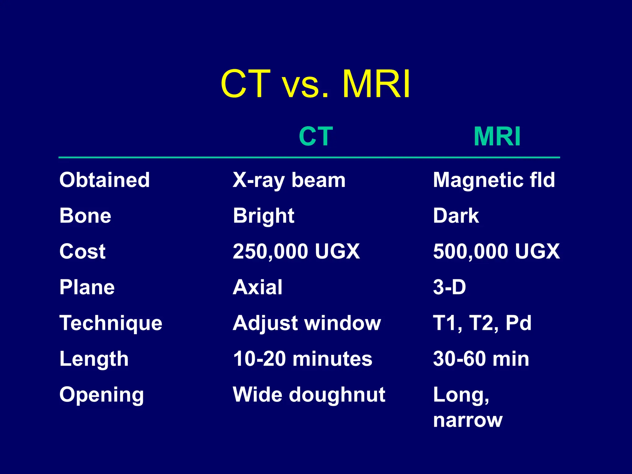
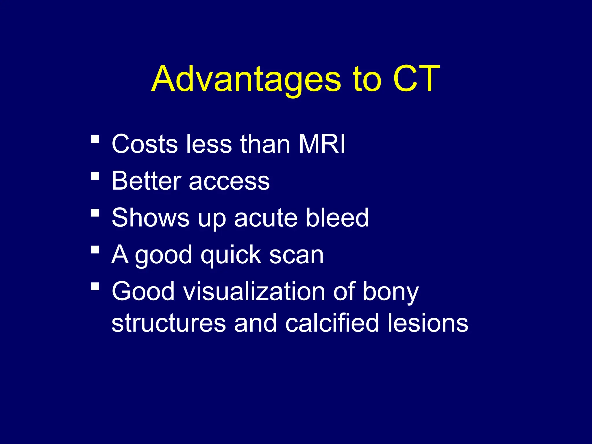
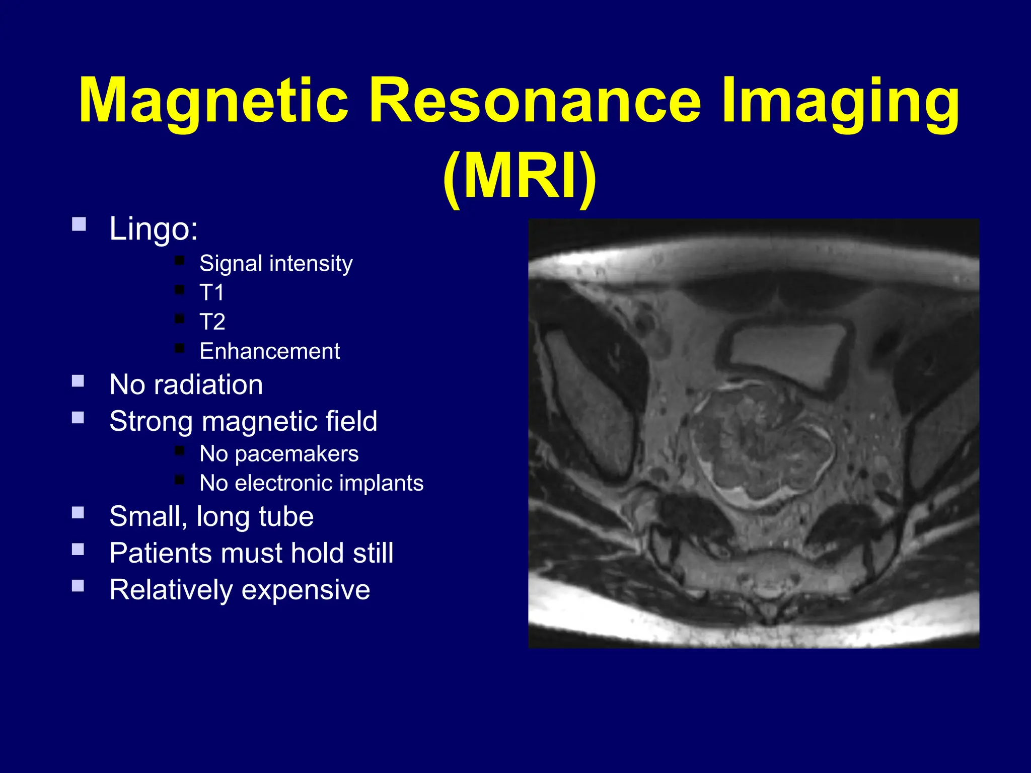
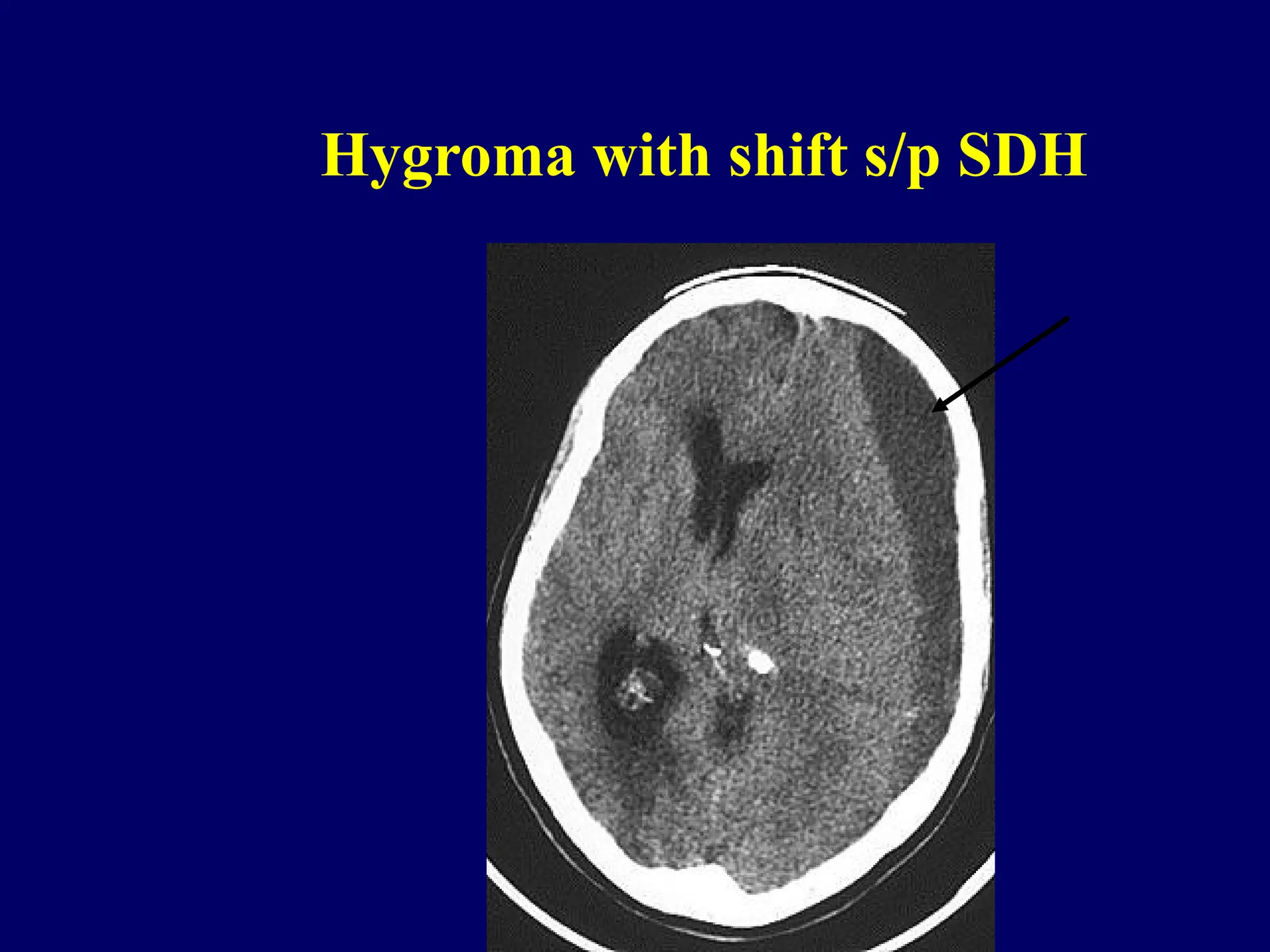
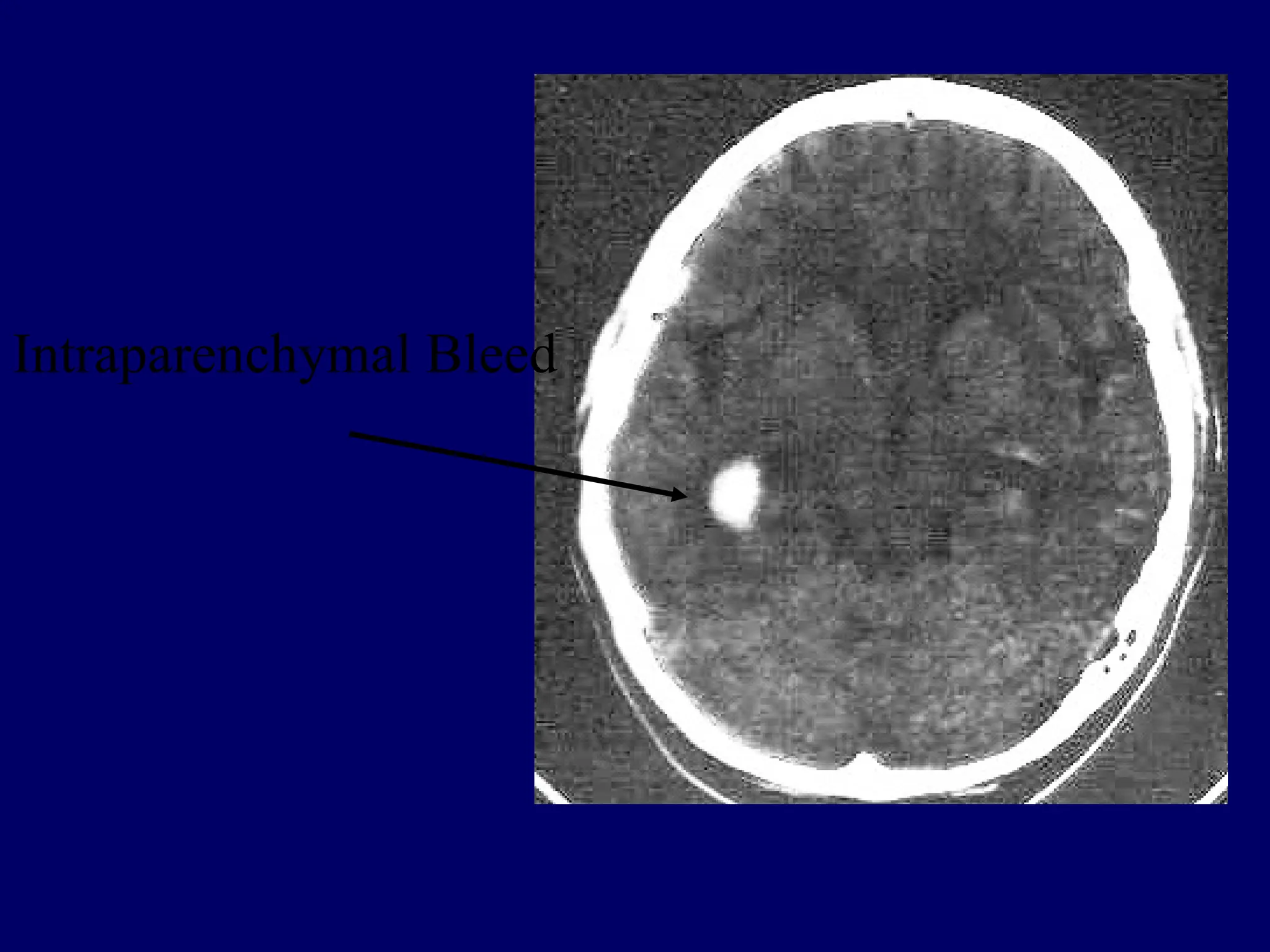
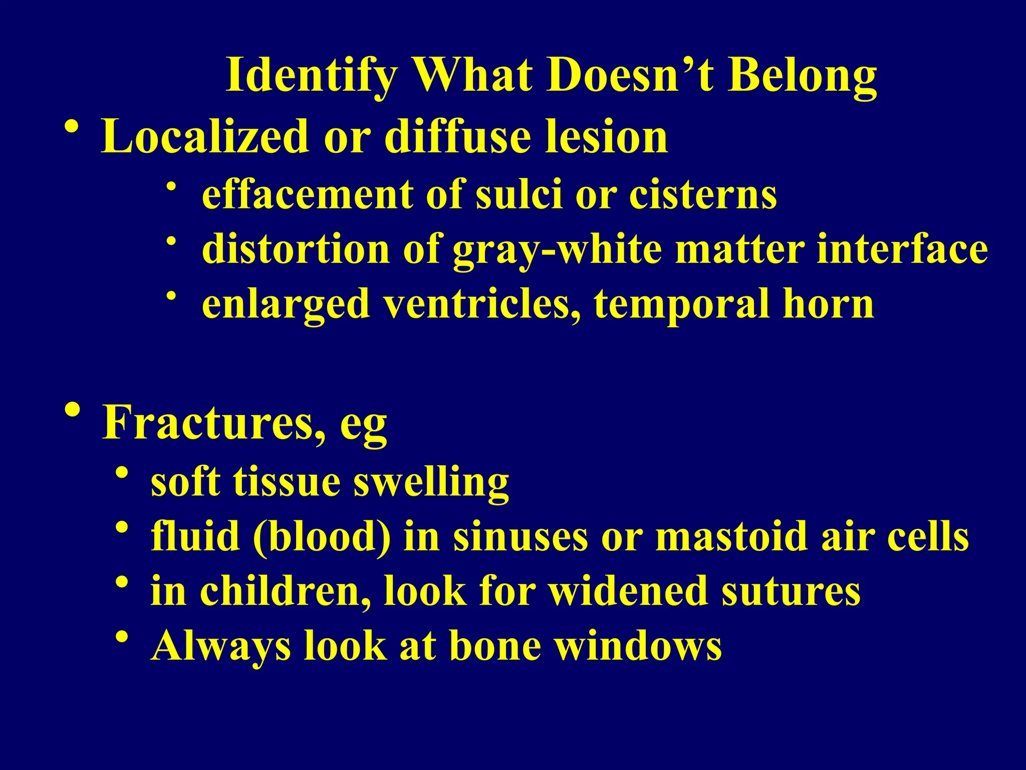
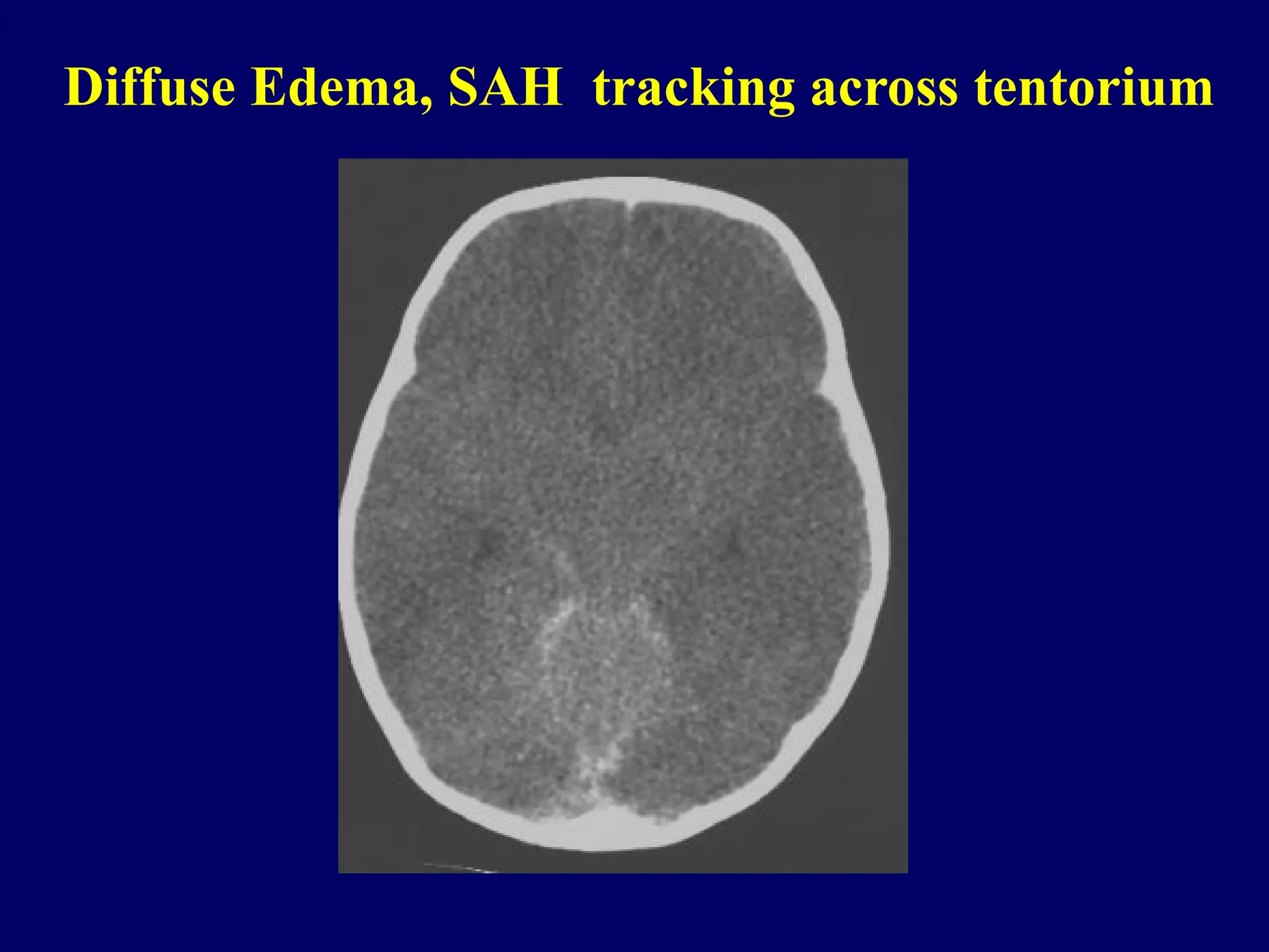
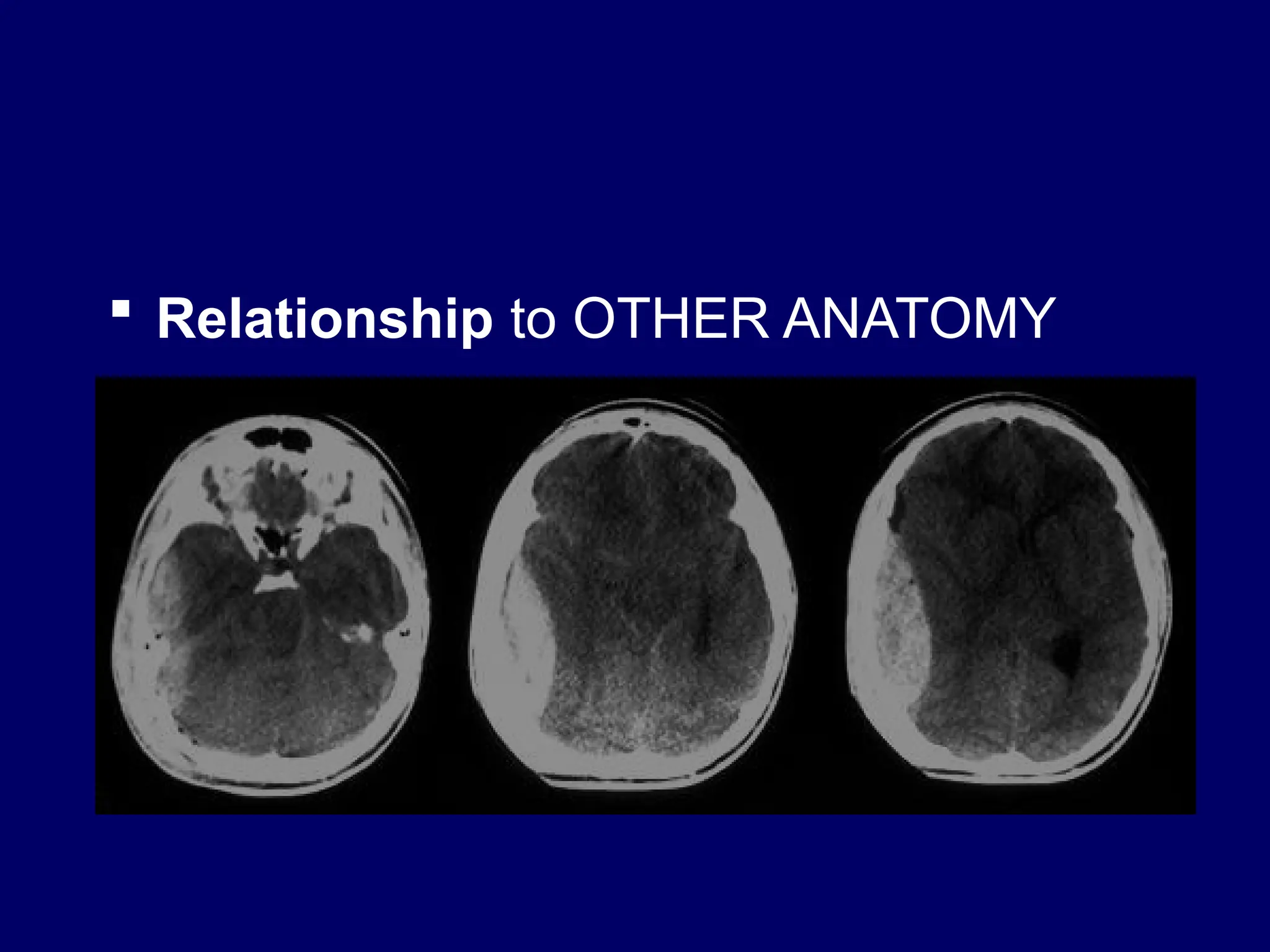
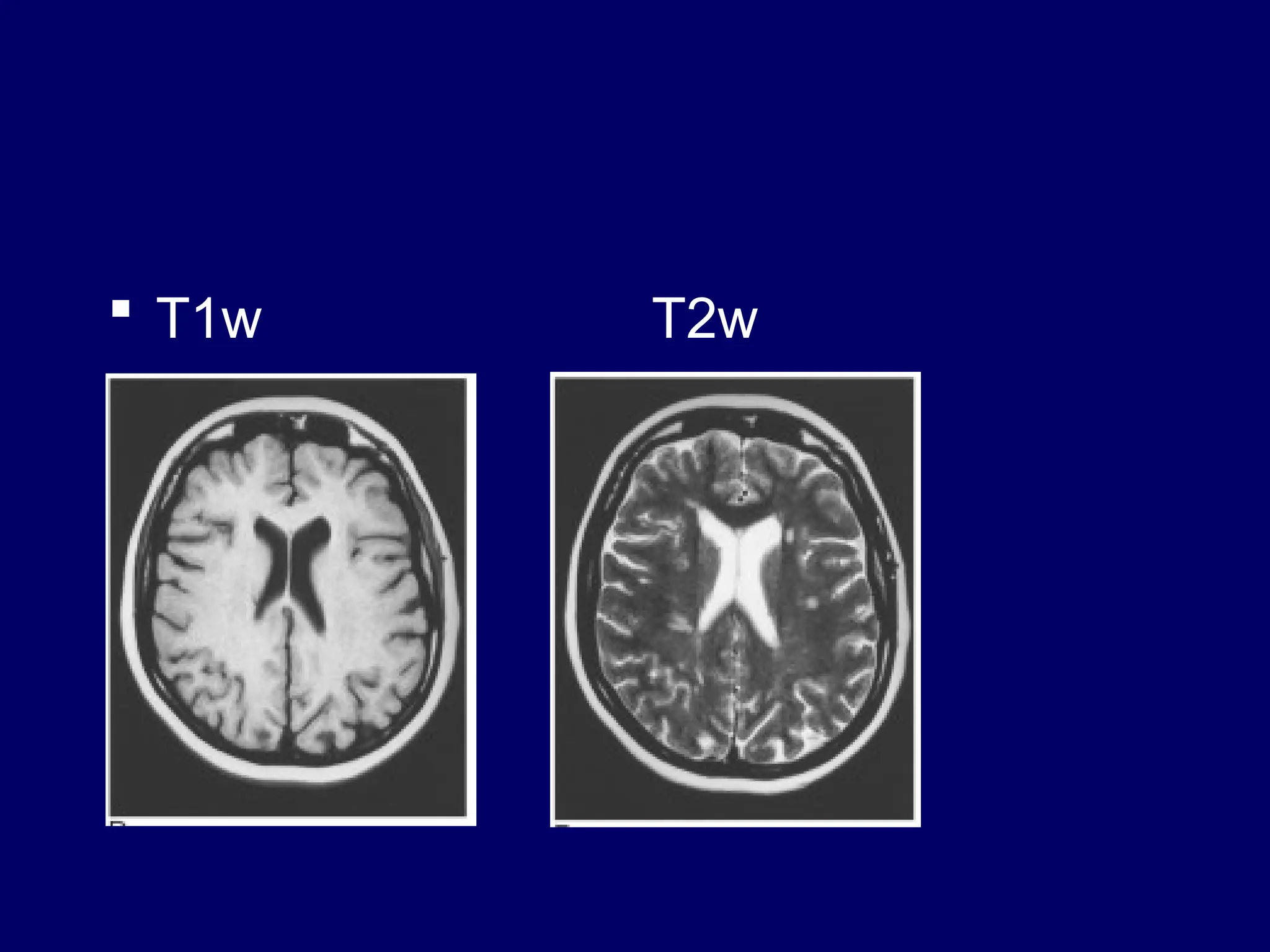
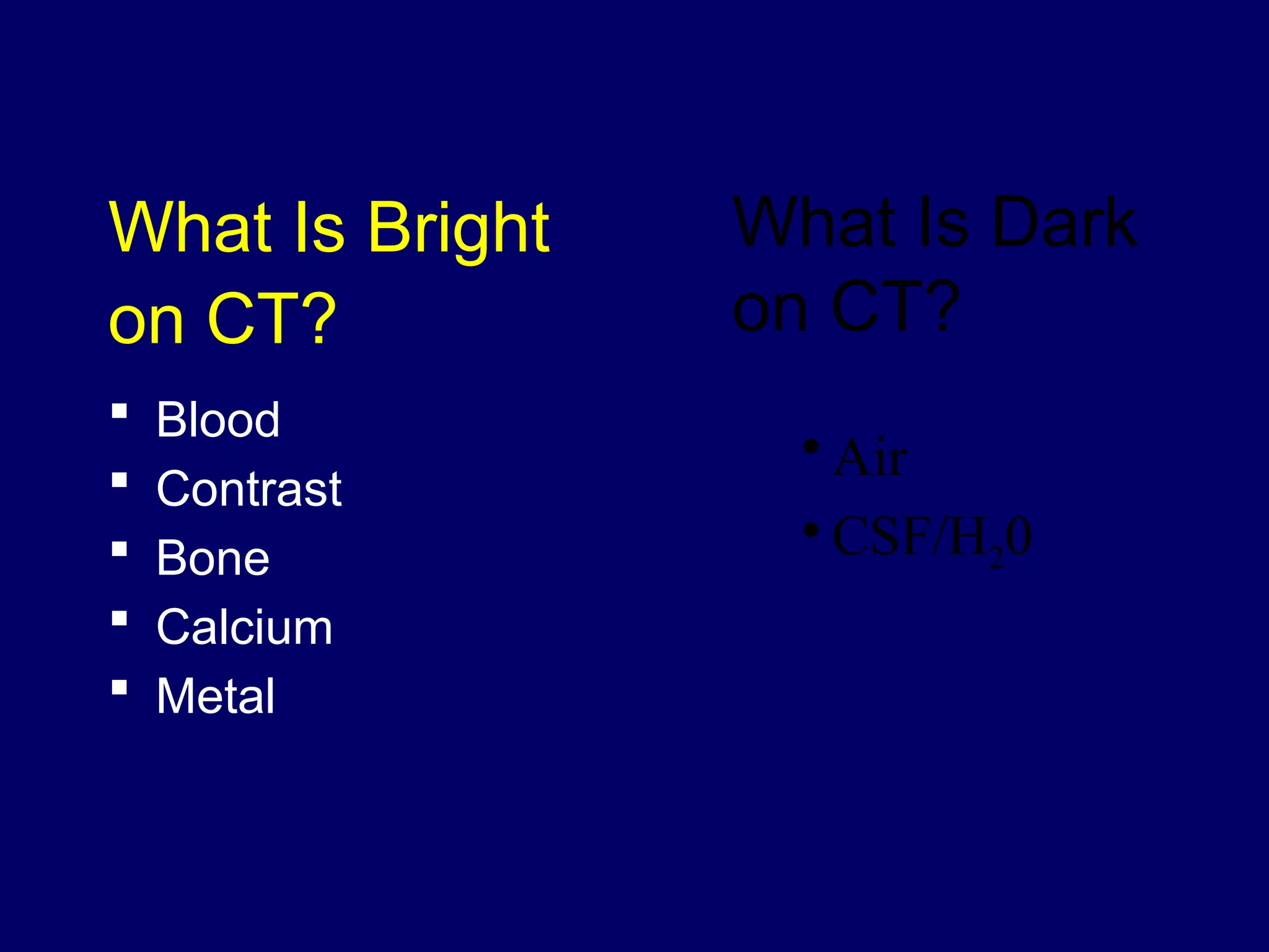
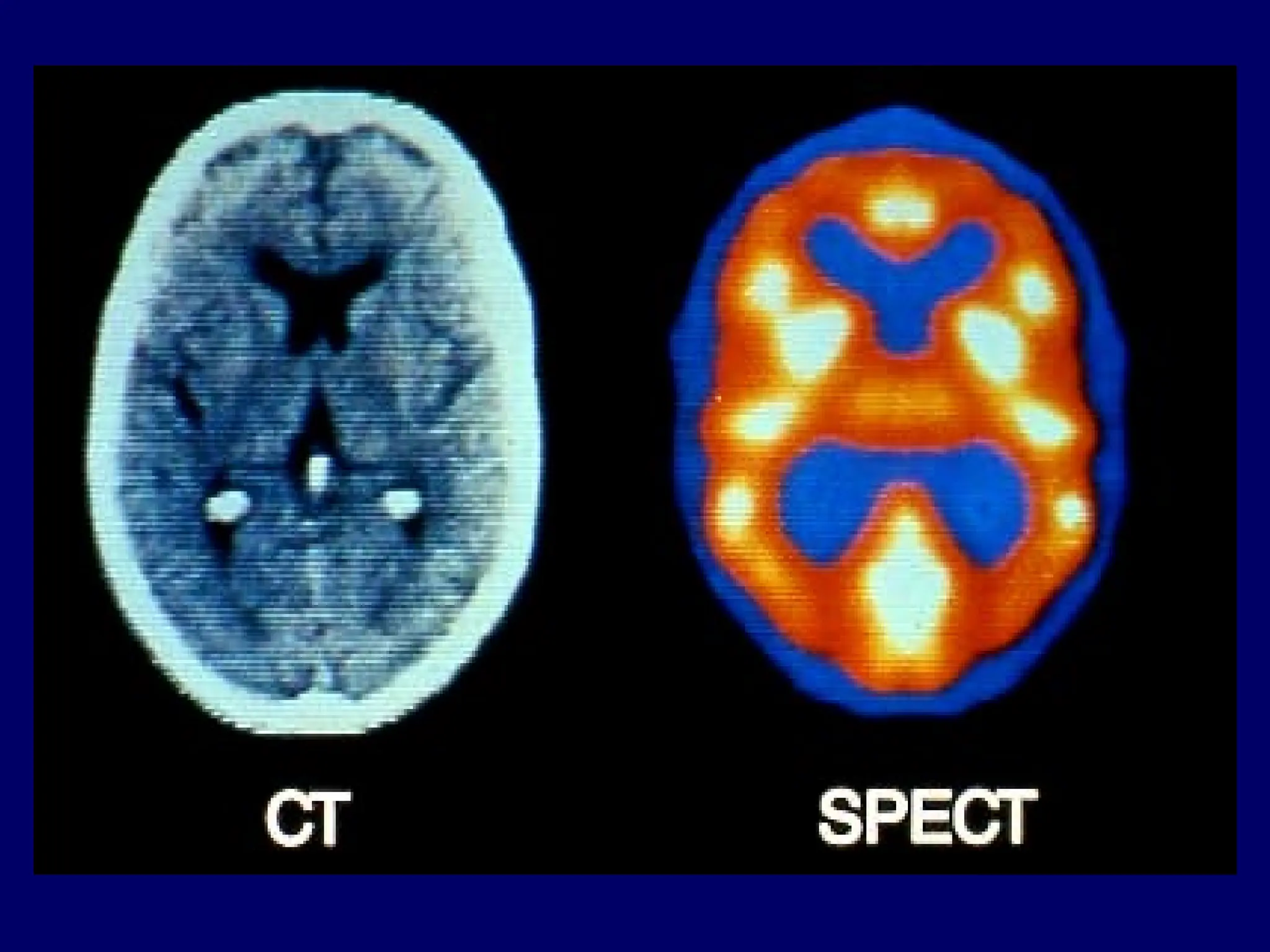
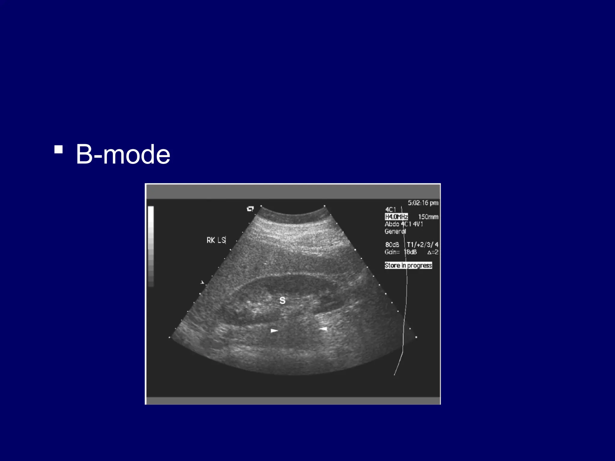
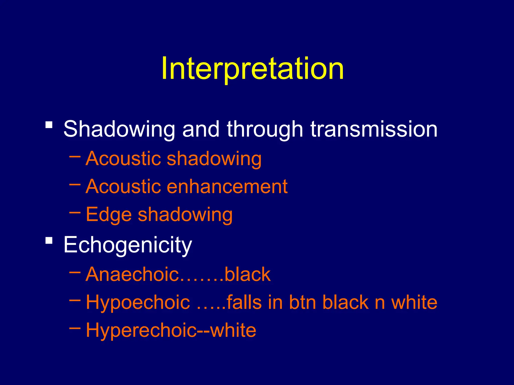
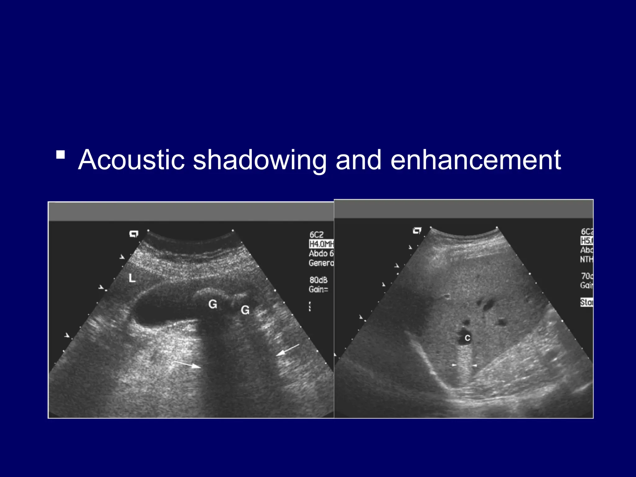
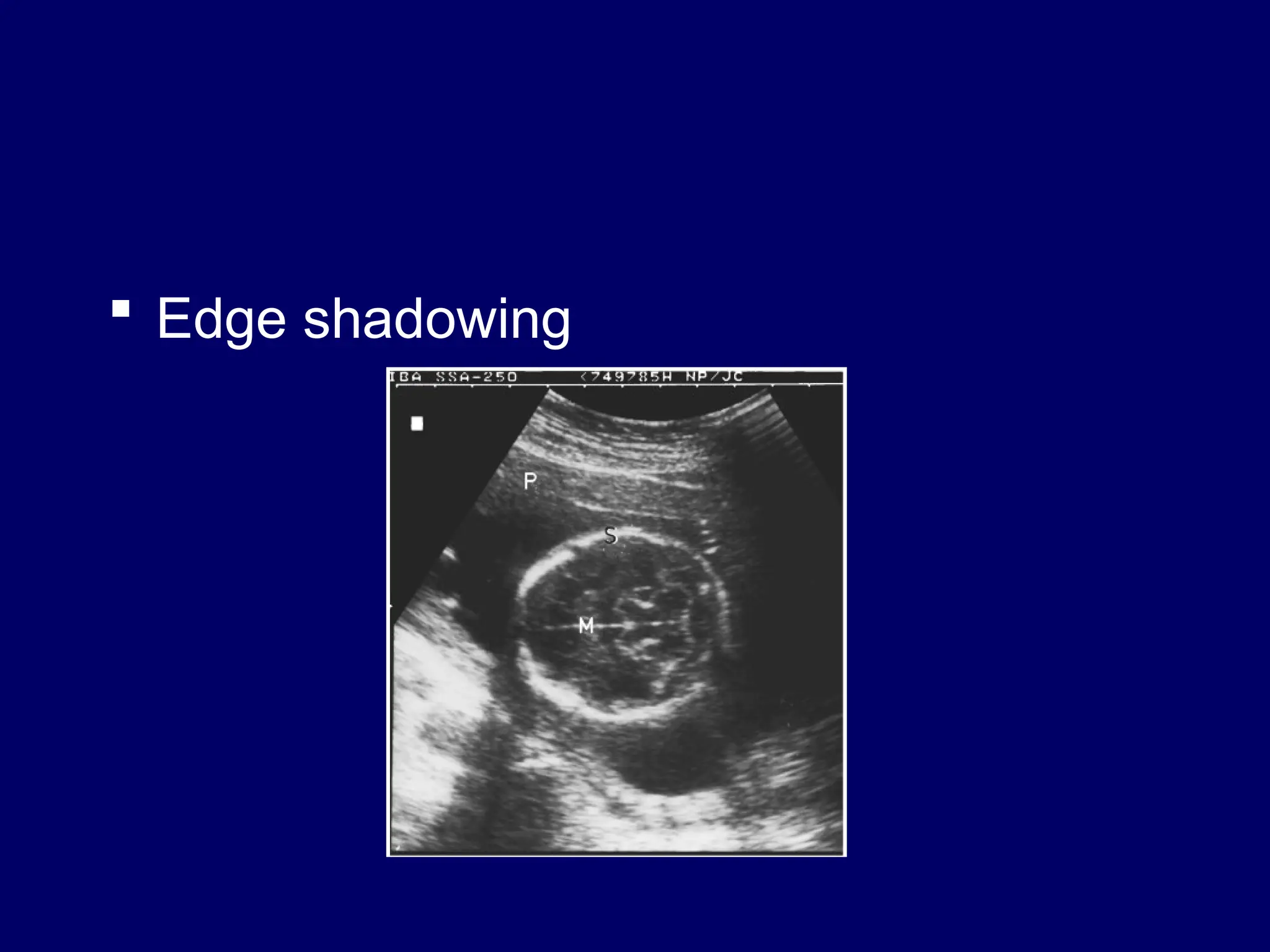
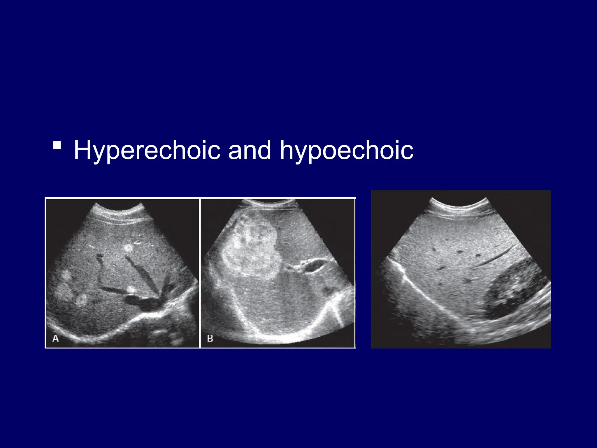
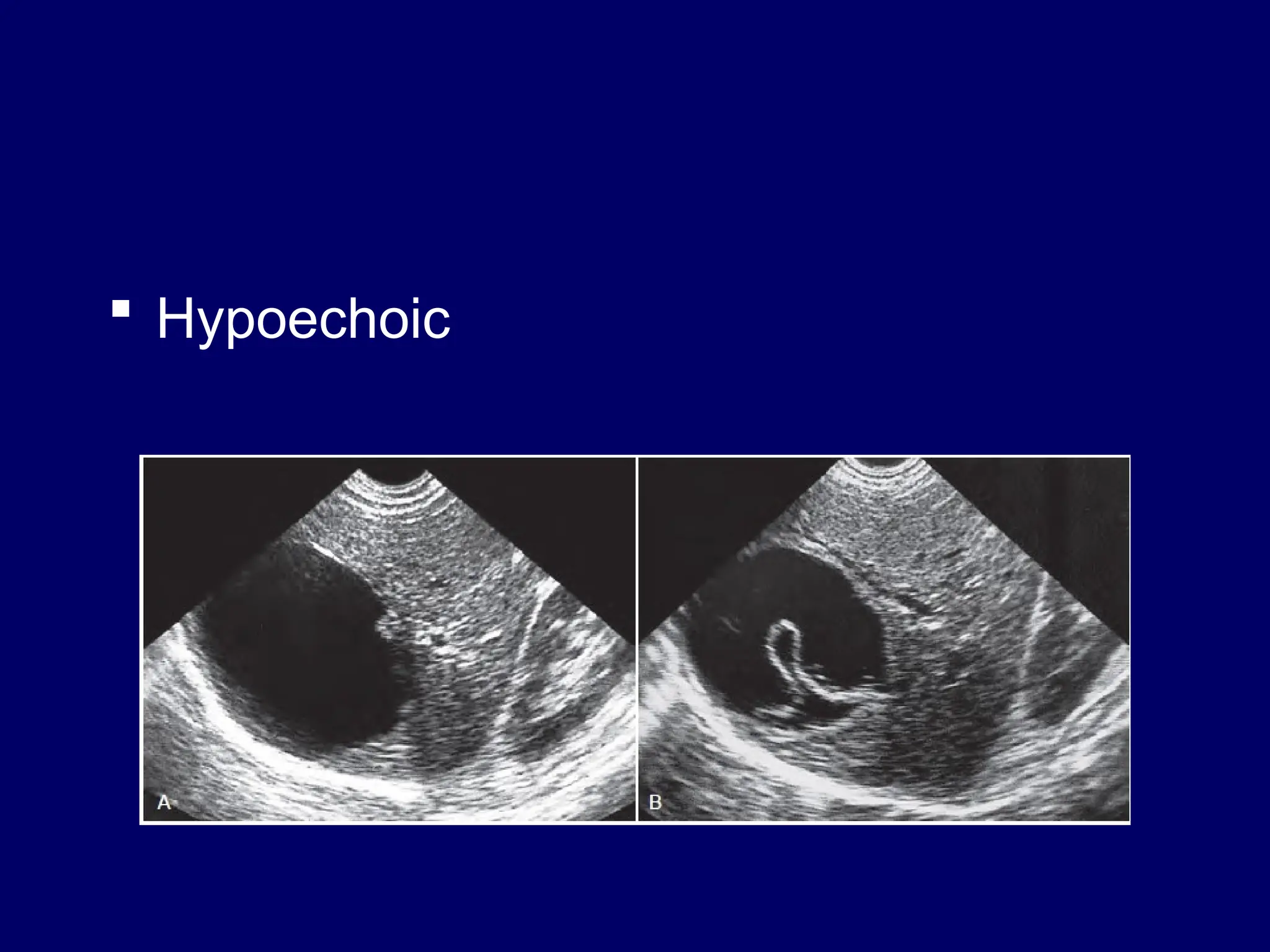
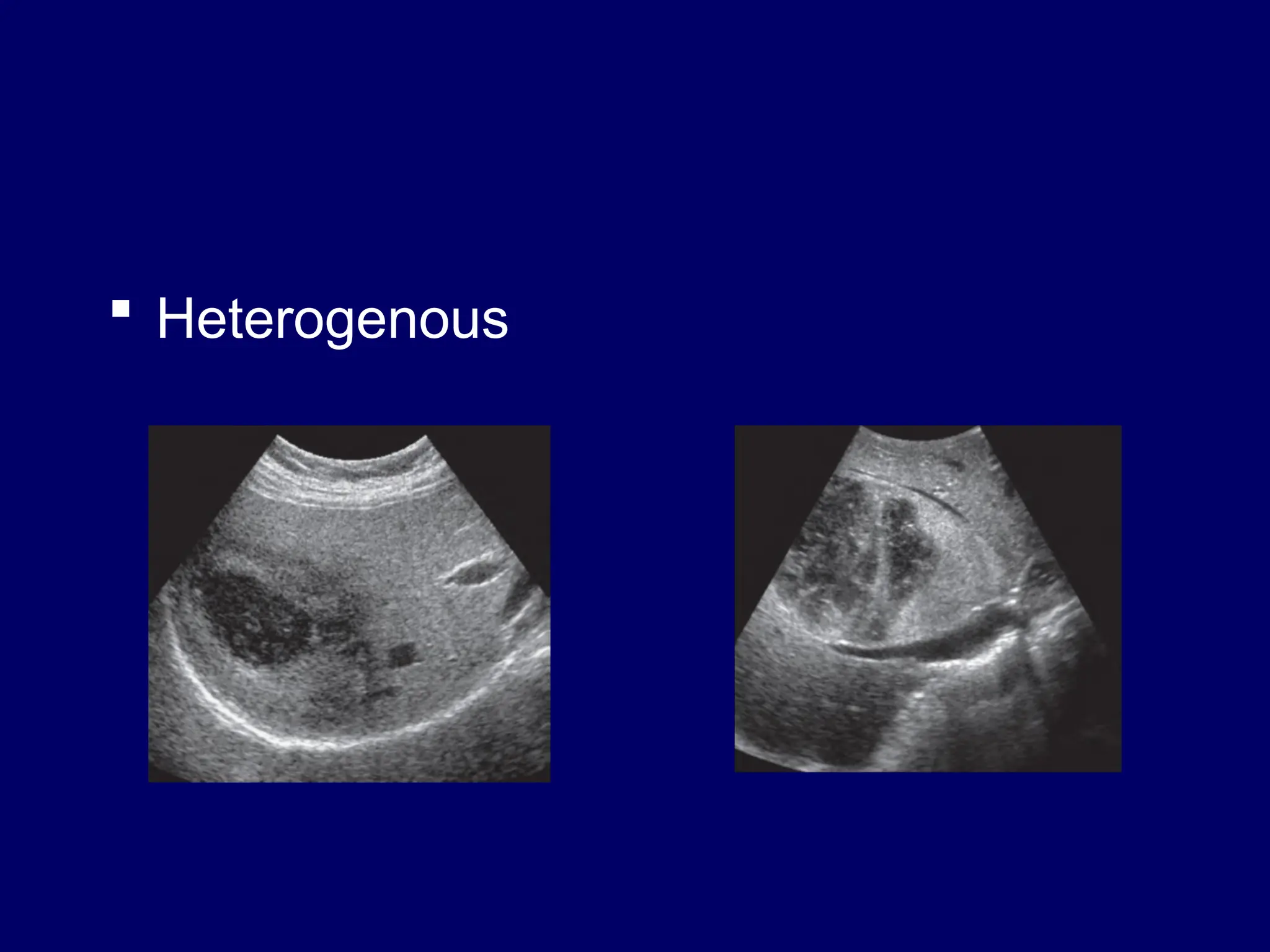
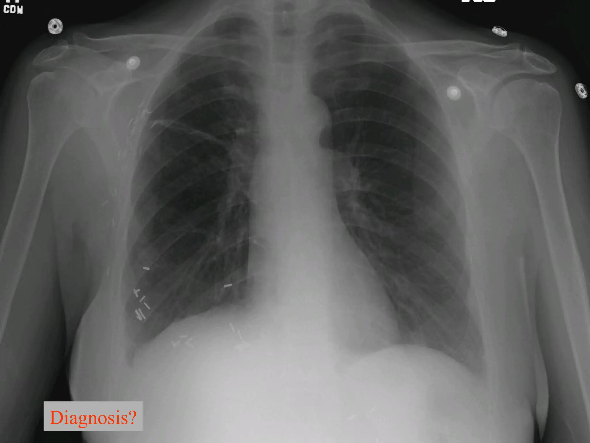
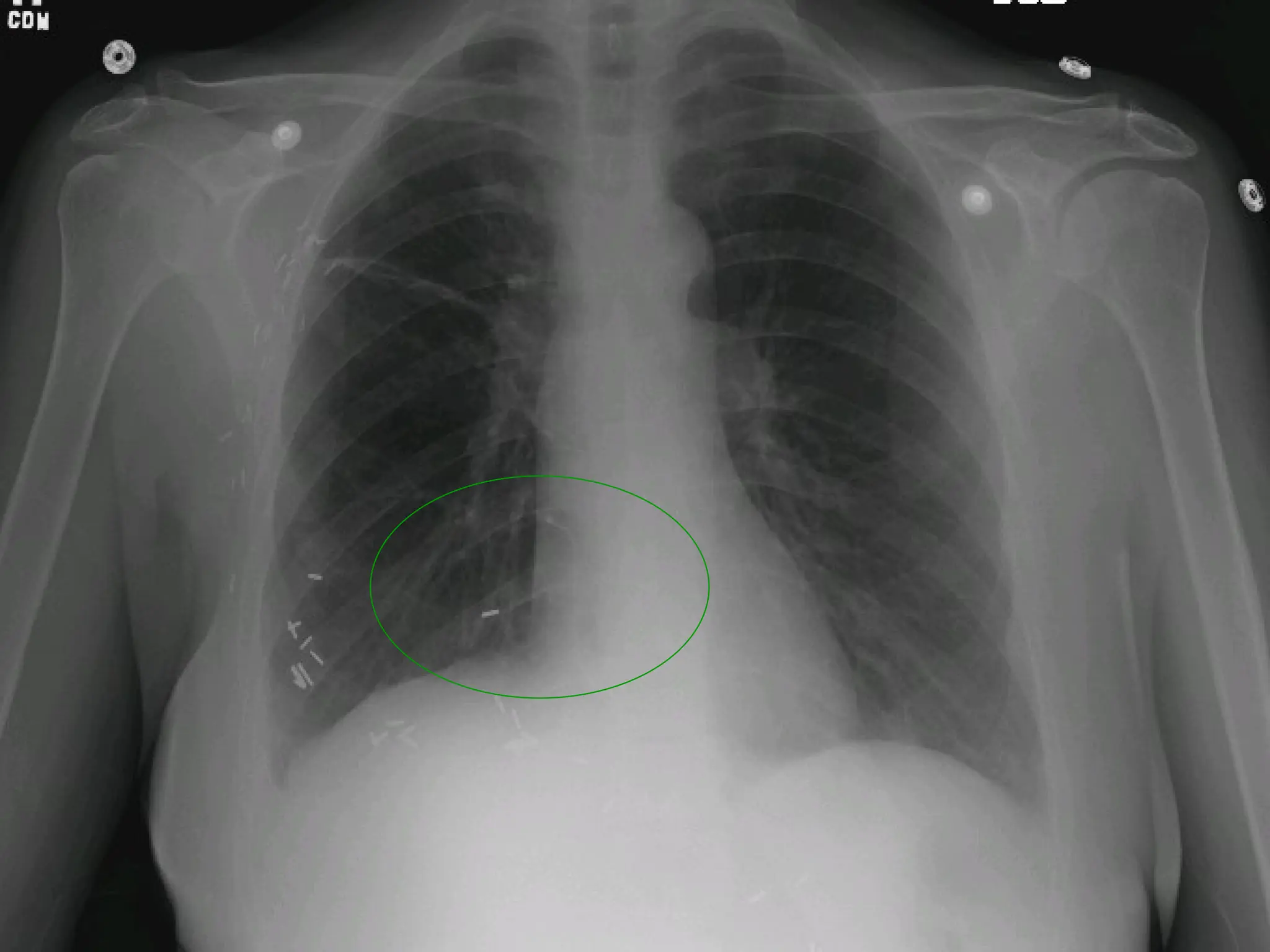
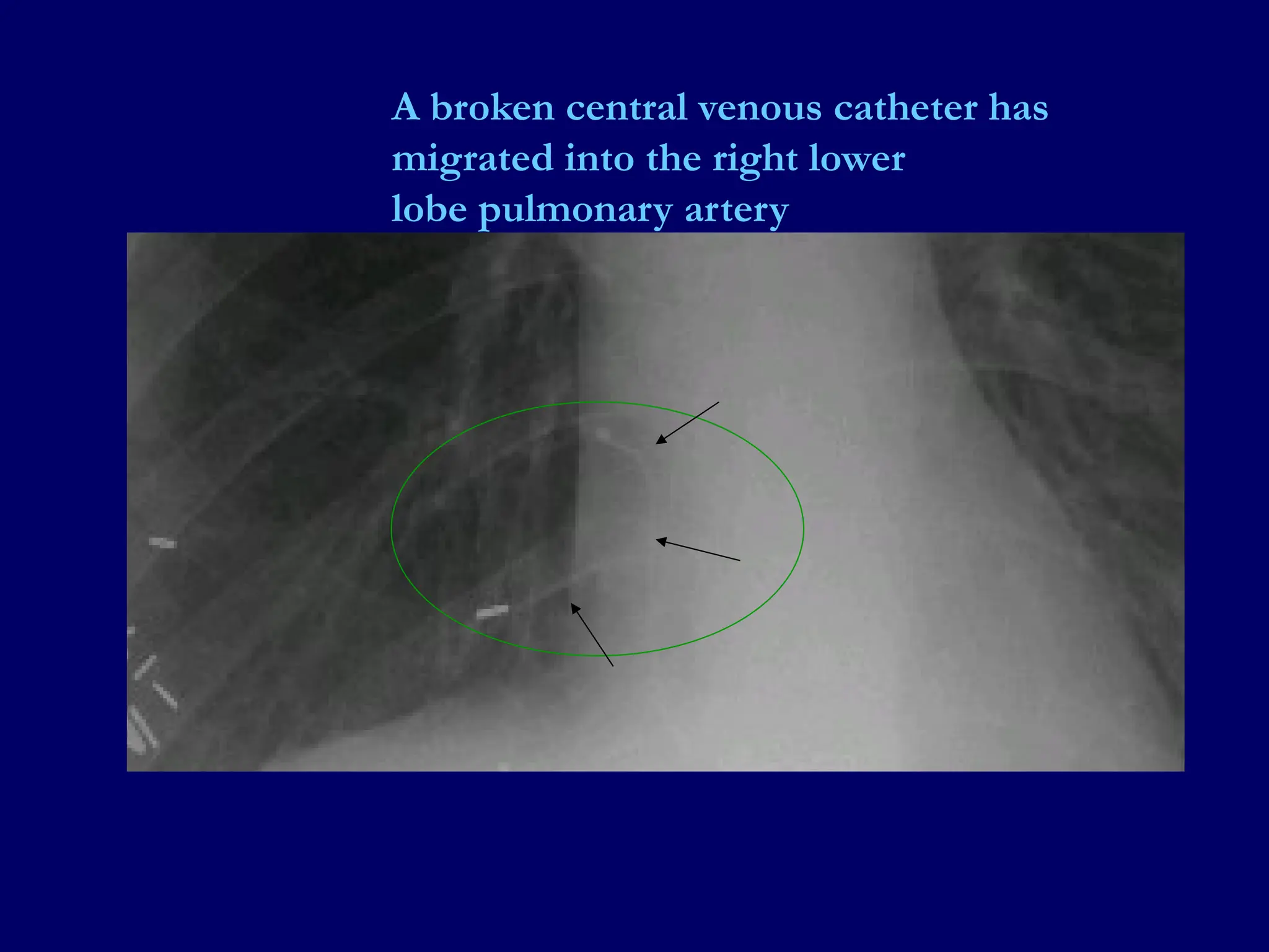
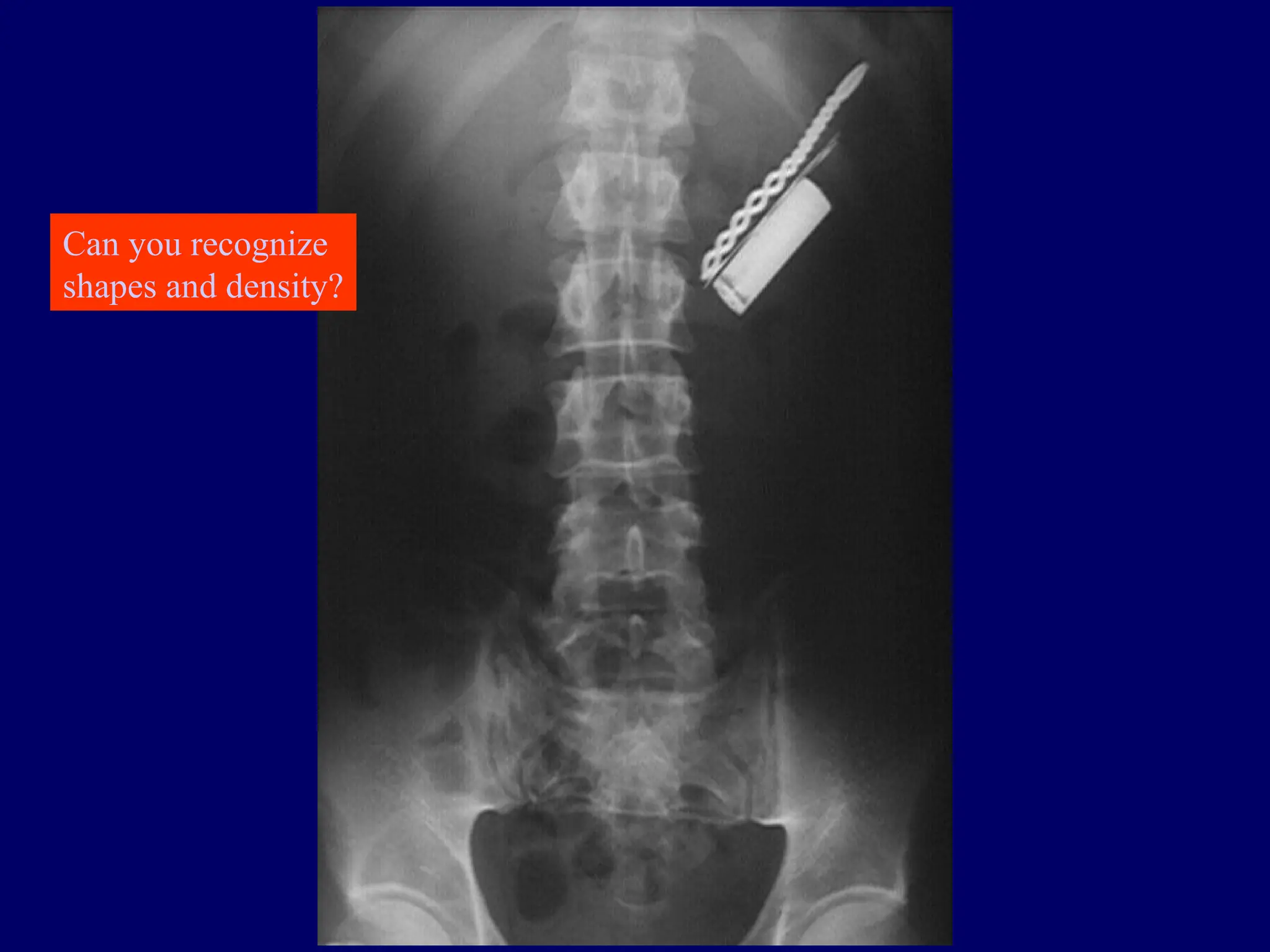
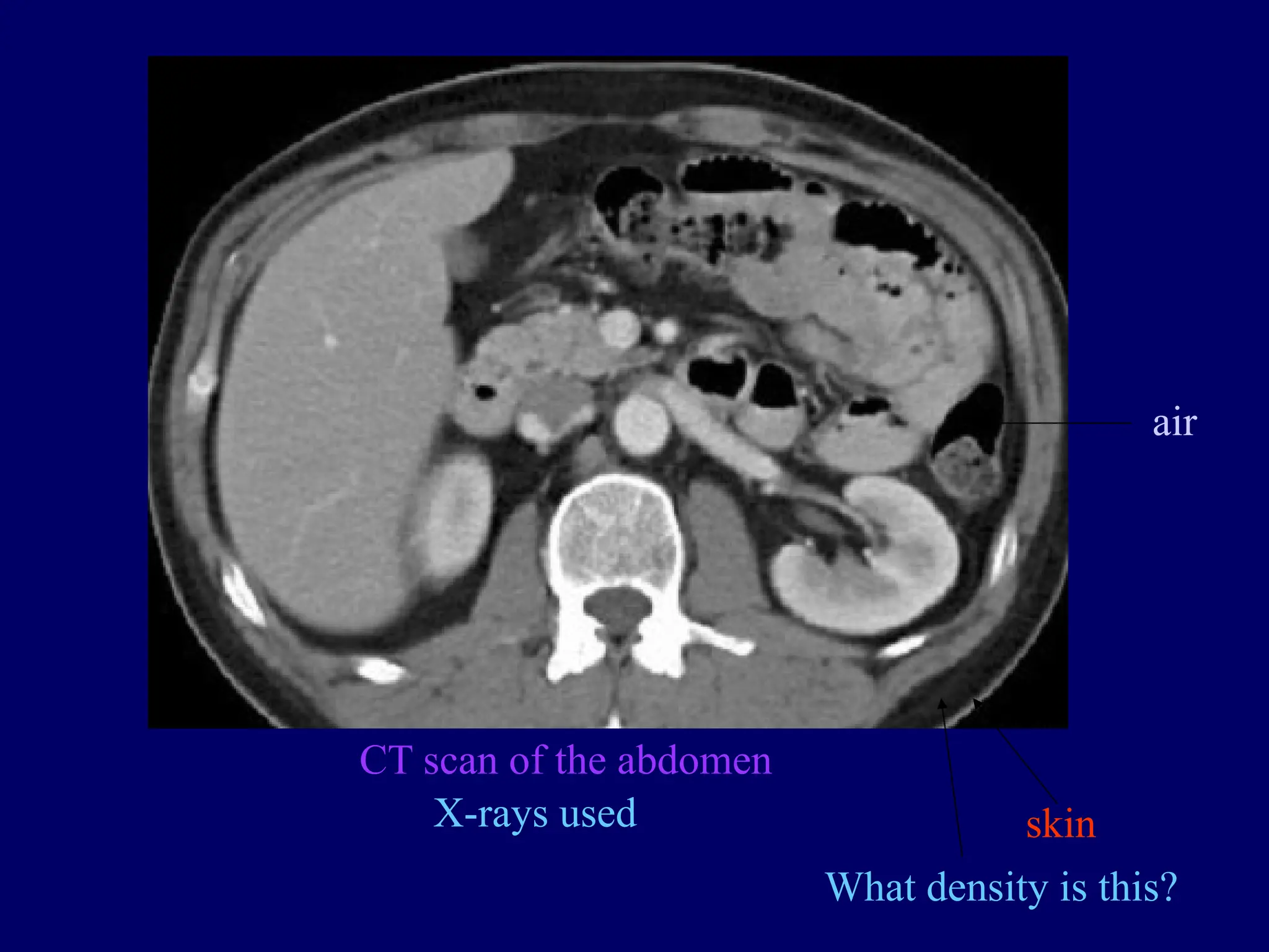
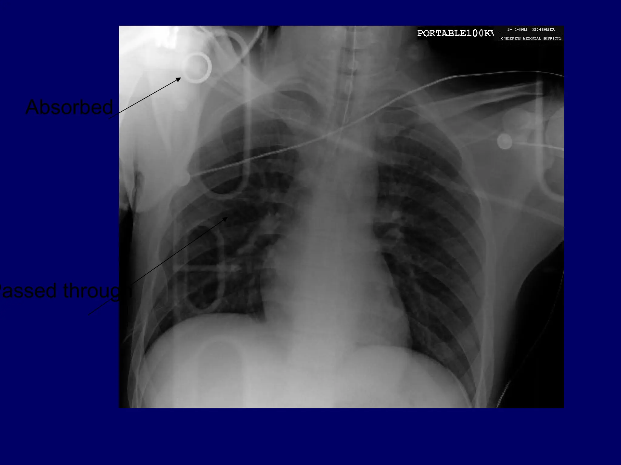
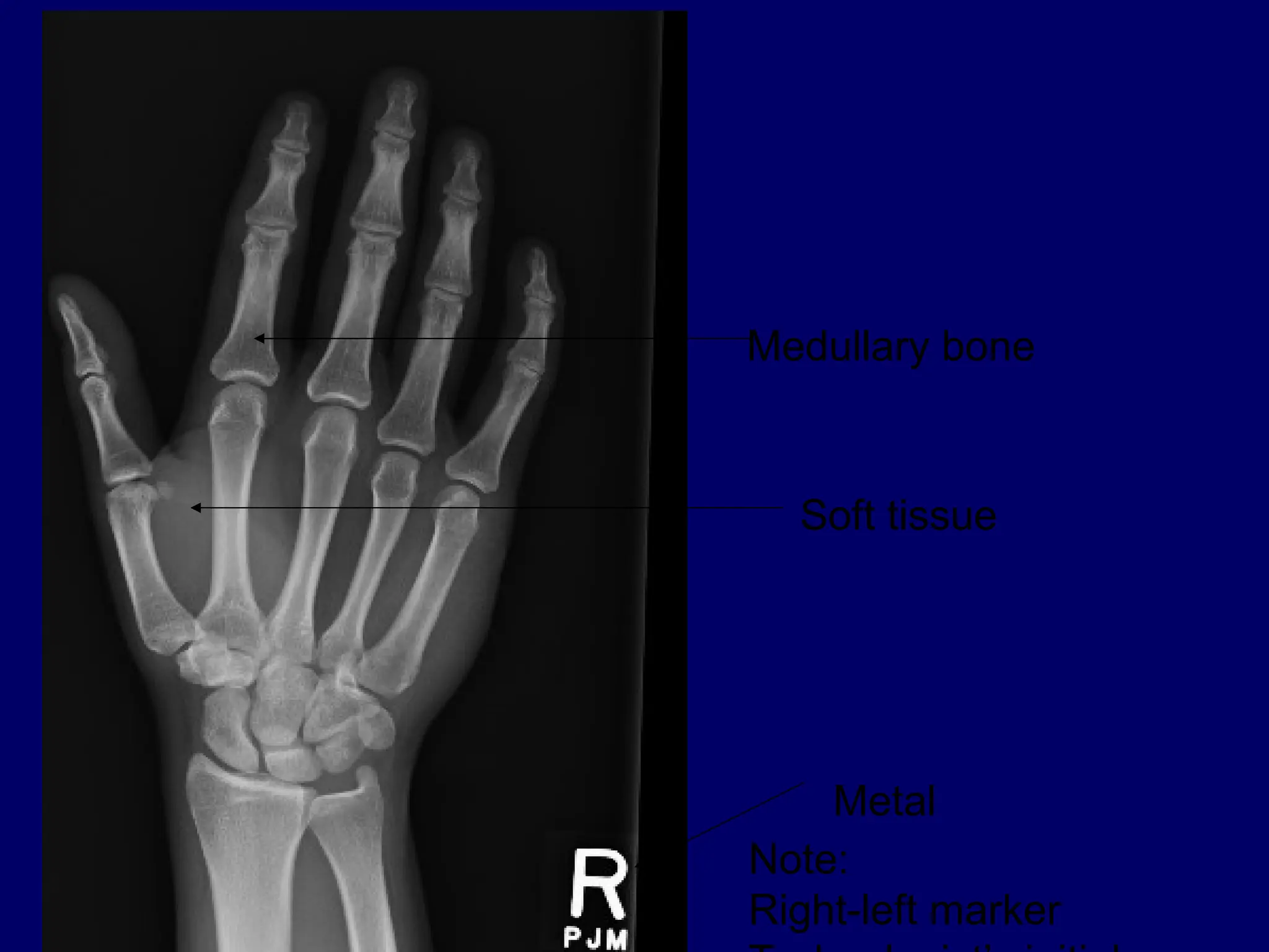
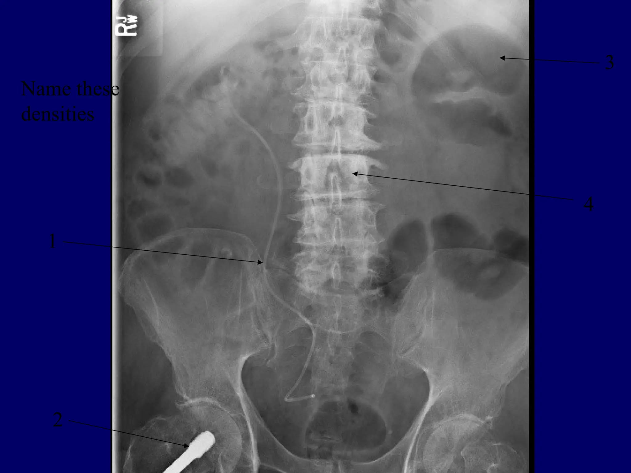
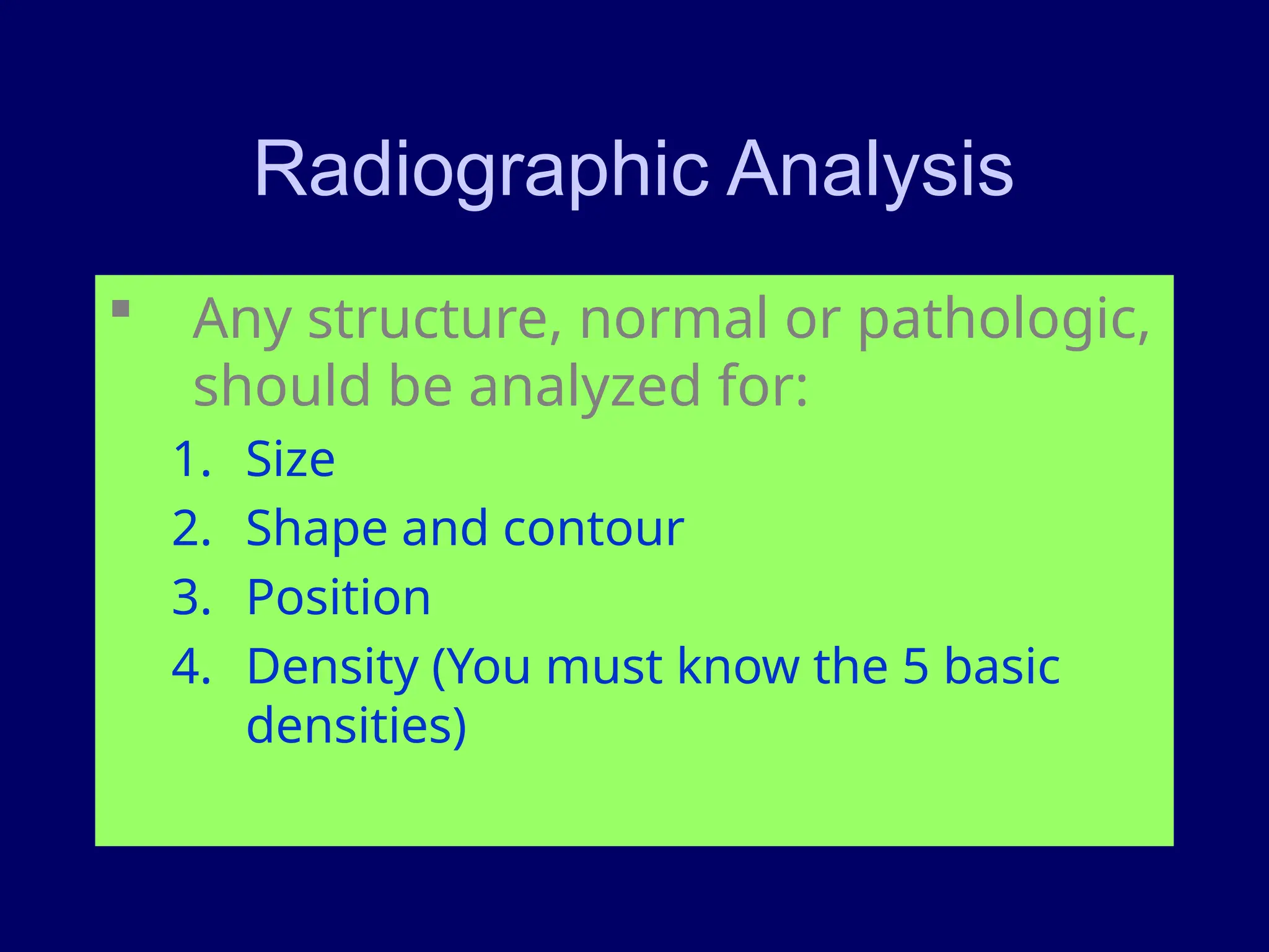
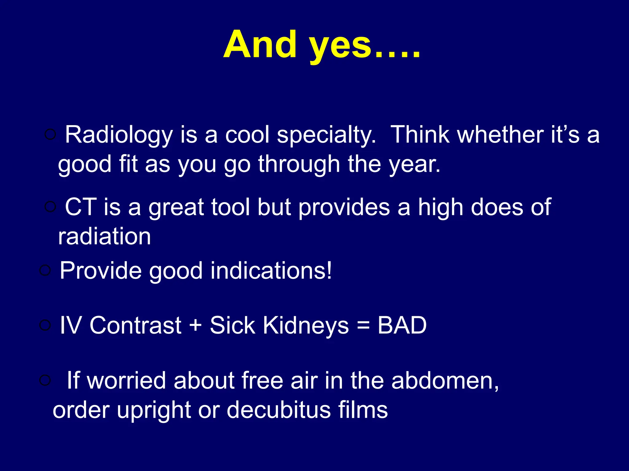
Principles of Radiological
Interpretation
Dr. Moses Acan
Department of Radiology
VIEWING
Where can you look at
images
 PACS (Picture Archiving and
Communication System) workstations
 ED
 ICUs
 WebCIS based PACS (java script)
 Web Based PACS
 Viewing boxes
Viewing the Radiographs
 Quite, dark room, no distractions
 At least 2 well lit view boxes
 A bright light illuminator for relatively over
exposed areas
 3 dimensional concept
– From a 2 dimensional image the 3rd
dimension must be constructed
– Orthogonal views
 Anatomy text, anatomical specimens,
notes
X-ray viewing station
Basics:
Looking at Imaging Studies:
 Adequate Study?
 Correctly labeled with patient’s name, MR#, and the date
of the study?
 Technically adequate?
 Systematic versus Focused look at a study:
 Radiologist does both!
 As the requesting clinician, you should also look at your
patient’s study (at least plain films), as well as follow up on
the final report.
 PTX, PNA, pleural effusions, SBO, free air
 Evaluate lines and tubes (especially the ones you placed!)
The Radiograph
 High technical quality – adequate
collimation, exposure, development and
are free of artifacts
 Is it normal, abnormal, artifactual?
Viewing the Radiographs
 Radiographs should be placed on the
view box the same way every time
 Laterals
 AP or PA
 Obliques
How do x-rays passing through
the body create an image?
 X-rays that pass through the body to the
film render the film dark (black)
 X-rays that are totally blocked do not reach
the film and render the film light (white)
 Air = low atomic # = x-rays get through =
image is dark
 Metal = high atomic # = x-rays blocked =
image is light (white)
5 Basic Radiographic
Densities
 Air
 Fat
 Soft tissue/fluid
 Mineral
 Metal
1.
2.
3.
4.
5.
Name these radiographic densities.
Radiographic Opacities
 Metal White
 Mineral – Bone Light grey
 Soft tissue – fluid Grey
 Fat Dark grey
 Air Black
Evaluating the Radiograph
 Take into account the patient history
and clinical signs
 Does an abnormality exist?
– Perception of abnormal
 Where exactly is the abnormality?
 Describe the roentgen signs
 Opinion
– Rule out list of prioritized differentials
Roentgen Signs
 Size
 Shape
 Margination
 Number
 Change in position
 Alteration in opacity
 Change in function of organs
– Roentgen signs should be correlated with history,
physical exam, disease patterns, pathogenesis
etc.
Other
 Summation – some opacity do not touch
but are superimposed  the opacity
where they overlap is increased
 Silhouette sign – structures of the same
opacity touch and their margins are lost
 Contrast Media
– GI, GU, neuro etc.
Pitfalls to Interpretation
 Do not get distracted by a large mass
for example and forget the rest of the
radiograph
 Tunnel vision
 Must use a systematic approach - every
time
Summary
 Properly displayed, Viewing
environment, technical quality
 Normal or abnormal – must know
anatomy
 Systematic evaluation and correlation
with clinical data
 Correct diagnosis or send to someone
for help
CT: A Little History...
 In the early 1970s, Hounsfield
developed a way of computerizing
Xrays to select certain densities for
viewing.
–Plain films: black, white or a few
shades of grey
–CT: thousands of shades of grey
based on the density of the tissue
(Hounsfield Units)
Computed Tomography (CT)
 Lingo:
 Attenuation
 Density
 Enhancement
 Hounsfield Units
 -1000 air ***
 -100 fat
 0 water ***
 20-80 soft tissues
 100’s bone/Ca/contrast
 >1000’s metal
 Large radiation dose
What We Need to Know
 Air is very black (less than -1000 HU)
 Water/CSF is black (near 0 HU)
 Bone is very dense/white (500-3000
HU)
 Blood is white (60-80 HU)
 Brain is gray 35-50 HU
CT vs. MRI
Wide doughnut
Opening
10-20 minutes
Length
Adjust window
Technique
Axial
Plane
250,000 UGX
Cost
Bright
Bone
Long,
narrow
30-60 min
T1, T2, Pd
3-D
500,000 UGX
Dark
Magnetic fld
X-ray beam
Obtained
MRI
CT
Advantages to CT
 Costs less than MRI
 Better access
 Shows up acute bleed
 A good quick scan
 Good visualization of bony
structures and calcified lesions
Disadvantages to CT
 Resolution
 Beam-hardening artifact
 Limited views of the posterior fossa
and poor visualization of white-
matter disease
Magnetic Resonance Imaging
(MRI)
 Lingo:
 Signal intensity
 T1
 T2
 Enhancement
 No radiation
 Strong magnetic field
 No pacemakers
 No electronic implants
 Small, long tube
 Patients must hold still
 Relatively expensive
Advantages to MRI
 No radiation exposure
 Gadolinium contrast is relatively
nontoxic
 Capacity for quantitative imaging, 3-D
reconstruction, angiography,
spectroscopy
Disadvantages of MRI
 Cost
 Some patients ineligible
because of pacemakers, other
metal
 Claustrophobia
 Long exam
 Access
ABNORMAL CTs and MRI
 Is the scan
– Contrast or noncontrast?
– Good quality?
 Describe the abnormality
Describe the abnormality
 Appearance of the lesion
– Hypodense, hyperdense, isodense or
mixed for CT.
– Hypointense, hyperintense, isointense or
mixed for MRI.
 Location
Epidural with fracture
 SHAPE round, oval, linear, saucer-shaped is a
lesion that starts at the periphery of the bone.
 BORDERS, poorly / well defined with
regular/irregular borders.
 SIZE in inches or mms Measure do not
estimate.
 SYMMETRY usually indicates a variant of
normal or an inherited condition.
On each slice look for:
 Anatomical Symmetry,
symmetry, symmetry
• E.g in the brain - sulci , cisterns
and ventricles, grey-white
differentiation
Identify What Doesn’t Belong
• Hyperdensities (whiter), e.g
• extra-axial hematomas (SDH, EDH)
• ICB or contusion
• SAH in sulci, cisterns or ventricles
• Hypodensities (darker)
• pneumocephaly (air is darker than CSF)
• infarction
Isodense Subdural Hematoma
Hygroma with shift s/p SDH
Acute on Chronic Subdural Hematoma
Intraparenchymal Bleed
Identify What Doesn’t Belong
• Localized or diffuse lesion
• effacement of sulci or cisterns
• distortion of gray-white matter interface
• enlarged ventricles, temporal horn
• Fractures, eg
• soft tissue swelling
• fluid (blood) in sinuses or mastoid air cells
• in children, look for widened sutures
• Always look at bone windows
Diffuse Edema, SAH tracking across tentorium
Blood: Acute blood is white (60-80 HU) on
CT, due to the density of hemoglobin.
As hgb breaks down, the HU decrease (i.e.
subacute and isodense hematomas)
 Relationship to OTHER ANATOMY
 T1w T2w
What Is Bright
on CT?
 Blood
 Contrast
 Bone
 Calcium
 Metal
What Is Dark
on CT?
• Air
• CSF/H20
Artifacts
 Beam
hardening
 Bone
 Foreign body
 Motion
• SPECT/ PET
•Brain scans
•Bone scans
• Cardiac scans
• Renal scans
• Lung scans
• Thyroid, parathyroid scans
•
Isotope scans
Uses for SPECT and PET
 Acute stroke
 Identify a seizure focus-increased
flow during sz and decreased
interictal flow
 Dementia-frontal pattern in FTLD,
temporo-parietal pattern in AD
 Ligand imaging in PD, others
ULTRASOUND SCAN –
INTERPRETATIVE
PRINCIPLES
 Is a very important technique for
imaging soft tissues
– Real time images
 Major applications: heart, abdomen and
pelvis as well as neck, breast,
peripheries, neonatal brain and
obstetrics.
 Limitations
– It cannot cross a tissue gas or tissue bone
interface
– Afticactual signals
– Operator dependant.
 Imaging methods
– A- mode (pulse echo)
– M- mode
– B- mode (grey scale display)
 A-mode M-mode
www.upei.ca/~vetrad
 B-mode
Interpretation
 Shadowing and through transmission
– Acoustic shadowing
– Acoustic enhancement
– Edge shadowing
 Echogenicity
– Anaechoic…….black
– Hypoechoic …..falls in btn black n white
– Hyperechoic--white
 Doppler ultrasound
– Colour Doppler
– Spectral Doppler
– 2D imaging + spectral Doppler.
 Acoustic shadowing and enhancement
 Edge shadowing
 Hyperechoic and hypoechoic
 Hypoechoic
 Heterogenous
A FEW CASES…
Diagnosis?
A broken central venous catheter has
migrated into the right lower
lobe pulmonary artery
Can you recognize
shapes and density?
D
i
Diagnosis?
History: 11 y/o twisting
injury of the foot
1.
2.
3.
4.
Please name these bones
Word bank:
Cuboid
Navicular
Medial cuneiform
Os naviculare
Proximal
Distal
1.
2.
3.
Word bank: epiphysis, metaphysis, diaphysis, cortex, medullary cavity
Naming the parts of a long bone
Summary:
 X-rays pass through the body to varying
degrees
 Higher atomic number structures block
x-rays better, example bone.
 Lower atomic number structures allow
x-rays to pass through, example: air in
the lungs.
Question: If x-rays were blocked to the same degree by all body
structures, could we see the internal parts of the body?
5 basic radiographic densities from
black to bright white
 Air
 Fat
 Soft tissue/fluid
 Bone/mineral
 Metal
What density
are the
lungs?
Why?
The list: air, fat, soft tissue, mineral and metal
CT scan of the abdomen
X-rays used skin
What density is this?
air
Absorbed
Passed through
Medullary bone
Soft tissue
Metal
Note:
Right-left marker
2
4
3
1
Name these
densities
What densit
is this?
Remember…..
The anatomical position
right left
Radiographic Analysis
 Any structure, normal or pathologic,
should be analyzed for:
1. Size
2. Shape and contour
3. Position
4. Density (You must know the 5 basic
densities)
And yes….
o CT is a great tool but provides a high does of
radiation
o Provide good indications!
o IV Contrast + Sick Kidneys = BAD
o If worried about free air in the abdomen,
order upright or decubitus films
o Radiology is a cool specialty. Think whether it’s a
good fit as you go through the year.
Questions for me?

LECTURE 2 Principles of Interpretation.pptx

  • 1.
    Principles of Radiological Interpretation Dr.Moses Acan Department of Radiology
  • 2.
  • 3.
    Where can youlook at images  PACS (Picture Archiving and Communication System) workstations  ED  ICUs  WebCIS based PACS (java script)  Web Based PACS  Viewing boxes
  • 4.
    Viewing the Radiographs Quite, dark room, no distractions  At least 2 well lit view boxes  A bright light illuminator for relatively over exposed areas  3 dimensional concept – From a 2 dimensional image the 3rd dimension must be constructed – Orthogonal views  Anatomy text, anatomical specimens, notes
  • 5.
  • 6.
    Basics: Looking at ImagingStudies:  Adequate Study?  Correctly labeled with patient’s name, MR#, and the date of the study?  Technically adequate?  Systematic versus Focused look at a study:  Radiologist does both!  As the requesting clinician, you should also look at your patient’s study (at least plain films), as well as follow up on the final report.  PTX, PNA, pleural effusions, SBO, free air  Evaluate lines and tubes (especially the ones you placed!)
  • 7.
    The Radiograph  Hightechnical quality – adequate collimation, exposure, development and are free of artifacts  Is it normal, abnormal, artifactual?
  • 8.
    Viewing the Radiographs Radiographs should be placed on the view box the same way every time  Laterals  AP or PA  Obliques
  • 9.
    How do x-rayspassing through the body create an image?  X-rays that pass through the body to the film render the film dark (black)  X-rays that are totally blocked do not reach the film and render the film light (white)  Air = low atomic # = x-rays get through = image is dark  Metal = high atomic # = x-rays blocked = image is light (white)
  • 10.
    5 Basic Radiographic Densities Air  Fat  Soft tissue/fluid  Mineral  Metal 1. 2. 3. 4. 5. Name these radiographic densities.
  • 11.
    Radiographic Opacities  MetalWhite  Mineral – Bone Light grey  Soft tissue – fluid Grey  Fat Dark grey  Air Black
  • 12.
    Evaluating the Radiograph Take into account the patient history and clinical signs  Does an abnormality exist? – Perception of abnormal  Where exactly is the abnormality?  Describe the roentgen signs  Opinion – Rule out list of prioritized differentials
  • 13.
    Roentgen Signs  Size Shape  Margination  Number  Change in position  Alteration in opacity  Change in function of organs – Roentgen signs should be correlated with history, physical exam, disease patterns, pathogenesis etc.
  • 14.
    Other  Summation –some opacity do not touch but are superimposed  the opacity where they overlap is increased  Silhouette sign – structures of the same opacity touch and their margins are lost  Contrast Media – GI, GU, neuro etc.
  • 15.
    Pitfalls to Interpretation Do not get distracted by a large mass for example and forget the rest of the radiograph  Tunnel vision  Must use a systematic approach - every time
  • 16.
    Summary  Properly displayed,Viewing environment, technical quality  Normal or abnormal – must know anatomy  Systematic evaluation and correlation with clinical data  Correct diagnosis or send to someone for help
  • 17.
    CT: A LittleHistory...  In the early 1970s, Hounsfield developed a way of computerizing Xrays to select certain densities for viewing. –Plain films: black, white or a few shades of grey –CT: thousands of shades of grey based on the density of the tissue (Hounsfield Units)
  • 18.
    Computed Tomography (CT) Lingo:  Attenuation  Density  Enhancement  Hounsfield Units  -1000 air ***  -100 fat  0 water ***  20-80 soft tissues  100’s bone/Ca/contrast  >1000’s metal  Large radiation dose
  • 19.
    What We Needto Know  Air is very black (less than -1000 HU)  Water/CSF is black (near 0 HU)  Bone is very dense/white (500-3000 HU)  Blood is white (60-80 HU)  Brain is gray 35-50 HU
  • 20.
    CT vs. MRI Widedoughnut Opening 10-20 minutes Length Adjust window Technique Axial Plane 250,000 UGX Cost Bright Bone Long, narrow 30-60 min T1, T2, Pd 3-D 500,000 UGX Dark Magnetic fld X-ray beam Obtained MRI CT
  • 21.
    Advantages to CT Costs less than MRI  Better access  Shows up acute bleed  A good quick scan  Good visualization of bony structures and calcified lesions
  • 22.
    Disadvantages to CT Resolution  Beam-hardening artifact  Limited views of the posterior fossa and poor visualization of white- matter disease
  • 23.
    Magnetic Resonance Imaging (MRI) Lingo:  Signal intensity  T1  T2  Enhancement  No radiation  Strong magnetic field  No pacemakers  No electronic implants  Small, long tube  Patients must hold still  Relatively expensive
  • 24.
    Advantages to MRI No radiation exposure  Gadolinium contrast is relatively nontoxic  Capacity for quantitative imaging, 3-D reconstruction, angiography, spectroscopy
  • 25.
    Disadvantages of MRI Cost  Some patients ineligible because of pacemakers, other metal  Claustrophobia  Long exam  Access
  • 26.
  • 27.
     Is thescan – Contrast or noncontrast? – Good quality?  Describe the abnormality
  • 28.
    Describe the abnormality Appearance of the lesion – Hypodense, hyperdense, isodense or mixed for CT. – Hypointense, hyperintense, isointense or mixed for MRI.  Location
  • 29.
  • 30.
     SHAPE round,oval, linear, saucer-shaped is a lesion that starts at the periphery of the bone.  BORDERS, poorly / well defined with regular/irregular borders.  SIZE in inches or mms Measure do not estimate.  SYMMETRY usually indicates a variant of normal or an inherited condition.
  • 31.
    On each slicelook for:  Anatomical Symmetry, symmetry, symmetry • E.g in the brain - sulci , cisterns and ventricles, grey-white differentiation
  • 32.
    Identify What Doesn’tBelong • Hyperdensities (whiter), e.g • extra-axial hematomas (SDH, EDH) • ICB or contusion • SAH in sulci, cisterns or ventricles • Hypodensities (darker) • pneumocephaly (air is darker than CSF) • infarction
  • 33.
  • 34.
  • 35.
    Acute on ChronicSubdural Hematoma
  • 36.
  • 37.
    Identify What Doesn’tBelong • Localized or diffuse lesion • effacement of sulci or cisterns • distortion of gray-white matter interface • enlarged ventricles, temporal horn • Fractures, eg • soft tissue swelling • fluid (blood) in sinuses or mastoid air cells • in children, look for widened sutures • Always look at bone windows
  • 38.
    Diffuse Edema, SAHtracking across tentorium
  • 39.
    Blood: Acute bloodis white (60-80 HU) on CT, due to the density of hemoglobin. As hgb breaks down, the HU decrease (i.e. subacute and isodense hematomas)
  • 40.
     Relationship toOTHER ANATOMY
  • 41.
  • 42.
    What Is Bright onCT?  Blood  Contrast  Bone  Calcium  Metal What Is Dark on CT? • Air • CSF/H20
  • 43.
  • 45.
    • SPECT/ PET •Brainscans •Bone scans • Cardiac scans • Renal scans • Lung scans • Thyroid, parathyroid scans • Isotope scans
  • 46.
    Uses for SPECTand PET  Acute stroke  Identify a seizure focus-increased flow during sz and decreased interictal flow  Dementia-frontal pattern in FTLD, temporo-parietal pattern in AD  Ligand imaging in PD, others
  • 47.
    ULTRASOUND SCAN – INTERPRETATIVE PRINCIPLES Is a very important technique for imaging soft tissues – Real time images  Major applications: heart, abdomen and pelvis as well as neck, breast, peripheries, neonatal brain and obstetrics.
  • 48.
     Limitations – Itcannot cross a tissue gas or tissue bone interface – Afticactual signals – Operator dependant.
  • 49.
     Imaging methods –A- mode (pulse echo) – M- mode – B- mode (grey scale display)
  • 50.
  • 51.
  • 52.
    Interpretation  Shadowing andthrough transmission – Acoustic shadowing – Acoustic enhancement – Edge shadowing  Echogenicity – Anaechoic…….black – Hypoechoic …..falls in btn black n white – Hyperechoic--white
  • 53.
     Doppler ultrasound –Colour Doppler – Spectral Doppler – 2D imaging + spectral Doppler.
  • 54.
     Acoustic shadowingand enhancement
  • 55.
  • 56.
  • 57.
  • 58.
  • 59.
  • 60.
  • 62.
    A broken centralvenous catheter has migrated into the right lower lobe pulmonary artery
  • 63.
  • 64.
  • 65.
    History: 11 y/otwisting injury of the foot
  • 67.
    1. 2. 3. 4. Please name thesebones Word bank: Cuboid Navicular Medial cuneiform Os naviculare
  • 68.
    Proximal Distal 1. 2. 3. Word bank: epiphysis,metaphysis, diaphysis, cortex, medullary cavity Naming the parts of a long bone
  • 69.
    Summary:  X-rays passthrough the body to varying degrees  Higher atomic number structures block x-rays better, example bone.  Lower atomic number structures allow x-rays to pass through, example: air in the lungs. Question: If x-rays were blocked to the same degree by all body structures, could we see the internal parts of the body?
  • 70.
    5 basic radiographicdensities from black to bright white  Air  Fat  Soft tissue/fluid  Bone/mineral  Metal
  • 71.
    What density are the lungs? Why? Thelist: air, fat, soft tissue, mineral and metal
  • 72.
    CT scan ofthe abdomen X-rays used skin What density is this? air
  • 73.
  • 74.
  • 75.
  • 76.
  • 77.
  • 78.
  • 79.
    Radiographic Analysis  Anystructure, normal or pathologic, should be analyzed for: 1. Size 2. Shape and contour 3. Position 4. Density (You must know the 5 basic densities)
  • 80.
    And yes…. o CTis a great tool but provides a high does of radiation o Provide good indications! o IV Contrast + Sick Kidneys = BAD o If worried about free air in the abdomen, order upright or decubitus films o Radiology is a cool specialty. Think whether it’s a good fit as you go through the year.
  • 81.

Editor's Notes

  • #10 Point out the different densities. Fluid is a soft tissue density. This means we can’t tell the difference between blood, pus, and simple fluid on an xray. Even though this is relatively inexpensive, a lot of information can be gained by x rays. In general, any osseous pathology is initially best evaluated by x-ray.
  • #18 Very useful modality, particularly as screening exam for brain pathology (bleed vs. no bleed) and intra-abdominal pathology -Exponentional increase in the use of CT 10% per year! -Not without its risk. High dose of radiation which has a not insiginficant risk of causing cancer…especially in younger patients! Think about other modalities (MR, ultrasound) with no radiation!
  • #23 -very useful exam for specific clinical questions -just because it’s the most expensive doesn’t mean its always the best -contraindications are metal (old aneurysm clips, pacemakers, shrapnel….) claustrophoia -difficult for peds patients b/c they have to hold still for extended periods of time and frequently require GA.