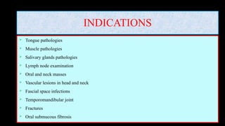
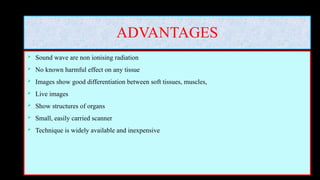
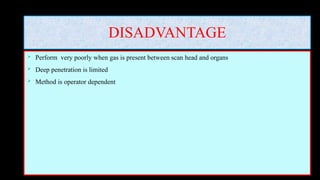
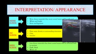
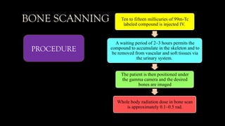
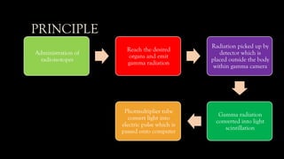
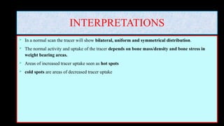
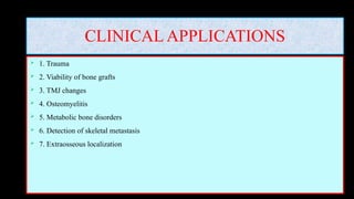
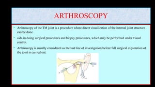
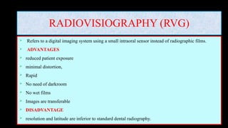
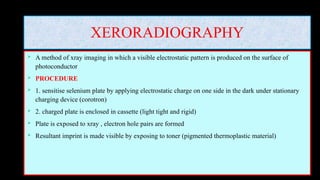
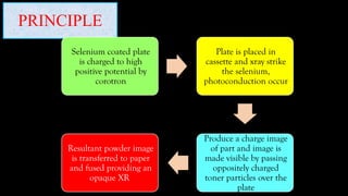
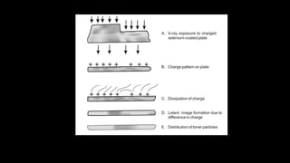
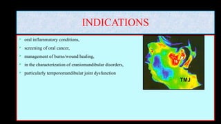
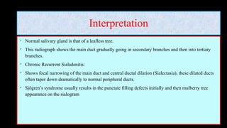
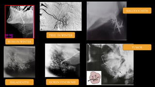
SPECIALIZED
IMAGING
MODALITIES
DR PRIYANKA
ORAL MEDICINE AND
RADIOLOGY
CONTENTS
MRI
ULTRASOUND
NUCLEAR MEDICINE
ARTHROGRAPHY
ARTHROSCOPY
THERMOGRAPHY
RADIOVISIOGRAPHY
SCANOGRAPHY
XERORADIOGRAPHY
SIALOGRAPHY
MAGNETIC RESONANCE IMAGING
 Non invasive imaging method
 Uses non ionising electromagnetic radiation i.e. radiofrequency waves
 MRI stands for
 Magnetic: require magnetic field
 Resonance : interaction between magnetic field and radiofrequency waves
 Imaging: to make images
INDICATIONS (soft tissue imaging)
1) Salivary gland pathologies
2) Temporomandibular joint pathologies (articular disc)
3) Intracranial pathologies
4) Benign and malignant tumors of jaw
5) Nerve disorders
6) Muscular pathologies
7) Lymph nodes evaluation
8) Vascular lesions
CONTRAINDICATIONS
 Cardiac pacemaker
 Cochlear implants
 Claustrophobic patients
ADVANTAGES
 Non ionising radiation
 Non invasive
 Contrast resolution
 Multiplanar images
 Image manipulation can be done
DISADVANTAGES
 Claustrophobia
 Expensive Equipment
 Image become distorted by metal
 Long scanning time and require patient cooperation
 Scanners are noisy
 Allergic reaction due to contrast agent
 Cannot distinguish between benign and malignant disease
PRECAUTIONS
 Should remove metal from their body
 Jewellery
 Watch
 Piercing coins
 Credit/debit cards
 wallet
PARTS OF MRI MACHINE
 MRI gantry (heavy magnet, transmitter, receiver and radiofrequency coil)
 Patient table
 Computer system
PROCEDURAL STEPS IN MRI
 STEP 1: patient into big magnetic field
 STEP 2: transmission of radiofrequency waves into subject from radiofrequency coils
 STEP 3: receive radio waves (retransmitted from subject by receivers)
 STEP 4: measured radiofrequency waves
 STEP 5: information from receiver send to computer for image reconstruction
TYPES OF IMAGES
 T1 / FAT IMAGES T2/ WATER IMAGES
 Useful for depicting small anatomic region used for inflammatory or pathological changes
 Fat appear brighter water appear brighter
 INTERPRETATION/ APPEARANCE
 HYPERINTENSE: white and bright
 ISOTENSE: grey
 HYPOINTENSE: dark, black and produce
weak signal
ULTRASOUND
 Imaging technique in which ultrasound waves are used
 Ultrasound is a sound with frequency greater than upper limit of human hearing
 PARTS OF ULTRASOUND MACHINE
 TRANSMITTER: transmit electric signal to transducer
 TRANSDUCER: convert electric signal to sound waves and vice versa
 RECEIVER: receives electric signal from transducer
 PROCESSOR: analyze the transmitted and received signals
 DISPLAY: display the image based on processor
HOW DOES IT WORK?
Transmission of electric
signal to transducer
Transducer convert
electric signal to high
frequency sound waves
(1-5 MHz) and this into
our body
Sound waves hit the
tissue and organs . Some
of them are partially
reflected, absorbed and
some return to
transducer
Transducer detect sound
waves and convert to
electric signal which are
received by receiver and
send to processor
Processor will evaluate
the change in electric
signal and based on data
given by processor, image
will seen on computer
INDICATIONS
 Tongue pathologies
 Muscle pathologies
 Salivary glands pathologies
 Lymph node examination
 Oral and neck masses
 Vascular lesions in head and neck
 Fascial space infections
 Temporomandibular joint
 Fractures
 Oral submucous fibrosis
ADVANTAGES
 Sound wave are non ionising radiation
 No known harmful effect on any tissue
 Images show good differentiation between soft tissues, muscles,
 Live images
 Show structures of organs
 Small, easily carried scanner
 Technique is widely available and inexpensive
DISADVANTAGE
 Perform very poorly when gas is present between scan head and organs
 Deep penetration is limited
 Method is operator dependent
INTERPRETATION/ APPEARANCE
• More dense materials thus more sound waves will be reflected
• White and bright
• Bone and ligament
• Have same density as surrounding structures
• Grey
• muscles
HYPER
ECHOIC
ISO
ECHOIC
HYPO
ECHOIC
• Less dense materials thus fewer sound waves will be reflected back
to receiver
• Dark, black
• Water, air
DISPLAY MODES/ SYSTEM
 A MODE: amplitude mode (only for ophthalmology)
 B MODE: bright mode
 GREY SCALE IMAGING: most commonly used
 M MODE: for moving structures
 D MODE or DOPPLER: for vascular lesion
 COLOR DOPPLER: blood flow is seen with color
 SPECTRAL DOPPLER: blood flow in graphical representation
 DUPLEX: combination of color and spectral
 3D & 4D
NUCLEAR MEDICINE
 Nuclear medicine is a diagnostic radiation science utilizing radioactive compounds or tracers having
affinities for particular tissues or organs in the body (iodine to the thyroid gland); these are termed as
target tissues
 The radioactive agents are administered to the patient either orally, intravenous
 These are detected and imaged by a variety of external detectors and imaging systems.
 This helps in studying the target tissue under static and dynamic conditions.
 Such studies are called scintigraphic scans, scintiscans or radionuclide scans
 When these are utilized for studying bone they referred to as bone scans and
 when performed for salivary gland they are termed as salivary gland scans.
RADIOPHARMACEUTICALS
 These are radioactive agents used in nuclear medicine procedures for imaging.
 They are produced using nuclear reactors and cyclotrons.
 Radiopharmaceuticals circulate and concentrate to varying degrees in different organs
throughout the body.
 The localized tracer uptake is dependent on changes in regional blood flow and areas of altered
bone physiology and pathology which show an invariable alteration in the osteoblastic and
osteoclastic activity.
 The organs that receive the greatest exposure are referred to as critical organs.
TYPES OF RADIOPHARMACEUTICALS
 1. Radioactive elements/compounds (Technetium-99m, Fluorine-18, Iodine-131)
 2. Non-radioactive carrier compound with a radioisotope (Gallium-67 labeled citrate, Iodine-125
labeled polyphosphate and Technetium labeled human serum albumin).
 Technetium-99m methylene diphosphonate (Tc-99m MDP) is the most widely used
radiopharmaceutical.
 It has a short half-life (6.5 h), hence there is minimal radiation exposure and has superior physical
and biological properties.
 MDP is localized in bone by chemiabsorption within newly formed inorganic matrix and cannot be
metabolized by the enzyme system.
BONE SCANNING Ten to fifteen millicuries of 99m-Tc
labeled compound is injected IV.
A waiting period of 2–3 hours permits the
compound to accumulate in the skeleton and to
be removed from vascular and soft tissues via
the urinary system.
The patient is then positioned under
the gamma camera and the desired
bones are imaged
Whole body radiation dose in bone scan
is approximately 0.1–0.5 rad.
PROCEDURE
PRINCIPLE
Administration of
radioisotopes
Reach the desired
organs and emit
gamma radiation
Radiation picked up by
detector which is
placed outside the body
within gamma camera
Gamma radiation
converted into light
scintillation
Photmultiplier tube
convert light into
electric pulse which is
passed onto computer
INTERPRETATIONS
 In a normal scan the tracer will show bilateral, uniform and symmetrical distribution.
 The normal activity and uptake of the tracer depends on bone mass/density and bone stress in
weight bearing areas.
 Areas of increased tracer uptake seen as hot spots
 cold spots are areas of decreased tracer uptake
CLINICAL APPLICATIONS
 1. Trauma
 2. Viability of bone grafts
 3. TMJ changes
 4. Osteomyelitis
 5. Metabolic bone disorders
 6. Detection of skeletal metastasis
 7. Extraosseous localization
Salivary Gland Scans
 Patient is advised to lie supine.
 Intravenously 99m-Tc pertechnetate is injected.
 For the next 25 minutes, sequential images are taken at 1 minute intervals.
 A large field of view gamma camera equipped with a low-energy, high-resolution, parallel-hole
collimator is used.
 Roughly, 15 minutes following the procedure, secretogogue like lemon juice may be given to
enhance salivary secretion.
 The effective equivalent radiation dose to the patient is 0.4–1.6 mSv.
CLINICAL APPLICATIONS
 Salivary scintigraphy may be used to
 assess obstructions in the salivary ducts with or without parenchymal damage,
 Sjogren’s syndrome,
 parenchymal impairment after radioiodine treatment in patients suffering from thyroid
ARTHROGRAPHY
 In arthrography, contrast agents are introduced into the joint spaces and then radiographed.
 Give information on soft tissue state of TMJ, (integrity and position of disk and its posterior
attachment)
 PROCEDURE : performed by catheterizing the upper and lower joint spaces with separate
injections and injecting 0.5 to 1 ml of radiographic contrast media (iodine) first into the lower space
and then into the upper space. A series of radiographs are taken following joint space opacification
with the jaws closed and in graded stages of opening.
 INTERPRETATION: The disk appears as a radiolucent void between two opaque areas of contrast
media. Opacification of both the joint spaces following injection of only one denotes a pathological
condition
INDICATIONS AND CONTRAINDICATIONS
• Long standing TMJ pain dysfunction unresponsive to simple treatments.
• Persistent history of locking.
• Limited opening of unknown etiology.
• Dynamic information on the position of the joint components and disk as they move in relation to one
another.
• Static images of the joint components with the mouth closed and with the mouth open. Any anterior or
anteromedial displacement of the disk can be observed.
• The integrity of the disk, i.e. presence of any perforations
CONTRAINDICATIONS: presence of infections in or near a joint and in patients who are allergic to
the contrast agents
DISADVANTAGES
• Very painful.
• Not indicated when the patient is hypersensitive to iodine or the contrast medium.
• Capsule may rupture if too much contrast media is injected.
• Very strict asepsis has to be maintained
ARTHROSCOPY
 Arthroscopy of the TM joint is a procedure where direct visualization of the internal joint structure
can be done.
 aids in doing surgical procedures and biopsy procedures, which may be performed under visual
control.
 Arthroscopy is usually considered as the last line of investigation before full surgical exploration of
the joint is carried out.
ADVANTAGES
• Safe procedure.
• Direct visualization.
• Minimal postoperative complications.
• Color change in inflammed tissues is clearly seen.
• Biopsy and surgical procedures can be easily visualized.
• It allows certain interventional procedures to be performed – Washing out the joint with saline –
Introduction of steroids directly into the joint – Division of adhesions – Removal of loose bodies from
within the joint
RADIOVISIOGRAPHY (RVG)
 Refers to a digital imaging system using a small intraoral sensor instead of radiographic films.
 ADVANTAGES
 reduced patient exposure
 minimal distortion,
 Rapid
 No need of darkroom
 No wet films
 Images are transferable
 DISADVANTAGE
 resolution and latitude are inferior to standard dental radiography.
XERORADIOGRAPHY
 A method of xray imaging in which a visible electrostatic pattern is produced on the surface of
photoconductor
 PROCEDURE
 1. sensitise selenium plate by applying electrostatic charge on one side in the dark under stationary
charging device (corotron)
 2. charged plate is enclosed in cassette (light tight and rigid)
 Plate is exposed to xray , electron hole pairs are formed
 Resultant imprint is made visible by exposing to toner (pigmented thermoplastic material)
PRINCIPLE
Selenium coated plate
is charged to high
positive potential by
corotron
Plate is placed in
cassette and xray strike
the selenium,
photoconduction occur
Produce a charge image
of part and image is
made visible by passing
oppositely charged
toner particles over the
plate
Resultant powder image
is transferred to paper
and fused providing an
opaque XR
ADVANTAGES
• No requirement of darkroom or solutions for processing
• The process is faster
• Dry and permanent image is formed
• The plates can be used for about 1000 exposures.
• Visible image of air, fat, water, cartilage, and bone can be seen
• Images have good detail or edge enhancement
• Xeroradiograph has better resolution
• The technique requires less radiation exposure
THERMOGRAPHY
 Thermography is a generic title for various methods of quantifying the temperature distribution from
body surfaces.
 There are three forms
 • Liquid crystal thermography (LCT)
 • Infrared thermography (IRT)
 • Microwave thermography (MCT).
INDICATIONS
 oral inflammatory conditions,
 screening of oral cancer,
 management of burns/wound healing,
 in the characterization of craniomandibular disorders,
 particularly temporomandibular joint dysfunction
SCANOGRAPHY
 This is a technique that uses a narrowly collimated, fan shaped beam of radiation to scan an area of
interest, sequentially projecting image data relative to this area onto a moving film (similar to that in
panoramic radiography).
 ADVANTAGES:
 higher contrast and greater details
 The Soredex Scanora is a commercially available X-ray unit capable of performing both rotational
and linear scanography.
ROTATIONAL SCANOGRAPHY
 the beam of radiation rotates about a fixed axis that is predetermined based on the area imaged.
 This results in the production of two to four scanograms, each made with the X-ray tube in a different
position, thus multiple images are made, which can be viewed on the stereoscope.
 This is found to be useful in the assessment of periodontal disease and detection of periapical lesions.
 IN LINEAR SCANOGRAPHY the X-ray beam and the film move in a linear fashion, scanning the
area of interest.
 Linear scanography can be thought of as panoramic radiography that has been “straightened out”.
 The Scanora system can make both posteroanterior and lateral linear scanning of the maxillofacial
complex
SIALOGRAPHY
 DEFINITION-
It is a specialized radiographic view taken by introduction of the radiopaque dye into the ductal
system of the major salivary glands, mainly parotid and submandibular
 INDICATIONS
1. Detection of Sialoliths both radiopaque and radiolucent.
2. Evaluation of the extent of irreversible ductal damage caused by infection.
3. Differentiation between the Sjögren’s syndrome, Sialosis and chronic sialadenitis.
4. Evaluation of diverticula, strictures and fistula.
5. It may be used as a dilating procedure for mild ductal stenosis.
 CONTRAINDICATIONS
1. Acute infection of the salivary gland.
2. Allergic reaction to any of the components of the radiopaque material to be used.
3. Thyroid disorders in patients due to the Iodine content of the contrast media.
 ARMAMENTERIUM
1. Sialographic Cannulas- Rabinov Cannulas-with tips ranging from 0.012 to 0.033 inches.
2. Most of these cannulas come with polyethylene flexible tube.
3. Lacrimal Dilators ranging from 0000 to caliber 0.
4. A 5 ml syringe (disposable)
5. Gauze sponge pads
6. Conray 420‚ Urograffin
7. Secretogogue such as fresh Lemon, Lemon extract or lemon concentrate.
8. Good dental lighting.
9. Magnifying glasses
METHOD
 Once the duct is cannulated the injection is made with hand pressure.
 The patient may complain of mild pain during at injection, however, a slow constant injection
technique usually can accomplish complete ductal filling without much patient discomfort.
 The patient’s sensation of glandular fullness is suggested by a sharp pain when the operator usually
stops and proceeds with radiography.
Interpretation
 Normal salivary gland is that of a leafless tree.
 This radiograph shows the main duct gradually going in secondary branches and then into tertiary
branches.
 Chronic Recurrent Sialadenitis:
 Shows focal narrowing of the main duct and central ductal dilation (Sialectasia), these dilated ducts
often taper down dramatically to normal peripheral ducts.
 Sjögren’s syndrome usually results in the punctate filling defects initially and then mulberry tree
appearance on the sialogram
BUSH IN WINTER
TREE IN WINTER
SIALODOCHITIS
SIALADENITIS SJOREN SYNDROME
TUMOR

SPECIAL IMAGING MODALITIES- RADIOGRAPHIC

  • 1.
  • 2.
  • 3.
    MAGNETIC RESONANCE IMAGING Non invasive imaging method  Uses non ionising electromagnetic radiation i.e. radiofrequency waves  MRI stands for  Magnetic: require magnetic field  Resonance : interaction between magnetic field and radiofrequency waves  Imaging: to make images
  • 4.
    INDICATIONS (soft tissueimaging) 1) Salivary gland pathologies 2) Temporomandibular joint pathologies (articular disc) 3) Intracranial pathologies 4) Benign and malignant tumors of jaw 5) Nerve disorders 6) Muscular pathologies 7) Lymph nodes evaluation 8) Vascular lesions
  • 5.
    CONTRAINDICATIONS  Cardiac pacemaker Cochlear implants  Claustrophobic patients
  • 6.
    ADVANTAGES  Non ionisingradiation  Non invasive  Contrast resolution  Multiplanar images  Image manipulation can be done
  • 7.
    DISADVANTAGES  Claustrophobia  ExpensiveEquipment  Image become distorted by metal  Long scanning time and require patient cooperation  Scanners are noisy  Allergic reaction due to contrast agent  Cannot distinguish between benign and malignant disease
  • 8.
    PRECAUTIONS  Should removemetal from their body  Jewellery  Watch  Piercing coins  Credit/debit cards  wallet
  • 9.
    PARTS OF MRIMACHINE  MRI gantry (heavy magnet, transmitter, receiver and radiofrequency coil)  Patient table  Computer system
  • 10.
    PROCEDURAL STEPS INMRI  STEP 1: patient into big magnetic field  STEP 2: transmission of radiofrequency waves into subject from radiofrequency coils  STEP 3: receive radio waves (retransmitted from subject by receivers)  STEP 4: measured radiofrequency waves  STEP 5: information from receiver send to computer for image reconstruction
  • 11.
    TYPES OF IMAGES T1 / FAT IMAGES T2/ WATER IMAGES  Useful for depicting small anatomic region used for inflammatory or pathological changes  Fat appear brighter water appear brighter  INTERPRETATION/ APPEARANCE  HYPERINTENSE: white and bright  ISOTENSE: grey  HYPOINTENSE: dark, black and produce weak signal
  • 12.
    ULTRASOUND  Imaging techniquein which ultrasound waves are used  Ultrasound is a sound with frequency greater than upper limit of human hearing  PARTS OF ULTRASOUND MACHINE  TRANSMITTER: transmit electric signal to transducer  TRANSDUCER: convert electric signal to sound waves and vice versa  RECEIVER: receives electric signal from transducer  PROCESSOR: analyze the transmitted and received signals  DISPLAY: display the image based on processor
  • 13.
    HOW DOES ITWORK? Transmission of electric signal to transducer Transducer convert electric signal to high frequency sound waves (1-5 MHz) and this into our body Sound waves hit the tissue and organs . Some of them are partially reflected, absorbed and some return to transducer Transducer detect sound waves and convert to electric signal which are received by receiver and send to processor Processor will evaluate the change in electric signal and based on data given by processor, image will seen on computer
  • 14.
    INDICATIONS  Tongue pathologies Muscle pathologies  Salivary glands pathologies  Lymph node examination  Oral and neck masses  Vascular lesions in head and neck  Fascial space infections  Temporomandibular joint  Fractures  Oral submucous fibrosis
  • 15.
    ADVANTAGES  Sound waveare non ionising radiation  No known harmful effect on any tissue  Images show good differentiation between soft tissues, muscles,  Live images  Show structures of organs  Small, easily carried scanner  Technique is widely available and inexpensive
  • 16.
    DISADVANTAGE  Perform verypoorly when gas is present between scan head and organs  Deep penetration is limited  Method is operator dependent
  • 17.
    INTERPRETATION/ APPEARANCE • Moredense materials thus more sound waves will be reflected • White and bright • Bone and ligament • Have same density as surrounding structures • Grey • muscles HYPER ECHOIC ISO ECHOIC HYPO ECHOIC • Less dense materials thus fewer sound waves will be reflected back to receiver • Dark, black • Water, air
  • 18.
    DISPLAY MODES/ SYSTEM A MODE: amplitude mode (only for ophthalmology)  B MODE: bright mode  GREY SCALE IMAGING: most commonly used  M MODE: for moving structures  D MODE or DOPPLER: for vascular lesion  COLOR DOPPLER: blood flow is seen with color  SPECTRAL DOPPLER: blood flow in graphical representation  DUPLEX: combination of color and spectral  3D & 4D
  • 19.
    NUCLEAR MEDICINE  Nuclearmedicine is a diagnostic radiation science utilizing radioactive compounds or tracers having affinities for particular tissues or organs in the body (iodine to the thyroid gland); these are termed as target tissues  The radioactive agents are administered to the patient either orally, intravenous  These are detected and imaged by a variety of external detectors and imaging systems.  This helps in studying the target tissue under static and dynamic conditions.  Such studies are called scintigraphic scans, scintiscans or radionuclide scans  When these are utilized for studying bone they referred to as bone scans and  when performed for salivary gland they are termed as salivary gland scans.
  • 20.
    RADIOPHARMACEUTICALS  These areradioactive agents used in nuclear medicine procedures for imaging.  They are produced using nuclear reactors and cyclotrons.  Radiopharmaceuticals circulate and concentrate to varying degrees in different organs throughout the body.  The localized tracer uptake is dependent on changes in regional blood flow and areas of altered bone physiology and pathology which show an invariable alteration in the osteoblastic and osteoclastic activity.  The organs that receive the greatest exposure are referred to as critical organs.
  • 21.
    TYPES OF RADIOPHARMACEUTICALS 1. Radioactive elements/compounds (Technetium-99m, Fluorine-18, Iodine-131)  2. Non-radioactive carrier compound with a radioisotope (Gallium-67 labeled citrate, Iodine-125 labeled polyphosphate and Technetium labeled human serum albumin).  Technetium-99m methylene diphosphonate (Tc-99m MDP) is the most widely used radiopharmaceutical.  It has a short half-life (6.5 h), hence there is minimal radiation exposure and has superior physical and biological properties.  MDP is localized in bone by chemiabsorption within newly formed inorganic matrix and cannot be metabolized by the enzyme system.
  • 22.
    BONE SCANNING Tento fifteen millicuries of 99m-Tc labeled compound is injected IV. A waiting period of 2–3 hours permits the compound to accumulate in the skeleton and to be removed from vascular and soft tissues via the urinary system. The patient is then positioned under the gamma camera and the desired bones are imaged Whole body radiation dose in bone scan is approximately 0.1–0.5 rad. PROCEDURE
  • 23.
    PRINCIPLE Administration of radioisotopes Reach thedesired organs and emit gamma radiation Radiation picked up by detector which is placed outside the body within gamma camera Gamma radiation converted into light scintillation Photmultiplier tube convert light into electric pulse which is passed onto computer
  • 24.
    INTERPRETATIONS  In anormal scan the tracer will show bilateral, uniform and symmetrical distribution.  The normal activity and uptake of the tracer depends on bone mass/density and bone stress in weight bearing areas.  Areas of increased tracer uptake seen as hot spots  cold spots are areas of decreased tracer uptake
  • 25.
    CLINICAL APPLICATIONS  1.Trauma  2. Viability of bone grafts  3. TMJ changes  4. Osteomyelitis  5. Metabolic bone disorders  6. Detection of skeletal metastasis  7. Extraosseous localization
  • 26.
    Salivary Gland Scans Patient is advised to lie supine.  Intravenously 99m-Tc pertechnetate is injected.  For the next 25 minutes, sequential images are taken at 1 minute intervals.  A large field of view gamma camera equipped with a low-energy, high-resolution, parallel-hole collimator is used.  Roughly, 15 minutes following the procedure, secretogogue like lemon juice may be given to enhance salivary secretion.  The effective equivalent radiation dose to the patient is 0.4–1.6 mSv.
  • 27.
    CLINICAL APPLICATIONS  Salivaryscintigraphy may be used to  assess obstructions in the salivary ducts with or without parenchymal damage,  Sjogren’s syndrome,  parenchymal impairment after radioiodine treatment in patients suffering from thyroid
  • 28.
    ARTHROGRAPHY  In arthrography,contrast agents are introduced into the joint spaces and then radiographed.  Give information on soft tissue state of TMJ, (integrity and position of disk and its posterior attachment)  PROCEDURE : performed by catheterizing the upper and lower joint spaces with separate injections and injecting 0.5 to 1 ml of radiographic contrast media (iodine) first into the lower space and then into the upper space. A series of radiographs are taken following joint space opacification with the jaws closed and in graded stages of opening.  INTERPRETATION: The disk appears as a radiolucent void between two opaque areas of contrast media. Opacification of both the joint spaces following injection of only one denotes a pathological condition
  • 30.
    INDICATIONS AND CONTRAINDICATIONS •Long standing TMJ pain dysfunction unresponsive to simple treatments. • Persistent history of locking. • Limited opening of unknown etiology. • Dynamic information on the position of the joint components and disk as they move in relation to one another. • Static images of the joint components with the mouth closed and with the mouth open. Any anterior or anteromedial displacement of the disk can be observed. • The integrity of the disk, i.e. presence of any perforations CONTRAINDICATIONS: presence of infections in or near a joint and in patients who are allergic to the contrast agents
  • 31.
    DISADVANTAGES • Very painful. •Not indicated when the patient is hypersensitive to iodine or the contrast medium. • Capsule may rupture if too much contrast media is injected. • Very strict asepsis has to be maintained
  • 32.
    ARTHROSCOPY  Arthroscopy ofthe TM joint is a procedure where direct visualization of the internal joint structure can be done.  aids in doing surgical procedures and biopsy procedures, which may be performed under visual control.  Arthroscopy is usually considered as the last line of investigation before full surgical exploration of the joint is carried out.
  • 33.
    ADVANTAGES • Safe procedure. •Direct visualization. • Minimal postoperative complications. • Color change in inflammed tissues is clearly seen. • Biopsy and surgical procedures can be easily visualized. • It allows certain interventional procedures to be performed – Washing out the joint with saline – Introduction of steroids directly into the joint – Division of adhesions – Removal of loose bodies from within the joint
  • 34.
    RADIOVISIOGRAPHY (RVG)  Refersto a digital imaging system using a small intraoral sensor instead of radiographic films.  ADVANTAGES  reduced patient exposure  minimal distortion,  Rapid  No need of darkroom  No wet films  Images are transferable  DISADVANTAGE  resolution and latitude are inferior to standard dental radiography.
  • 35.
    XERORADIOGRAPHY  A methodof xray imaging in which a visible electrostatic pattern is produced on the surface of photoconductor  PROCEDURE  1. sensitise selenium plate by applying electrostatic charge on one side in the dark under stationary charging device (corotron)  2. charged plate is enclosed in cassette (light tight and rigid)  Plate is exposed to xray , electron hole pairs are formed  Resultant imprint is made visible by exposing to toner (pigmented thermoplastic material)
  • 36.
    PRINCIPLE Selenium coated plate ischarged to high positive potential by corotron Plate is placed in cassette and xray strike the selenium, photoconduction occur Produce a charge image of part and image is made visible by passing oppositely charged toner particles over the plate Resultant powder image is transferred to paper and fused providing an opaque XR
  • 38.
    ADVANTAGES • No requirementof darkroom or solutions for processing • The process is faster • Dry and permanent image is formed • The plates can be used for about 1000 exposures. • Visible image of air, fat, water, cartilage, and bone can be seen • Images have good detail or edge enhancement • Xeroradiograph has better resolution • The technique requires less radiation exposure
  • 39.
    THERMOGRAPHY  Thermography isa generic title for various methods of quantifying the temperature distribution from body surfaces.  There are three forms  • Liquid crystal thermography (LCT)  • Infrared thermography (IRT)  • Microwave thermography (MCT).
  • 40.
    INDICATIONS  oral inflammatoryconditions,  screening of oral cancer,  management of burns/wound healing,  in the characterization of craniomandibular disorders,  particularly temporomandibular joint dysfunction
  • 41.
    SCANOGRAPHY  This isa technique that uses a narrowly collimated, fan shaped beam of radiation to scan an area of interest, sequentially projecting image data relative to this area onto a moving film (similar to that in panoramic radiography).  ADVANTAGES:  higher contrast and greater details  The Soredex Scanora is a commercially available X-ray unit capable of performing both rotational and linear scanography.
  • 42.
    ROTATIONAL SCANOGRAPHY  thebeam of radiation rotates about a fixed axis that is predetermined based on the area imaged.  This results in the production of two to four scanograms, each made with the X-ray tube in a different position, thus multiple images are made, which can be viewed on the stereoscope.  This is found to be useful in the assessment of periodontal disease and detection of periapical lesions.  IN LINEAR SCANOGRAPHY the X-ray beam and the film move in a linear fashion, scanning the area of interest.  Linear scanography can be thought of as panoramic radiography that has been “straightened out”.  The Scanora system can make both posteroanterior and lateral linear scanning of the maxillofacial complex
  • 43.
    SIALOGRAPHY  DEFINITION- It isa specialized radiographic view taken by introduction of the radiopaque dye into the ductal system of the major salivary glands, mainly parotid and submandibular  INDICATIONS 1. Detection of Sialoliths both radiopaque and radiolucent. 2. Evaluation of the extent of irreversible ductal damage caused by infection. 3. Differentiation between the Sjögren’s syndrome, Sialosis and chronic sialadenitis. 4. Evaluation of diverticula, strictures and fistula. 5. It may be used as a dilating procedure for mild ductal stenosis.
  • 44.
     CONTRAINDICATIONS 1. Acuteinfection of the salivary gland. 2. Allergic reaction to any of the components of the radiopaque material to be used. 3. Thyroid disorders in patients due to the Iodine content of the contrast media.  ARMAMENTERIUM 1. Sialographic Cannulas- Rabinov Cannulas-with tips ranging from 0.012 to 0.033 inches. 2. Most of these cannulas come with polyethylene flexible tube. 3. Lacrimal Dilators ranging from 0000 to caliber 0. 4. A 5 ml syringe (disposable) 5. Gauze sponge pads 6. Conray 420‚ Urograffin 7. Secretogogue such as fresh Lemon, Lemon extract or lemon concentrate. 8. Good dental lighting. 9. Magnifying glasses
  • 45.
    METHOD  Once theduct is cannulated the injection is made with hand pressure.  The patient may complain of mild pain during at injection, however, a slow constant injection technique usually can accomplish complete ductal filling without much patient discomfort.  The patient’s sensation of glandular fullness is suggested by a sharp pain when the operator usually stops and proceeds with radiography.
  • 46.
    Interpretation  Normal salivarygland is that of a leafless tree.  This radiograph shows the main duct gradually going in secondary branches and then into tertiary branches.  Chronic Recurrent Sialadenitis:  Shows focal narrowing of the main duct and central ductal dilation (Sialectasia), these dilated ducts often taper down dramatically to normal peripheral ducts.  Sjögren’s syndrome usually results in the punctate filling defects initially and then mulberry tree appearance on the sialogram
  • 47.
    BUSH IN WINTER TREEIN WINTER SIALODOCHITIS SIALADENITIS SJOREN SYNDROME TUMOR