SlideShare a Scribd company logo
Medical Image Processing
G R Sinha, PhD
IEEE Senior Member, ACM Distinguished Speaker, IEEE Distinguished Speaker
Professor, Myanmar Institute of Information Technology Mandalay
Recipient of ISTE National Award, TCS Award, IEI Award, Expert Engineer Award, Young Engineer Award, Young Scientist Award
Email: drgrsinha@ieee.org, ganeshsinha@acm.com, gr_sinha@miit.edu.mm
2
 Image Processing and CAD
 Breast Cancer- A Case Study
 Health Informatics
Lecture Outline
Medical Image Processing G R Sinha
Image Processing and CAD (Computer-aided Diagnosis)
3Medical Image Processing G R Sinha
4
Medical Image Processing G R Sinha
Digital Image
5
 Pre-processing
 Image Enhancement
 Image Transformation
 Image Restoration
 Color Image Processing
 Image Compression
 Image Segmentation, Representation and Description
 Content-based Image Retrieval
 Pattern Recognition
Digital Image Processing
Medical Image Processing G R Sinha
6
Analog signal
X(t)
Sampling
Quantization
Sampled signal
X(k)
Discrete signal
Digital signal
Encoding
Image processing is a set of tools which involves converting an
analogue or continuous image into digital form.
 Then performing some operations upon it so that the image
quality may be enhanced or some useful information could be
extracted.
Digital Signal
Medical Image Processing G R Sinha
Conventional Medical Diagnosis System
 Human expertise is a scarce resource
 Human gets tired and forget
 Humans are inconsistent in their day to day decisions for the same set of data
 Human can lie, die, and hide
 Screening Challenges: Complex image interpretation; high volume and small viewing
time
7
Medical Image Processing G R Sinha
Radiologist CAD Radiologist + CAD
Detected Marked Detected
Missed
Missed
Oversight
Missed
CAD + Radiologist
8
Medical Image Processing G R Sinha
CAD (Computer-aided Diagnosis)
 CAD Combines creation, recognition, representation, collection, organization,
transformation, communication, evaluation and control of information.
 Art, science, engineering and human dimensions
 Diagnose; monitor; analyze; interpret; plan; design; instruct; clarify and Learn Efficiently
9Medical Image Processing G R Sinha
Imaging Process
Raw data
Reconstruction
123……………
2346…………..
65789…………
6578…………..Quantitative output
Processing
Analysis
Filtering
“Raw data”
Signal
acquisition
10
Medical Image Processing G R Sinha
 Body Region, Organ, Tissue, Cell
 Energy sources and Detectors
 Image formation and Display
 User Interface
 Connection to other Systems
11
Components of Medical Imaging
Medical Image Processing G R Sinha
Converting an image into data
 Qualitative and quantitative features
Examination
Level: Feature 1
Feature 2
Feature 3
.
.
Finding: Feature 1
Feature 2
.
.
12
Medical Image Processing G R Sinha
CAD helps in
 Visualization: Enhancement for visual analysis.
 Detection: Detect the presence of disease manifestation/abnormality.
 Localization and Segmentation: Localize or segment the spatial regions.
 Detection of the abnormality and Classification of likelihood that the abnormality
represents a malignancy’
13
Medical Image Processing G R Sinha
CAD for Breast Cancer
 A mammogram is an X-ray of breast tissue used for detection of lumps, changes in breast
tissue or calcifications.
 Abnormal tissues are generally dense white.
14Medical Image Processing G R Sinha
X-ray physics
 X-rays are form of electromagnetic energy which travel at the speed of light; and can pass all the way
through the body; be deflected or scattered or be absorbed
 Depends on the energy of the x-ray and the atomic number of the tissue.
 Higher energy x-rays: More likely to pass through
 Higher atomic number: More likely to absorb the x-ray
15
Medical Image Processing G R Sinha
Radiographic Densities
1. Air
2. Fat
3. Soft tissue/fluid
4. Mineral
5. Metal
1.
2.
3.
4.
5.
16
Medical Image Processing G R Sinha
Detection
Diagnosis: “Yes”I think my dog
swallowed something
17
Medical Image Processing G R Sinha
Ultrasound
 Non-invasive
 Abdominal problems, measurement of blood flow and detection of constrictions in arteries and
veins.
 Also used in non-destructive testing in industry: e.g., cracks in structures.
18
Medical Image Processing G R Sinha
 High-frequency sound (ultrasonic) waves to produce images of structures within the human body
 Piezoelectric crystal creates sound waves
 Still images or a moving picture
 Commonly used to examine fetuses to ascertain size, position, or abnormalities; also for heart, liver,
kidneys, gallbladder, breast, eye, and major blood vessels
contd..
19
Medical Image Processing G R Sinha
MRI and CT
 Computed tomography (CT) also known as computed axial tomography (CAT) that uses X-rays
as ionizing radiation to acquire images for examining tissue such as bone and calcifications
(calcium based) within the body (carbon based flesh), or of structures (vessels, bowel).
 MRI uses non-ionizing radio frequency (RF) signals to acquire its images and is best suited for
non-calcified tissue.
20
Medical Image Processing G R Sinha
Mammography
 Uses a low-dose x-ray system to examine breasts.
 Mammography replaces x-ray film by solid-state detectors that convert x-rays into
electrical signals which are used to produce images.
 Mammography can show changes in the breast up to two years before a physician
can feel.
21Medical Image Processing G R Sinha
Detection of Malignant Masses
malignant benign
22
Medical Image Processing G R Sinha
Difficult Case
 Heterogeneously dense breast
 Fibro-glandular tissue (white areas) may hide the tumor
 Breasts of younger women contain more glands and ligaments
resulting in dense breast tissue
23
Medical Image Processing G R Sinha
Easier Case
 With age, breast tissue becomes fattier and has less number
of glands
 Cancer is relatively easy to detect in this type of breast tissue
24
Medical Image Processing G R Sinha
CAD Characterization of a CT
25Medical Image Processing G R Sinha
Challenges in computer-aided characterization
 Large number of training samples and features: dimensionality
 Variation in Nodule size and boundaries
 Different types of imaging acquisition parameters
 Clinical evaluation: observer performance studies require collaboration with medical
experts or hospitals
26Medical Image Processing G R Sinha
Selection of Features
 SNR, PSNR, MSE and Entropy
 Shape Features: Euclidian distance, perimeter, convex perimeter, major and minor axis,
rectangularity, convexity, solidity etc.
 Texture Features: mean, variance, skewness etc.
Major axis
Minor axis
27
Medical Image Processing G R Sinha
Evaluation Parameters
 True Positive (TP): A case when the suspected abnormality is malignant i.e. the prediction is true.
 True Negative (TN): If there is no detection of abnormality in healthy person. A case where no
symptoms were found truly.
 False Positives (FP): Indicates that detection of abnormality is found in healthy person. The prediction
of presence of abnormality is not true.
 False Negatives (FN): No detection of malignant lesion is found, proves to be false.
28
Medical Image Processing G R Sinha
Breast Cancer Detection-A Case Study
29Medical Image Processing G R Sinha
Breast Anatomy and Micro-calcifications
30
 Micro-calcifications: Tiny deposits of calcium.
 Most women have one or more areas of micro-calcifications of various sizes.
 Majority of calcium deposits are harmless and a small percentage may be precancerous or cancerous
(benign).
 Some of the cells begin growing abnormally and may spread through the breast, to the lymph or to
other parts of the body (malignant).
 Common type of breast cancer begins in the milk-production ducts, but cancer may also occur in the
lobules or in other breast tissue.
Medical Image Processing G R Sinha
31
Author Method Findings Limitations
Naseera et al.
(2016)
Adaptive histogram equalization
technique followed by Water
shed Segmentation
Better classification is realized
for benign and malignant
tissues.
Size and area of masses are not calculated
Singh et al.
(2015)
Max-Mean and Least-Variance
technique
Segmenting the Cancer Region Manual selection of threshold parameter and
size of averaging filter.
Hu et al. (2014) Adaptive global & local
threshold
segmentation.
Suspicious Lesions are detected
and analyzed.
 Other combinations of lesion feature are not
considered.
 Higher False negative rate is found.
Massich et al.
(2014)
Gaussian and
Seed region growing method.
Lesion are identified and
segmented.
Does not address the problem of segmentation
other non-lesion structures.
Quintana et al.
(2013)
Histogram techniques and
Canny algorithm.
Automatic micro calcification is
recognized in mammographic
images.
Does not perform satisfactorily due to non
uniform background.
 Higher FP rate is achieved.
Few Important Contributions
Medical Image Processing G R Sinha
32
Author Method Findings Limitations
Minavathi et al.
(2012)
Gaussian and
Anisotropic diffusion
filter.
Mass in breast are identified and
Classified .
Blurred images are obtained.
Incorrect identification of boundary obtained.
Lei et al.
(2011)
Watershed transform Masses are segmented. Over-segmentation.
Jumaat et al.
(2011)
Median filter and
Balloon snake
Cancers or non-cancerous masses are
classified and their boundaries are
identified.
Lower degree of accuracy.
 Higher false negative rate.
Maitra et al.
(2011)
Divide and Conquer,
seeded Region growing
Algorithms
Abnormal Masses are detected. No mathematical background involved to measure the
mass, shape, size, position and density of masses.
Mohideen et
al. (2011)
Multi wavelet method Microcalcifications and suspicious
structures are detected.
Improvement of local details required in low contrast
regions.
Luo et al.
(2010)
Support vector machine. Breast Masses are segmented and
diagnosed.
Density of mass is not properly achieved.
contd..
Medical Image Processing G R Sinha
Author Segmentation
Feature Extraction
(TP,FP,TN, TP)
Size and shape
define
Cancer Stage
Identification
Accuracy
(Sensitivity
Specificity)
Kai Hu et al.(2014) Yes Yes No No Yes
Kowal et al. (2011) Yes No Yes No No
Maitra et al.(2011) Yes No No No No
Nithya, et al. (2011) No Yes No No Yes
Shekhar et al. (2011) No Yes yes Yes No
Quintana et al. (2011) Yes Yes No No Yes
Liu et al. (2010) Yes Yes No No Yes
Bick et al. (2009) Yes No No No No
Bator et al. (2009) Yes Yes No No No
Yuji Ikedo et al. (2007) No Yes No No Yes
Kom et al (2007) Yes No No No No
Madabhushi et al. (2003) Yes Yes No No No
Lei et al. (2001) Yes Yes No No Yes
33
Comparison
Medical Image Processing G R Sinha
34
 Breast positioning.
 Underexposure of images.
 Blurring and speckle noise
 Fatty breasts.
 Smaller malignancies.
 Boundary identification.
Problems
Medical Image Processing G R Sinha
Input Image
De-noise the image
Perform Segmentation
Refine the Segmentation
Obtained Resulting
Mammographic Image
Size and shape identification
Feature Extraction
Feature Classification
Pre-processing Step
Segmentation Step
Post-processing Step
(CAD Evaluation)
35
Work-Flow
Medical Image Processing G R Sinha
 Low Pass Filter(LPF)
 High Pass Filter(HPF)
 Histogram Equalization(HE)
 Adaptive Histogram Equalization(AHE)
 Global Histogram Equalization(GHE)
 Local Histogram Equalization(LHE)
 Contrast Limited Adaptive Histogram Equalization(CLAHE)
 Gray Level Grouping(GLG)
36
Pre-processing
Medical Image Processing G R Sinha
37
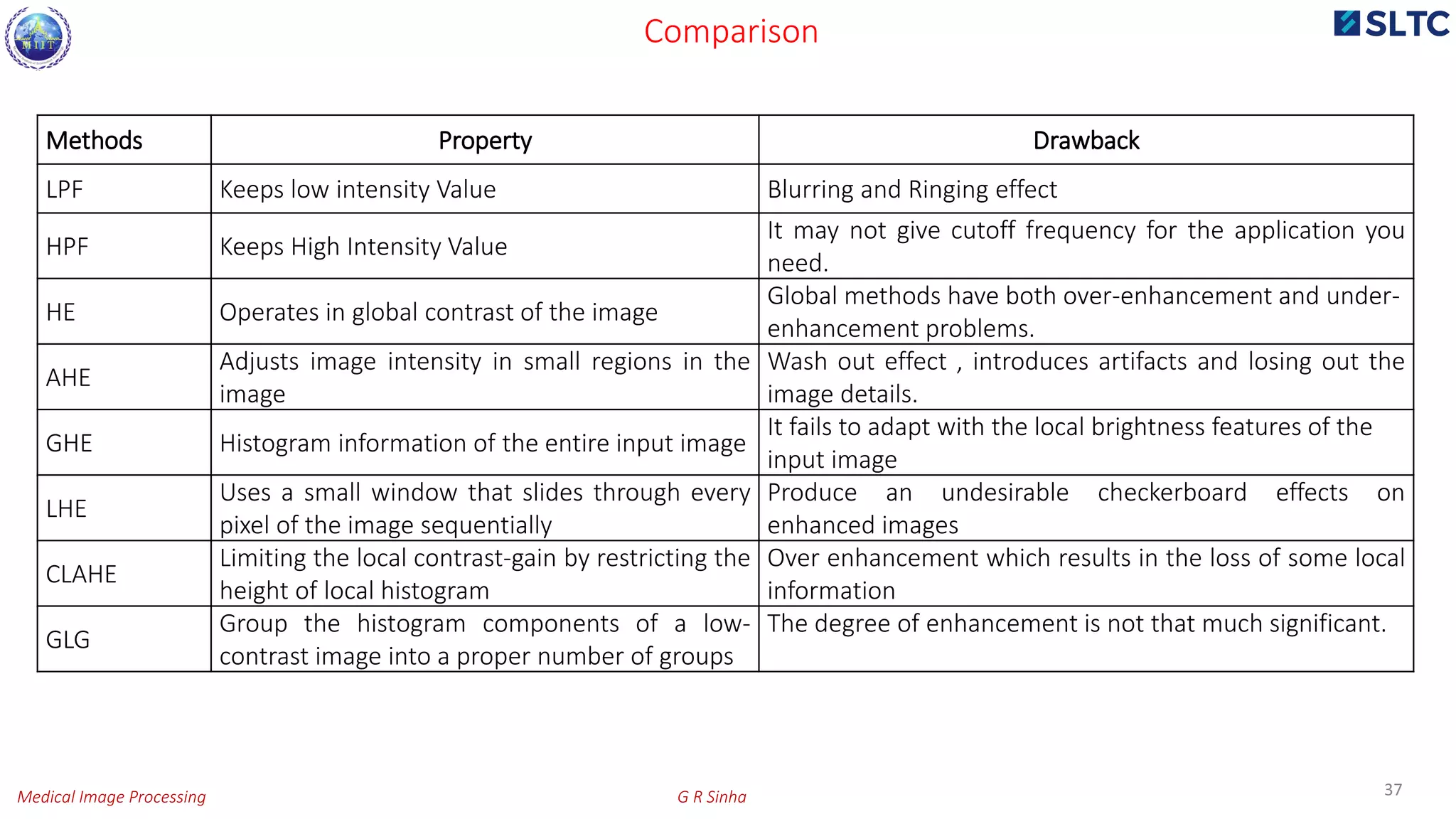
Methods Property Drawback
LPF Keeps low intensity Value Blurring and Ringing effect
HPF Keeps High Intensity Value
It may not give cutoff frequency for the application you
need.
HE Operates in global contrast of the image
Global methods have both over-enhancement and under-
enhancement problems.
AHE
Adjusts image intensity in small regions in the
image
Wash out effect , introduces artifacts and losing out the
image details.
GHE Histogram information of the entire input image
It fails to adapt with the local brightness features of the
input image
LHE
Uses a small window that slides through every
pixel of the image sequentially
Produce an undesirable checkerboard effects on
enhanced images
CLAHE
Limiting the local contrast-gain by restricting the
height of local histogram
Over enhancement which results in the loss of some local
information
GLG
Group the histogram components of a low-
contrast image into a proper number of groups
The degree of enhancement is not that much significant.
Comparison
Medical Image Processing G R Sinha
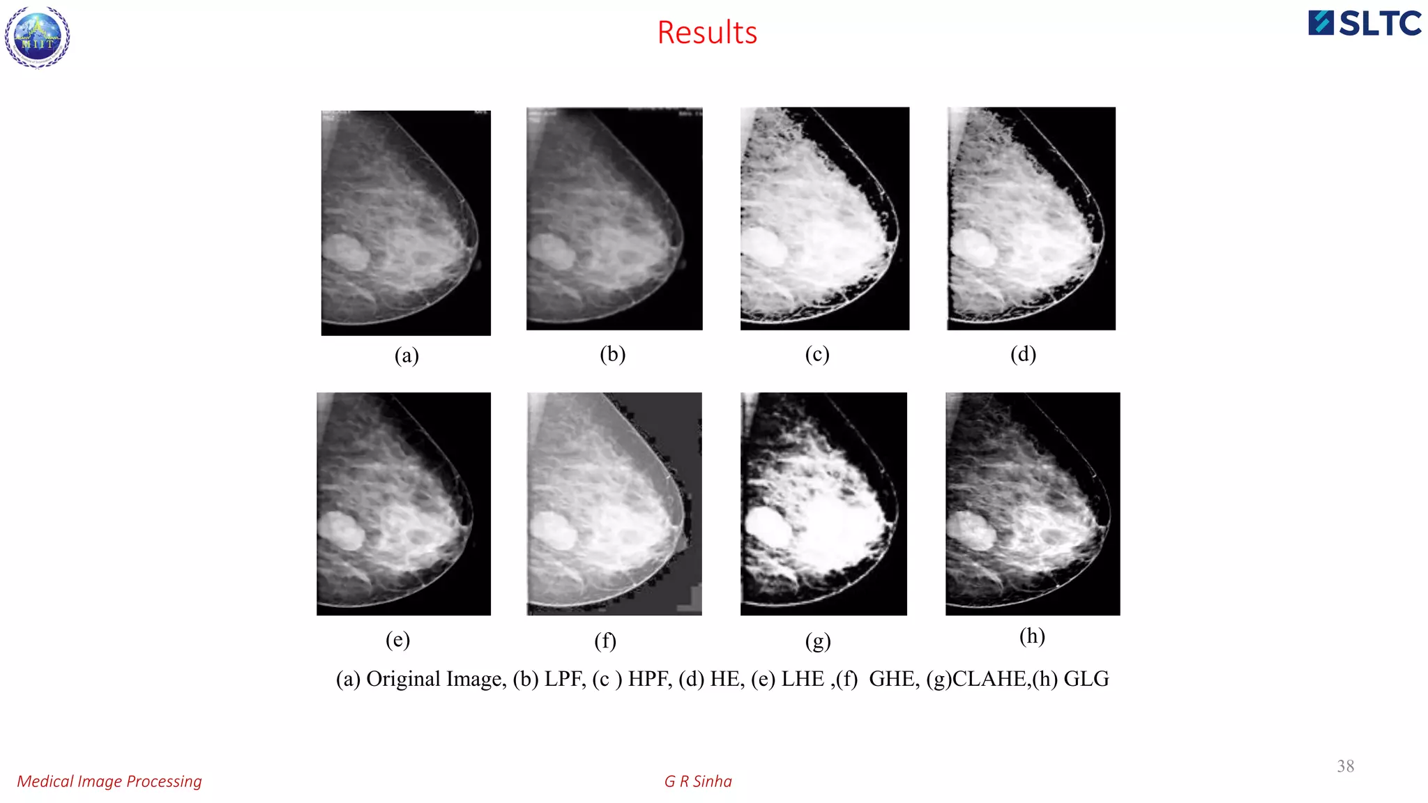
(a) Original Image, (b) LPF, (c ) HPF, (d) HE, (e) LHE ,(f) GHE, (g)CLAHE,(h) GLG
(a) (b) (c) (d)
(f) (g) (h)
38
(e)
Results
Medical Image Processing G R Sinha
 Mass segmentation
 Pixel based segmentation methods
 K-means clustering
 Threshold based
 Adaptive thresholding
 Region based segmentation
 Split and merge
 Region growing
39
Segmentation
Medical Image Processing G R Sinha
(a)Original image, (b) K-Means, (c) Thresholding ,(d) Adaptive thresholding ,(e) Split and Merge, (f) Region Growing .
(a) (c)(b)
(d) (e) (f)
40
Results
Medical Image Processing G R Sinha
41
(a) (b) (c)
(a) circular, (b) lobular and (c) speculated.
Masses
Medical Image Processing G R Sinha
42
TX: The mass cannot be assessed.
T0: Evidence of tumor is absent.
Tis: The cancer may be.
T1: The mass is 2 cm or smaller in diameter.
T2: The mass is 2-5 cm in diameter.
T3: The mass is more than 5 cm in diameter.
Cancer Stage
Medical Image Processing G R Sinha
43
Left Group
Gray Level 0 1 2 3 4 5 6 7
Amplitude
× 6 × 1 3 5 × 9
× 6 × 4 5 × 9
× 6 × 9 × 9
× 15 × × 9
× 24 × ×
Right Group
0 1 2 3 4 5 6 7
× 6 × 1 3 5 × 9
× 6 × 4 5 × 9
× 6 × 9 × 9
× × 15 × 9
× × × 24
6
1
3
5
9
0
2
4
6
8
10
0 1 2 3 4 5 6 7
Ampltude
Gray Level
15
4 4
9
24
0
5
10
15
20
25
30
0 1 2 3 4 5 6 7
Ampltude
Gray Level
Modified GLC-CE
Medical Image Processing G R Sinha
Methods Statistical Parameter
Non Cancerous Images
GRSDB-1 GRSDB-8 GRSDB-14
AMF
PSNR 16.889 16.645 15.396
RMSE 07.527 08.402 06.701
CNR 23.244 23.476 24.735
NAE 00.113 00.061 0.0764
HBF
PSNR 18.645 17.477 17.888
RMSE 08.556 07.479 07.841
CNR 29.485 30.653 30.242
NAE 00.088 00.064 00.062
CLAHE
PSNR 23.136 23.334 22.516
RMSE 08.372 09.868 08.027
CNR 34.994 33.796 33.614
NAE 00.278 00.203 00.221
GLC
PSNR 26.243 27.524 27.432
RMSE 08.235 08.984 08.342
CNR 37.241 36.862 37.563
NAE 00.354 00.284 00.431
GLC-CE (α=0.6)
PSNR 25.776 25.117 25.202
RMSE 07.907 08.332 07.267
CNR 35.534 35.365 35.185
NAE 00.245 00.189 00.223
44
Comparison
Medical Image Processing G R Sinha
45
Methods Statistical Parameter
Cancerous Images
GRSDB-19 GRSDB-23 GRSDB-31
AMF
PSNR 25.525 24.768 25.426
RMSE 06.764 07.657 08.514
CNR 26.605 25.568 26.364
NAE 00.048 00.065 00.086
HBF
PSNR 32.674 33.546 33.736
RMSE 06.756 06.486 07.253
CNR 16.763 17.342 16.341
NAE 00.047 00.075 00.094
CLAHE
PSNR 39.352 38.413 39.697
RMSE 08.257 08.873 07.364
CNR 40.596 39.654 40.974
NAE 00.278 00.263 00.254
GLC
PSNR 42.342 42.624 42.285
RMSE 08.534 08.874 07.543
CNR 43.126 43.075 44.453
NAE 00.325 00.267 00.287
GLC-CE (α=0.6)
PSNR 40.112 40.193 41.285
RMSE 07.883 08.115 07.142
CNR 41.262 40.052 42.184
NAE 00.217 00.213 00.231
contd..
Medical Image Processing G R Sinha
46
Method Used /
Database
GRSDB-19 GRSDB-23 GRSDB- 31
Original
Image
K-Means
ATS
RGS
GLC-CE Subjected to Segmentation
Medical Image Processing G R Sinha
Extracted Masses from GRSDB-14 (Non-cancerous Image)
47
Mass Segmentation (GLC-CE &ATS)
Medical Image Processing G R Sinha
Extracted mass from GRSDB-14 (Cancerous Image)
48
Contd..
Medical Image Processing G R Sinha
ATS with GLC-CE for GRSDB-14
S.N. Mass # Mean
Result obtained by CAD
Area (mm2) Perimeter (mm) Diameter (mm)
1. 15 252.9 0. 668 2.332 0.90
2. 23 253.0 0.352 2.353 0.64
3. 26 224.9 0.159 2.341 0.48
4. 61 253.0 0.344 2.469 0.53
5. 64 237.3 0.662 2.499 0.91
6. 81 236.6 0.412 2.610 0.77
7. 82 252.7 0.227 2.581 0.47
49
S.N. Mass # Mean
Result obtained by CAD
Area (mm2) Perimeter (mm) Diameter (mm)
1. 5 253.3 0.125 1.889 0.36
2. 7 252.7 0.102 1.929 0.34
3. 8 252.6 0.364 2.067 0.67
4. 10 253.0 0.366 2.256 0.62
5. 11 253.0 0.384 2.335 0.81
6. 14 252.5 0.131 2.344 0.38
7. 47 241.8 0.216 2.603 0.55
ATS with CLAHE for GRSDB-14
Features
Medical Image Processing G R Sinha
S.
N.
Mass # Mean
Result obtained by CAD Result by Radiologist Area
Diff.
[A-B]
Area (mm2)
[A]
Perimeter
(mm)
Diameter
(mm)
Area (mm2)
[B]
Peri- meter
(mm)
Diameter
(mm)
1. 5 244.3 7.90 23.7 12.74 7.1 23.2 12.30 + 0.80
2. 8 248.5 11.3 33.9 18.23 11.5 34.5 18.76 - 0.20
3. 9 253.2 8.33 24.99 13.44 8.53 25.32 13.75 - 0.20
4. 32 252.0 5.23 15.69 8.44 5.2 15.46 8.28 + 0.03
5. 36 253.5 4.40 13.2 7.10 4.21 13.64 7.41 + 0.19
6. 56 210.2 2.43 7.29 3.92 2.33 7.68 4.32 + 0.10
7. 59 245.9 3.32 9.96 5.35 3.41 9.84 5.22 - 0.09
8. 64 252.5 3.50 10.5 5.65 3.46 10.72 5.81 + 0.04
9. 78 252.9 6.31 18.93 10.18 6.35 18.60 10.02 - 0.04
10. 83 225.0 2.67 8.01 4.31 2.6 8.27 4.57 + 0.07
50
ATS with GLC-CE for GRSDB-19
contd..
Medical Image Processing G R Sinha
51
Image
Preprocessing
Methods
Segmentation
Methods
Range of area difference (mm2)
Positive Negative
GRSDB-17
CLAHE
ATS 0.23 - 1.89 0.14 - 0.23
RGS 0.54 - 3.69 0.17 - 2.32
GLC-CE
ATS 0.18 - 1.25 0.15 - 0.22
RGS 0.75 - 2.63 0.65 - 2.74
GRSDB-18
CLAHE
ATS 0.29 - 1.65 0.15 - 0.29
RGS 0.76 - 3.54 0.19 - 2.62
GLC-CE
ATS 0.17 - 1.45 0.13 - 0.21
RGS 0.83 - 2.42 0.86 - 2.69
GRSDB-29
CLAHE
ATS 0.23 - 1.43 0.14 - 0.32
RGS 0.61 - 2.69 0.11 - 2.17
GLC-CE
ATS 0.19 - 0.93 0.09 - 0.28
RGS 0.31 - 2.54 0.41 - 1.73
Comparison
Medical Image Processing G R Sinha
Image
Result
by CAD
Result by
Radiologist
Image
Result by
CAD
Result by
Radiologist
GRSDB-1 Tx T0 GRSDB-22 T1 T1
GRSDB-2 T0 T0 GRSDB-23 T2 T2
GRSDB-3 T0 Tx GRSDB-24 T1 T1
GRSDB-4 Tx Tx GRSDB-25 T1 T1
GRSDB-5 T0 T0 GRSDB-26 T1 T1
GRSDB-17 T1 T1 GRSDB-27 T1 T1
GRSDB-18 T1 T1 GRSDB-28 T2 T2
GRSDB-19 T1 T2 GRSDB-29 T2 T1
GRSDB-20 T2 T1 GRSDB-30 T2 T1
GRSDB-21 T1 T1 GRSDB-31 T2 T1
52
Cancer Stage
Medical Image Processing G R Sinha
53
Database TP FP FN TN Sensitivity% Specificity% Accuracy%
GRSDB-17 52 3 2 39 94.55 95.12 94.79
GRSDB-18 49 2 1 41 96.08 97.62 96.77
GRSDB-19 38 1 2 40 97.44 95.24 96.30
GRSDB-20 44 2 2 37 95.65 94.87 95.29
GRSDB-21 40 1 2 50 97.56 96.15 96.77
GRSDB-22 42 3 1 38 93.33 97.44 95.24
GRSDB-23 37 1 2 53 97.37 96.36 96.77
GRSDB-24 38 2 2 42 95.00 95.45 95.24
GRSDB-25 43 2 2 47 95.56 95.92 95.74
GRSDB-26 41 1 3 49 97.62 94.23 95.74
GRSDB-27 45 2 1 45 95.74 97.83 96.77
GRSDB-28 44 2 2 46 95.65 95.83 95.74
GRSDB-29 51 3 2 39 94.44 95.12 94.74
GRSDB-30 47 2 2 43 95.92 95.56 95.74
CAD Performance
Medical Image Processing G R Sinha
Authors Methods Sensitivity % Specificity % Accuracy %
Lei et al. (2001) AI with DWT 97.3 96 95.4
Jumaat et
al.(2010) Balloon Snake Not Calculated Not Calculated 95.53
Rangayyan et al.
(2007)
Gabot filter and curvilinear
structures 92 95.4 86.7
Mencattini et
al. [(2010) Luminance-region based approach 92.6 96.6 95.12
Bator et al.
(2009) Linear Structure method 90-95 88-92 Not
Calculated
Kai Hu et al.
(2011)
Adaptive global and local
thresholding segmentation method 91.73 89.8 94.5
Jun Liu et al.
(2010)
Anisotropic diffusion filter and
watershed method 95 94 96
Minavathi et al
(2012)
Gaussian smoothing and Active
contour method 92.7 90.3 94
Madabhushi et
al . (2014)
Second-order butterworth filter and
region growing method 80 82 81.4
Proposed CAD
system GLC-CE-ATS 97.55 96.37 96.75
54
Validation
Medical Image Processing G R Sinha
Health Informatics
55Medical Image Processing G R Sinha
 Health informatics, also known as medical informatics, deals with the methods or devices
that are used to acquire, store, retrieve, and use information in the health care sector.
 Technology enables a health care provider to keep electronic medical records for billing,
scheduling, and research.
Health Informatics
56
Medical Image Processing G R Sinha
 The use of hand-held or portable devices to assist the healthcare provider with data
entry or medical decision.
 ‘mHealth’ is a developing technology in the health informatics field.
contd…
57
Medical Image Processing G R Sinha
 Clinical informatics focuses on computer applications for all types of medical data and
knowledge that may be collected, organized, analyzed, stored , and used in a medical
clinic.
 Digitalized images are used in the practices of cardiology, dermatology, surgery,
obstetrics, gynecology and pathology.
58
contd..
Medical Image Processing G R Sinha
Feature Extraction
Similarity Retrieval
Image FeaturesImage Database
Query Image
Query Results
Feedback
Algorithm
User Evaluation
59
CBMIR
Medical Image Processing G R Sinha
60
Inspiring Equation
E= mc2
E = Excellence m= Motivation c=Commitment
Example: c= 0.5 (half hearted), E= ¼ & c= 2 (doubly committed), E= 4
Thank you, any queries please!
Medical Image Processing G R Sinha

More Related Content

What's hot

Image Registration (Digital Image Processing)
Image Registration (Digital Image Processing)Image Registration (Digital Image Processing)
Image Registration (Digital Image Processing)
VARUN KUMAR
 
Image filtering in Digital image processing
Image filtering in Digital image processingImage filtering in Digital image processing
Image filtering in Digital image processing
Abinaya B
 
Applications of Digital image processing in Medical Field
Applications of Digital image processing in Medical FieldApplications of Digital image processing in Medical Field
Applications of Digital image processing in Medical Field
Ashwani Srivastava
 
Image pre processing - local processing
Image pre processing - local processingImage pre processing - local processing
Image pre processing - local processing
Ashish Kumar
 
image restoration.ppt
image restoration.pptimage restoration.ppt
image restoration.ppt
RajarajeswariKannan
 
Image transforms
Image transformsImage transforms
Image transforms
11mr11mahesh
 
Introduction to Image Processing:Image Modalities
Introduction to Image Processing:Image ModalitiesIntroduction to Image Processing:Image Modalities
Introduction to Image Processing:Image Modalities
Kalyan Acharjya
 
Image enhancement
Image enhancementImage enhancement
Image enhancement
Dr INBAMALAR T M
 
Digital Image Fundamentals
Digital Image FundamentalsDigital Image Fundamentals
Digital Image Fundamentals
A B Shinde
 
Image Restoration
Image RestorationImage Restoration
Image Restoration
Srishti Kakade
 
Image Sampling and Quantization.pptx
Image Sampling and Quantization.pptxImage Sampling and Quantization.pptx
Image Sampling and Quantization.pptx
RUBIN (A) JEBIN
 
camera calibration
 camera calibration camera calibration
camera calibration
Sandeep Sasidharan
 
Role of artificial intellegence (a.i) in radiology department nitish virmani
Role of artificial intellegence (a.i) in radiology department nitish virmaniRole of artificial intellegence (a.i) in radiology department nitish virmani
Role of artificial intellegence (a.i) in radiology department nitish virmani
Nitish Virmani
 
5. gray level transformation
5. gray level transformation5. gray level transformation
5. gray level transformation
MdFazleRabbi18
 
Histogram processing
Histogram processingHistogram processing
Image enhancement techniques
Image enhancement techniques Image enhancement techniques
Image enhancement techniques
Arshad khan
 
Lec2: Digital Images and Medical Imaging Modalities
Lec2: Digital Images and Medical Imaging ModalitiesLec2: Digital Images and Medical Imaging Modalities
Lec2: Digital Images and Medical Imaging Modalities
Ulaş Bağcı
 
3D visualisation of medical images
3D visualisation of medical images3D visualisation of medical images
3D visualisation of medical images
Shashank
 
Color Image Processing: Basics
Color Image Processing: BasicsColor Image Processing: Basics
Color Image Processing: Basics
A B Shinde
 
Image Enhancement in Spatial Domain
Image Enhancement in Spatial DomainImage Enhancement in Spatial Domain
Image Enhancement in Spatial Domain
DEEPASHRI HK
 

What's hot (20)

Image Registration (Digital Image Processing)
Image Registration (Digital Image Processing)Image Registration (Digital Image Processing)
Image Registration (Digital Image Processing)
 
Image filtering in Digital image processing
Image filtering in Digital image processingImage filtering in Digital image processing
Image filtering in Digital image processing
 
Applications of Digital image processing in Medical Field
Applications of Digital image processing in Medical FieldApplications of Digital image processing in Medical Field
Applications of Digital image processing in Medical Field
 
Image pre processing - local processing
Image pre processing - local processingImage pre processing - local processing
Image pre processing - local processing
 
image restoration.ppt
image restoration.pptimage restoration.ppt
image restoration.ppt
 
Image transforms
Image transformsImage transforms
Image transforms
 
Introduction to Image Processing:Image Modalities
Introduction to Image Processing:Image ModalitiesIntroduction to Image Processing:Image Modalities
Introduction to Image Processing:Image Modalities
 
Image enhancement
Image enhancementImage enhancement
Image enhancement
 
Digital Image Fundamentals
Digital Image FundamentalsDigital Image Fundamentals
Digital Image Fundamentals
 
Image Restoration
Image RestorationImage Restoration
Image Restoration
 
Image Sampling and Quantization.pptx
Image Sampling and Quantization.pptxImage Sampling and Quantization.pptx
Image Sampling and Quantization.pptx
 
camera calibration
 camera calibration camera calibration
camera calibration
 
Role of artificial intellegence (a.i) in radiology department nitish virmani
Role of artificial intellegence (a.i) in radiology department nitish virmaniRole of artificial intellegence (a.i) in radiology department nitish virmani
Role of artificial intellegence (a.i) in radiology department nitish virmani
 
5. gray level transformation
5. gray level transformation5. gray level transformation
5. gray level transformation
 
Histogram processing
Histogram processingHistogram processing
Histogram processing
 
Image enhancement techniques
Image enhancement techniques Image enhancement techniques
Image enhancement techniques
 
Lec2: Digital Images and Medical Imaging Modalities
Lec2: Digital Images and Medical Imaging ModalitiesLec2: Digital Images and Medical Imaging Modalities
Lec2: Digital Images and Medical Imaging Modalities
 
3D visualisation of medical images
3D visualisation of medical images3D visualisation of medical images
3D visualisation of medical images
 
Color Image Processing: Basics
Color Image Processing: BasicsColor Image Processing: Basics
Color Image Processing: Basics
 
Image Enhancement in Spatial Domain
Image Enhancement in Spatial DomainImage Enhancement in Spatial Domain
Image Enhancement in Spatial Domain
 

Similar to Medical image processing

Optimizing Problem of Brain Tumor Detection using Image Processing
Optimizing Problem of Brain Tumor Detection using Image ProcessingOptimizing Problem of Brain Tumor Detection using Image Processing
Optimizing Problem of Brain Tumor Detection using Image Processing
IRJET Journal
 
Bt36430432
Bt36430432Bt36430432
Bt36430432
IJERA Editor
 
IRJET- Brain Tumor Detection and Identification using Support Vector Machine
IRJET- Brain Tumor Detection and Identification using Support Vector MachineIRJET- Brain Tumor Detection and Identification using Support Vector Machine
IRJET- Brain Tumor Detection and Identification using Support Vector Machine
IRJET Journal
 
Artificial neural network for cervical abnormalities detection on computed to...
Artificial neural network for cervical abnormalities detection on computed to...Artificial neural network for cervical abnormalities detection on computed to...
Artificial neural network for cervical abnormalities detection on computed to...
IAESIJAI
 
A Progressive Review on Early Stage Breast Cancer Detection
A Progressive Review on Early Stage Breast Cancer DetectionA Progressive Review on Early Stage Breast Cancer Detection
A Progressive Review on Early Stage Breast Cancer Detection
IRJET Journal
 
Comparative Study on Cancer Images using Watershed Transformation
Comparative Study on Cancer Images using Watershed TransformationComparative Study on Cancer Images using Watershed Transformation
Comparative Study on Cancer Images using Watershed Transformation
ijtsrd
 
IRJET - Machine Learning Applications on Cancer Prognosis and Prediction
IRJET - Machine Learning Applications on Cancer Prognosis and PredictionIRJET - Machine Learning Applications on Cancer Prognosis and Prediction
IRJET - Machine Learning Applications on Cancer Prognosis and Prediction
IRJET Journal
 
IRJET- A Study on Brain Tumor Detection Algorithms for MRI Images
IRJET- A Study on Brain Tumor Detection Algorithms for MRI ImagesIRJET- A Study on Brain Tumor Detection Algorithms for MRI Images
IRJET- A Study on Brain Tumor Detection Algorithms for MRI Images
IRJET Journal
 
A Review of Super Resolution and Tumor Detection Techniques in Medical Imaging
A Review of Super Resolution and Tumor Detection Techniques in Medical ImagingA Review of Super Resolution and Tumor Detection Techniques in Medical Imaging
A Review of Super Resolution and Tumor Detection Techniques in Medical Imaging
ijtsrd
 
IMAGE SEGMENTATION USING FCM ALGORITM | J4RV3I12021
IMAGE SEGMENTATION USING FCM ALGORITM | J4RV3I12021IMAGE SEGMENTATION USING FCM ALGORITM | J4RV3I12021
IMAGE SEGMENTATION USING FCM ALGORITM | J4RV3I12021
Journal For Research
 
Ieee brain tumor
Ieee brain tumorIeee brain tumor
Ieee brain tumor
Prakash Mhaske
 
Automated Intracranial Neoplasm Detection Using Convolutional Neural Networks
Automated Intracranial Neoplasm Detection Using Convolutional Neural NetworksAutomated Intracranial Neoplasm Detection Using Convolutional Neural Networks
Automated Intracranial Neoplasm Detection Using Convolutional Neural Networks
IRJET Journal
 
medical imaging esraa-multimedia-presentation.pptx
medical imaging esraa-multimedia-presentation.pptxmedical imaging esraa-multimedia-presentation.pptx
medical imaging esraa-multimedia-presentation.pptx
PrincessSaro
 
IRJET- Brain Tumor Detection using Convolutional Neural Network
IRJET- Brain Tumor Detection using Convolutional Neural NetworkIRJET- Brain Tumor Detection using Convolutional Neural Network
IRJET- Brain Tumor Detection using Convolutional Neural Network
IRJET Journal
 
IRJET- Novel Approach for Detection of Brain Tumor :A Review
IRJET-  	  Novel Approach for Detection of Brain Tumor :A ReviewIRJET-  	  Novel Approach for Detection of Brain Tumor :A Review
IRJET- Novel Approach for Detection of Brain Tumor :A Review
IRJET Journal
 
Objective Quality Assessment of Image Enhancement Methods in Digital Mammogra...
Objective Quality Assessment of Image Enhancement Methods in Digital Mammogra...Objective Quality Assessment of Image Enhancement Methods in Digital Mammogra...
Objective Quality Assessment of Image Enhancement Methods in Digital Mammogra...
sipij
 
IRJET - Brain Tumor Detection using Image Processing, ML & NLP
IRJET - Brain Tumor Detection using Image Processing, ML & NLPIRJET - Brain Tumor Detection using Image Processing, ML & NLP
IRJET - Brain Tumor Detection using Image Processing, ML & NLP
IRJET Journal
 
IRJET- Brain Tumor Detection using Image Processing, ML & NLP
IRJET- Brain Tumor Detection using Image Processing, ML & NLPIRJET- Brain Tumor Detection using Image Processing, ML & NLP
IRJET- Brain Tumor Detection using Image Processing, ML & NLP
IRJET Journal
 
BRAIN TUMOR DETECTION USING CNN & ML TECHNIQUES
BRAIN TUMOR DETECTION USING CNN & ML TECHNIQUESBRAIN TUMOR DETECTION USING CNN & ML TECHNIQUES
BRAIN TUMOR DETECTION USING CNN & ML TECHNIQUES
IRJET Journal
 
journals public
journals publicjournals public
journals public
rikaseorika
 

Similar to Medical image processing (20)

Optimizing Problem of Brain Tumor Detection using Image Processing
Optimizing Problem of Brain Tumor Detection using Image ProcessingOptimizing Problem of Brain Tumor Detection using Image Processing
Optimizing Problem of Brain Tumor Detection using Image Processing
 
Bt36430432
Bt36430432Bt36430432
Bt36430432
 
IRJET- Brain Tumor Detection and Identification using Support Vector Machine
IRJET- Brain Tumor Detection and Identification using Support Vector MachineIRJET- Brain Tumor Detection and Identification using Support Vector Machine
IRJET- Brain Tumor Detection and Identification using Support Vector Machine
 
Artificial neural network for cervical abnormalities detection on computed to...
Artificial neural network for cervical abnormalities detection on computed to...Artificial neural network for cervical abnormalities detection on computed to...
Artificial neural network for cervical abnormalities detection on computed to...
 
A Progressive Review on Early Stage Breast Cancer Detection
A Progressive Review on Early Stage Breast Cancer DetectionA Progressive Review on Early Stage Breast Cancer Detection
A Progressive Review on Early Stage Breast Cancer Detection
 
Comparative Study on Cancer Images using Watershed Transformation
Comparative Study on Cancer Images using Watershed TransformationComparative Study on Cancer Images using Watershed Transformation
Comparative Study on Cancer Images using Watershed Transformation
 
IRJET - Machine Learning Applications on Cancer Prognosis and Prediction
IRJET - Machine Learning Applications on Cancer Prognosis and PredictionIRJET - Machine Learning Applications on Cancer Prognosis and Prediction
IRJET - Machine Learning Applications on Cancer Prognosis and Prediction
 
IRJET- A Study on Brain Tumor Detection Algorithms for MRI Images
IRJET- A Study on Brain Tumor Detection Algorithms for MRI ImagesIRJET- A Study on Brain Tumor Detection Algorithms for MRI Images
IRJET- A Study on Brain Tumor Detection Algorithms for MRI Images
 
A Review of Super Resolution and Tumor Detection Techniques in Medical Imaging
A Review of Super Resolution and Tumor Detection Techniques in Medical ImagingA Review of Super Resolution and Tumor Detection Techniques in Medical Imaging
A Review of Super Resolution and Tumor Detection Techniques in Medical Imaging
 
IMAGE SEGMENTATION USING FCM ALGORITM | J4RV3I12021
IMAGE SEGMENTATION USING FCM ALGORITM | J4RV3I12021IMAGE SEGMENTATION USING FCM ALGORITM | J4RV3I12021
IMAGE SEGMENTATION USING FCM ALGORITM | J4RV3I12021
 
Ieee brain tumor
Ieee brain tumorIeee brain tumor
Ieee brain tumor
 
Automated Intracranial Neoplasm Detection Using Convolutional Neural Networks
Automated Intracranial Neoplasm Detection Using Convolutional Neural NetworksAutomated Intracranial Neoplasm Detection Using Convolutional Neural Networks
Automated Intracranial Neoplasm Detection Using Convolutional Neural Networks
 
medical imaging esraa-multimedia-presentation.pptx
medical imaging esraa-multimedia-presentation.pptxmedical imaging esraa-multimedia-presentation.pptx
medical imaging esraa-multimedia-presentation.pptx
 
IRJET- Brain Tumor Detection using Convolutional Neural Network
IRJET- Brain Tumor Detection using Convolutional Neural NetworkIRJET- Brain Tumor Detection using Convolutional Neural Network
IRJET- Brain Tumor Detection using Convolutional Neural Network
 
IRJET- Novel Approach for Detection of Brain Tumor :A Review
IRJET-  	  Novel Approach for Detection of Brain Tumor :A ReviewIRJET-  	  Novel Approach for Detection of Brain Tumor :A Review
IRJET- Novel Approach for Detection of Brain Tumor :A Review
 
Objective Quality Assessment of Image Enhancement Methods in Digital Mammogra...
Objective Quality Assessment of Image Enhancement Methods in Digital Mammogra...Objective Quality Assessment of Image Enhancement Methods in Digital Mammogra...
Objective Quality Assessment of Image Enhancement Methods in Digital Mammogra...
 
IRJET - Brain Tumor Detection using Image Processing, ML & NLP
IRJET - Brain Tumor Detection using Image Processing, ML & NLPIRJET - Brain Tumor Detection using Image Processing, ML & NLP
IRJET - Brain Tumor Detection using Image Processing, ML & NLP
 
IRJET- Brain Tumor Detection using Image Processing, ML & NLP
IRJET- Brain Tumor Detection using Image Processing, ML & NLPIRJET- Brain Tumor Detection using Image Processing, ML & NLP
IRJET- Brain Tumor Detection using Image Processing, ML & NLP
 
BRAIN TUMOR DETECTION USING CNN & ML TECHNIQUES
BRAIN TUMOR DETECTION USING CNN & ML TECHNIQUESBRAIN TUMOR DETECTION USING CNN & ML TECHNIQUES
BRAIN TUMOR DETECTION USING CNN & ML TECHNIQUES
 
journals public
journals publicjournals public
journals public
 

More from Dr G R Sinha

Modern signal processing is dead without machine learning! 5th july 2020
Modern signal processing is dead without machine learning! 5th july 2020Modern signal processing is dead without machine learning! 5th july 2020
Modern signal processing is dead without machine learning! 5th july 2020
Dr G R Sinha
 
5 g and IoT for healthcare
5 g and IoT  for healthcare 5 g and IoT  for healthcare
5 g and IoT for healthcare
Dr G R Sinha
 
Current trends in cognitive science and brain computing research 18th june 2020
Current trends in cognitive science and brain computing research 18th june 2020Current trends in cognitive science and brain computing research 18th june 2020
Current trends in cognitive science and brain computing research 18th june 2020
Dr G R Sinha
 
Research based leraning for developing employabilty skills in young minds
Research based leraning for developing employabilty skills in young mindsResearch based leraning for developing employabilty skills in young minds
Research based leraning for developing employabilty skills in young minds
Dr G R Sinha
 
Writing research grant proposal
Writing research grant proposalWriting research grant proposal
Writing research grant proposal
Dr G R Sinha
 
Writing book proposal
Writing book proposalWriting book proposal
Writing book proposal
Dr G R Sinha
 
Mapping learning outcomes with learning objectives to achieve real impact of OBE
Mapping learning outcomes with learning objectives to achieve real impact of OBEMapping learning outcomes with learning objectives to achieve real impact of OBE
Mapping learning outcomes with learning objectives to achieve real impact of OBE
Dr G R Sinha
 
Mapping learning outcomes with learning objectives to achieve real impact of obe
Mapping learning outcomes with learning objectives to achieve real impact of obeMapping learning outcomes with learning objectives to achieve real impact of obe
Mapping learning outcomes with learning objectives to achieve real impact of obe
Dr G R Sinha
 
ICT enabled agriculture for sustainable rural economy
ICT enabled agriculture for sustainable rural economyICT enabled agriculture for sustainable rural economy
ICT enabled agriculture for sustainable rural economy
Dr G R Sinha
 
Why research!
Why research!Why research!
Why research!
Dr G R Sinha
 
Developing employability skills using ICT tools
Developing employability skills using ICT toolsDeveloping employability skills using ICT tools
Developing employability skills using ICT tools
Dr G R Sinha
 
Study of assessment of cognitive ability
Study of assessment of cognitive abilityStudy of assessment of cognitive ability
Study of assessment of cognitive ability
Dr G R Sinha
 
Cognitive ability of human brain and soft computing techniques
Cognitive ability of human brain and soft computing techniquesCognitive ability of human brain and soft computing techniques
Cognitive ability of human brain and soft computing techniques
Dr G R Sinha
 
Research based skills towards acdemic excellence
Research based skills towards acdemic excellenceResearch based skills towards acdemic excellence
Research based skills towards acdemic excellence
Dr G R Sinha
 
Learning research based skills towards academic excellence
Learning research based skills towards academic excellenceLearning research based skills towards academic excellence
Learning research based skills towards academic excellence
Dr G R Sinha
 
Thevenin's theorem and application
Thevenin's theorem and applicationThevenin's theorem and application
Thevenin's theorem and application
Dr G R Sinha
 

More from Dr G R Sinha (16)

Modern signal processing is dead without machine learning! 5th july 2020
Modern signal processing is dead without machine learning! 5th july 2020Modern signal processing is dead without machine learning! 5th july 2020
Modern signal processing is dead without machine learning! 5th july 2020
 
5 g and IoT for healthcare
5 g and IoT  for healthcare 5 g and IoT  for healthcare
5 g and IoT for healthcare
 
Current trends in cognitive science and brain computing research 18th june 2020
Current trends in cognitive science and brain computing research 18th june 2020Current trends in cognitive science and brain computing research 18th june 2020
Current trends in cognitive science and brain computing research 18th june 2020
 
Research based leraning for developing employabilty skills in young minds
Research based leraning for developing employabilty skills in young mindsResearch based leraning for developing employabilty skills in young minds
Research based leraning for developing employabilty skills in young minds
 
Writing research grant proposal
Writing research grant proposalWriting research grant proposal
Writing research grant proposal
 
Writing book proposal
Writing book proposalWriting book proposal
Writing book proposal
 
Mapping learning outcomes with learning objectives to achieve real impact of OBE
Mapping learning outcomes with learning objectives to achieve real impact of OBEMapping learning outcomes with learning objectives to achieve real impact of OBE
Mapping learning outcomes with learning objectives to achieve real impact of OBE
 
Mapping learning outcomes with learning objectives to achieve real impact of obe
Mapping learning outcomes with learning objectives to achieve real impact of obeMapping learning outcomes with learning objectives to achieve real impact of obe
Mapping learning outcomes with learning objectives to achieve real impact of obe
 
ICT enabled agriculture for sustainable rural economy
ICT enabled agriculture for sustainable rural economyICT enabled agriculture for sustainable rural economy
ICT enabled agriculture for sustainable rural economy
 
Why research!
Why research!Why research!
Why research!
 
Developing employability skills using ICT tools
Developing employability skills using ICT toolsDeveloping employability skills using ICT tools
Developing employability skills using ICT tools
 
Study of assessment of cognitive ability
Study of assessment of cognitive abilityStudy of assessment of cognitive ability
Study of assessment of cognitive ability
 
Cognitive ability of human brain and soft computing techniques
Cognitive ability of human brain and soft computing techniquesCognitive ability of human brain and soft computing techniques
Cognitive ability of human brain and soft computing techniques
 
Research based skills towards acdemic excellence
Research based skills towards acdemic excellenceResearch based skills towards acdemic excellence
Research based skills towards acdemic excellence
 
Learning research based skills towards academic excellence
Learning research based skills towards academic excellenceLearning research based skills towards academic excellence
Learning research based skills towards academic excellence
 
Thevenin's theorem and application
Thevenin's theorem and applicationThevenin's theorem and application
Thevenin's theorem and application
 

Recently uploaded

Software Engineering and Project Management - Introduction, Modeling Concepts...
Software Engineering and Project Management - Introduction, Modeling Concepts...Software Engineering and Project Management - Introduction, Modeling Concepts...
Software Engineering and Project Management - Introduction, Modeling Concepts...
Prakhyath Rai
 
Design and optimization of ion propulsion drone
Design and optimization of ion propulsion droneDesign and optimization of ion propulsion drone
Design and optimization of ion propulsion drone
bjmsejournal
 
Redefining brain tumor segmentation: a cutting-edge convolutional neural netw...
Redefining brain tumor segmentation: a cutting-edge convolutional neural netw...Redefining brain tumor segmentation: a cutting-edge convolutional neural netw...
Redefining brain tumor segmentation: a cutting-edge convolutional neural netw...
IJECEIAES
 
An Introduction to the Compiler Designss
An Introduction to the Compiler DesignssAn Introduction to the Compiler Designss
An Introduction to the Compiler Designss
ElakkiaU
 
22CYT12-Unit-V-E Waste and its Management.ppt
22CYT12-Unit-V-E Waste and its Management.ppt22CYT12-Unit-V-E Waste and its Management.ppt
22CYT12-Unit-V-E Waste and its Management.ppt
KrishnaveniKrishnara1
 
2008 BUILDING CONSTRUCTION Illustrated - Ching Chapter 02 The Building.pdf
2008 BUILDING CONSTRUCTION Illustrated - Ching Chapter 02 The Building.pdf2008 BUILDING CONSTRUCTION Illustrated - Ching Chapter 02 The Building.pdf
2008 BUILDING CONSTRUCTION Illustrated - Ching Chapter 02 The Building.pdf
Yasser Mahgoub
 
Data Driven Maintenance | UReason Webinar
Data Driven Maintenance | UReason WebinarData Driven Maintenance | UReason Webinar
Data Driven Maintenance | UReason Webinar
UReason
 
Mechanical Engineering on AAI Summer Training Report-003.pdf
Mechanical Engineering on AAI Summer Training Report-003.pdfMechanical Engineering on AAI Summer Training Report-003.pdf
Mechanical Engineering on AAI Summer Training Report-003.pdf
21UME003TUSHARDEB
 
哪里办理(csu毕业证书)查尔斯特大学毕业证硕士学历原版一模一样
哪里办理(csu毕业证书)查尔斯特大学毕业证硕士学历原版一模一样哪里办理(csu毕业证书)查尔斯特大学毕业证硕士学历原版一模一样
哪里办理(csu毕业证书)查尔斯特大学毕业证硕士学历原版一模一样
insn4465
 
4. Mosca vol I -Fisica-Tipler-5ta-Edicion-Vol-1.pdf
4. Mosca vol I -Fisica-Tipler-5ta-Edicion-Vol-1.pdf4. Mosca vol I -Fisica-Tipler-5ta-Edicion-Vol-1.pdf
4. Mosca vol I -Fisica-Tipler-5ta-Edicion-Vol-1.pdf
Gino153088
 
Advanced control scheme of doubly fed induction generator for wind turbine us...
Advanced control scheme of doubly fed induction generator for wind turbine us...Advanced control scheme of doubly fed induction generator for wind turbine us...
Advanced control scheme of doubly fed induction generator for wind turbine us...
IJECEIAES
 
Engineering Drawings Lecture Detail Drawings 2014.pdf
Engineering Drawings Lecture Detail Drawings 2014.pdfEngineering Drawings Lecture Detail Drawings 2014.pdf
Engineering Drawings Lecture Detail Drawings 2014.pdf
abbyasa1014
 
Generative AI leverages algorithms to create various forms of content
Generative AI leverages algorithms to create various forms of contentGenerative AI leverages algorithms to create various forms of content
Generative AI leverages algorithms to create various forms of content
Hitesh Mohapatra
 
Properties Railway Sleepers and Test.pptx
Properties Railway Sleepers and Test.pptxProperties Railway Sleepers and Test.pptx
Properties Railway Sleepers and Test.pptx
MDSABBIROJJAMANPAYEL
 
CompEx~Manual~1210 (2).pdf COMPEX GAS AND VAPOURS
CompEx~Manual~1210 (2).pdf COMPEX GAS AND VAPOURSCompEx~Manual~1210 (2).pdf COMPEX GAS AND VAPOURS
CompEx~Manual~1210 (2).pdf COMPEX GAS AND VAPOURS
RamonNovais6
 
spirit beverages ppt without graphics.pptx
spirit beverages ppt without graphics.pptxspirit beverages ppt without graphics.pptx
spirit beverages ppt without graphics.pptx
Madan Karki
 
Electric vehicle and photovoltaic advanced roles in enhancing the financial p...
Electric vehicle and photovoltaic advanced roles in enhancing the financial p...Electric vehicle and photovoltaic advanced roles in enhancing the financial p...
Electric vehicle and photovoltaic advanced roles in enhancing the financial p...
IJECEIAES
 
Embedded machine learning-based road conditions and driving behavior monitoring
Embedded machine learning-based road conditions and driving behavior monitoringEmbedded machine learning-based road conditions and driving behavior monitoring
Embedded machine learning-based road conditions and driving behavior monitoring
IJECEIAES
 
Unit-III-ELECTROCHEMICAL STORAGE DEVICES.ppt
Unit-III-ELECTROCHEMICAL STORAGE DEVICES.pptUnit-III-ELECTROCHEMICAL STORAGE DEVICES.ppt
Unit-III-ELECTROCHEMICAL STORAGE DEVICES.ppt
KrishnaveniKrishnara1
 
cnn.pptx Convolutional neural network used for image classication
cnn.pptx Convolutional neural network used for image classicationcnn.pptx Convolutional neural network used for image classication
cnn.pptx Convolutional neural network used for image classication
SakkaravarthiShanmug
 

Recently uploaded (20)

Software Engineering and Project Management - Introduction, Modeling Concepts...
Software Engineering and Project Management - Introduction, Modeling Concepts...Software Engineering and Project Management - Introduction, Modeling Concepts...
Software Engineering and Project Management - Introduction, Modeling Concepts...
 
Design and optimization of ion propulsion drone
Design and optimization of ion propulsion droneDesign and optimization of ion propulsion drone
Design and optimization of ion propulsion drone
 
Redefining brain tumor segmentation: a cutting-edge convolutional neural netw...
Redefining brain tumor segmentation: a cutting-edge convolutional neural netw...Redefining brain tumor segmentation: a cutting-edge convolutional neural netw...
Redefining brain tumor segmentation: a cutting-edge convolutional neural netw...
 
An Introduction to the Compiler Designss
An Introduction to the Compiler DesignssAn Introduction to the Compiler Designss
An Introduction to the Compiler Designss
 
22CYT12-Unit-V-E Waste and its Management.ppt
22CYT12-Unit-V-E Waste and its Management.ppt22CYT12-Unit-V-E Waste and its Management.ppt
22CYT12-Unit-V-E Waste and its Management.ppt
 
2008 BUILDING CONSTRUCTION Illustrated - Ching Chapter 02 The Building.pdf
2008 BUILDING CONSTRUCTION Illustrated - Ching Chapter 02 The Building.pdf2008 BUILDING CONSTRUCTION Illustrated - Ching Chapter 02 The Building.pdf
2008 BUILDING CONSTRUCTION Illustrated - Ching Chapter 02 The Building.pdf
 
Data Driven Maintenance | UReason Webinar
Data Driven Maintenance | UReason WebinarData Driven Maintenance | UReason Webinar
Data Driven Maintenance | UReason Webinar
 
Mechanical Engineering on AAI Summer Training Report-003.pdf
Mechanical Engineering on AAI Summer Training Report-003.pdfMechanical Engineering on AAI Summer Training Report-003.pdf
Mechanical Engineering on AAI Summer Training Report-003.pdf
 
哪里办理(csu毕业证书)查尔斯特大学毕业证硕士学历原版一模一样
哪里办理(csu毕业证书)查尔斯特大学毕业证硕士学历原版一模一样哪里办理(csu毕业证书)查尔斯特大学毕业证硕士学历原版一模一样
哪里办理(csu毕业证书)查尔斯特大学毕业证硕士学历原版一模一样
 
4. Mosca vol I -Fisica-Tipler-5ta-Edicion-Vol-1.pdf
4. Mosca vol I -Fisica-Tipler-5ta-Edicion-Vol-1.pdf4. Mosca vol I -Fisica-Tipler-5ta-Edicion-Vol-1.pdf
4. Mosca vol I -Fisica-Tipler-5ta-Edicion-Vol-1.pdf
 
Advanced control scheme of doubly fed induction generator for wind turbine us...
Advanced control scheme of doubly fed induction generator for wind turbine us...Advanced control scheme of doubly fed induction generator for wind turbine us...
Advanced control scheme of doubly fed induction generator for wind turbine us...
 
Engineering Drawings Lecture Detail Drawings 2014.pdf
Engineering Drawings Lecture Detail Drawings 2014.pdfEngineering Drawings Lecture Detail Drawings 2014.pdf
Engineering Drawings Lecture Detail Drawings 2014.pdf
 
Generative AI leverages algorithms to create various forms of content
Generative AI leverages algorithms to create various forms of contentGenerative AI leverages algorithms to create various forms of content
Generative AI leverages algorithms to create various forms of content
 
Properties Railway Sleepers and Test.pptx
Properties Railway Sleepers and Test.pptxProperties Railway Sleepers and Test.pptx
Properties Railway Sleepers and Test.pptx
 
CompEx~Manual~1210 (2).pdf COMPEX GAS AND VAPOURS
CompEx~Manual~1210 (2).pdf COMPEX GAS AND VAPOURSCompEx~Manual~1210 (2).pdf COMPEX GAS AND VAPOURS
CompEx~Manual~1210 (2).pdf COMPEX GAS AND VAPOURS
 
spirit beverages ppt without graphics.pptx
spirit beverages ppt without graphics.pptxspirit beverages ppt without graphics.pptx
spirit beverages ppt without graphics.pptx
 
Electric vehicle and photovoltaic advanced roles in enhancing the financial p...
Electric vehicle and photovoltaic advanced roles in enhancing the financial p...Electric vehicle and photovoltaic advanced roles in enhancing the financial p...
Electric vehicle and photovoltaic advanced roles in enhancing the financial p...
 
Embedded machine learning-based road conditions and driving behavior monitoring
Embedded machine learning-based road conditions and driving behavior monitoringEmbedded machine learning-based road conditions and driving behavior monitoring
Embedded machine learning-based road conditions and driving behavior monitoring
 
Unit-III-ELECTROCHEMICAL STORAGE DEVICES.ppt
Unit-III-ELECTROCHEMICAL STORAGE DEVICES.pptUnit-III-ELECTROCHEMICAL STORAGE DEVICES.ppt
Unit-III-ELECTROCHEMICAL STORAGE DEVICES.ppt
 
cnn.pptx Convolutional neural network used for image classication
cnn.pptx Convolutional neural network used for image classicationcnn.pptx Convolutional neural network used for image classication
cnn.pptx Convolutional neural network used for image classication
 

Medical image processing

  • 1. Medical Image Processing G R Sinha, PhD IEEE Senior Member, ACM Distinguished Speaker, IEEE Distinguished Speaker Professor, Myanmar Institute of Information Technology Mandalay Recipient of ISTE National Award, TCS Award, IEI Award, Expert Engineer Award, Young Engineer Award, Young Scientist Award Email: drgrsinha@ieee.org, ganeshsinha@acm.com, gr_sinha@miit.edu.mm
  • 2. 2  Image Processing and CAD  Breast Cancer- A Case Study  Health Informatics Lecture Outline Medical Image Processing G R Sinha
  • 3. Image Processing and CAD (Computer-aided Diagnosis) 3Medical Image Processing G R Sinha
  • 4. 4 Medical Image Processing G R Sinha Digital Image
  • 5. 5  Pre-processing  Image Enhancement  Image Transformation  Image Restoration  Color Image Processing  Image Compression  Image Segmentation, Representation and Description  Content-based Image Retrieval  Pattern Recognition Digital Image Processing Medical Image Processing G R Sinha
  • 6. 6 Analog signal X(t) Sampling Quantization Sampled signal X(k) Discrete signal Digital signal Encoding Image processing is a set of tools which involves converting an analogue or continuous image into digital form.  Then performing some operations upon it so that the image quality may be enhanced or some useful information could be extracted. Digital Signal Medical Image Processing G R Sinha
  • 7. Conventional Medical Diagnosis System  Human expertise is a scarce resource  Human gets tired and forget  Humans are inconsistent in their day to day decisions for the same set of data  Human can lie, die, and hide  Screening Challenges: Complex image interpretation; high volume and small viewing time 7 Medical Image Processing G R Sinha
  • 8. Radiologist CAD Radiologist + CAD Detected Marked Detected Missed Missed Oversight Missed CAD + Radiologist 8 Medical Image Processing G R Sinha
  • 9. CAD (Computer-aided Diagnosis)  CAD Combines creation, recognition, representation, collection, organization, transformation, communication, evaluation and control of information.  Art, science, engineering and human dimensions  Diagnose; monitor; analyze; interpret; plan; design; instruct; clarify and Learn Efficiently 9Medical Image Processing G R Sinha
  • 10. Imaging Process Raw data Reconstruction 123…………… 2346………….. 65789………… 6578…………..Quantitative output Processing Analysis Filtering “Raw data” Signal acquisition 10 Medical Image Processing G R Sinha
  • 11.  Body Region, Organ, Tissue, Cell  Energy sources and Detectors  Image formation and Display  User Interface  Connection to other Systems 11 Components of Medical Imaging Medical Image Processing G R Sinha
  • 12. Converting an image into data  Qualitative and quantitative features Examination Level: Feature 1 Feature 2 Feature 3 . . Finding: Feature 1 Feature 2 . . 12 Medical Image Processing G R Sinha
  • 13. CAD helps in  Visualization: Enhancement for visual analysis.  Detection: Detect the presence of disease manifestation/abnormality.  Localization and Segmentation: Localize or segment the spatial regions.  Detection of the abnormality and Classification of likelihood that the abnormality represents a malignancy’ 13 Medical Image Processing G R Sinha
  • 14. CAD for Breast Cancer  A mammogram is an X-ray of breast tissue used for detection of lumps, changes in breast tissue or calcifications.  Abnormal tissues are generally dense white. 14Medical Image Processing G R Sinha
  • 15. X-ray physics  X-rays are form of electromagnetic energy which travel at the speed of light; and can pass all the way through the body; be deflected or scattered or be absorbed  Depends on the energy of the x-ray and the atomic number of the tissue.  Higher energy x-rays: More likely to pass through  Higher atomic number: More likely to absorb the x-ray 15 Medical Image Processing G R Sinha
  • 16. Radiographic Densities 1. Air 2. Fat 3. Soft tissue/fluid 4. Mineral 5. Metal 1. 2. 3. 4. 5. 16 Medical Image Processing G R Sinha
  • 17. Detection Diagnosis: “Yes”I think my dog swallowed something 17 Medical Image Processing G R Sinha
  • 18. Ultrasound  Non-invasive  Abdominal problems, measurement of blood flow and detection of constrictions in arteries and veins.  Also used in non-destructive testing in industry: e.g., cracks in structures. 18 Medical Image Processing G R Sinha
  • 19.  High-frequency sound (ultrasonic) waves to produce images of structures within the human body  Piezoelectric crystal creates sound waves  Still images or a moving picture  Commonly used to examine fetuses to ascertain size, position, or abnormalities; also for heart, liver, kidneys, gallbladder, breast, eye, and major blood vessels contd.. 19 Medical Image Processing G R Sinha
  • 20. MRI and CT  Computed tomography (CT) also known as computed axial tomography (CAT) that uses X-rays as ionizing radiation to acquire images for examining tissue such as bone and calcifications (calcium based) within the body (carbon based flesh), or of structures (vessels, bowel).  MRI uses non-ionizing radio frequency (RF) signals to acquire its images and is best suited for non-calcified tissue. 20 Medical Image Processing G R Sinha
  • 21. Mammography  Uses a low-dose x-ray system to examine breasts.  Mammography replaces x-ray film by solid-state detectors that convert x-rays into electrical signals which are used to produce images.  Mammography can show changes in the breast up to two years before a physician can feel. 21Medical Image Processing G R Sinha
  • 22. Detection of Malignant Masses malignant benign 22 Medical Image Processing G R Sinha
  • 23. Difficult Case  Heterogeneously dense breast  Fibro-glandular tissue (white areas) may hide the tumor  Breasts of younger women contain more glands and ligaments resulting in dense breast tissue 23 Medical Image Processing G R Sinha
  • 24. Easier Case  With age, breast tissue becomes fattier and has less number of glands  Cancer is relatively easy to detect in this type of breast tissue 24 Medical Image Processing G R Sinha
  • 25. CAD Characterization of a CT 25Medical Image Processing G R Sinha
  • 26. Challenges in computer-aided characterization  Large number of training samples and features: dimensionality  Variation in Nodule size and boundaries  Different types of imaging acquisition parameters  Clinical evaluation: observer performance studies require collaboration with medical experts or hospitals 26Medical Image Processing G R Sinha
  • 27. Selection of Features  SNR, PSNR, MSE and Entropy  Shape Features: Euclidian distance, perimeter, convex perimeter, major and minor axis, rectangularity, convexity, solidity etc.  Texture Features: mean, variance, skewness etc. Major axis Minor axis 27 Medical Image Processing G R Sinha
  • 28. Evaluation Parameters  True Positive (TP): A case when the suspected abnormality is malignant i.e. the prediction is true.  True Negative (TN): If there is no detection of abnormality in healthy person. A case where no symptoms were found truly.  False Positives (FP): Indicates that detection of abnormality is found in healthy person. The prediction of presence of abnormality is not true.  False Negatives (FN): No detection of malignant lesion is found, proves to be false. 28 Medical Image Processing G R Sinha
  • 29. Breast Cancer Detection-A Case Study 29Medical Image Processing G R Sinha
  • 30. Breast Anatomy and Micro-calcifications 30  Micro-calcifications: Tiny deposits of calcium.  Most women have one or more areas of micro-calcifications of various sizes.  Majority of calcium deposits are harmless and a small percentage may be precancerous or cancerous (benign).  Some of the cells begin growing abnormally and may spread through the breast, to the lymph or to other parts of the body (malignant).  Common type of breast cancer begins in the milk-production ducts, but cancer may also occur in the lobules or in other breast tissue. Medical Image Processing G R Sinha
  • 31. 31 Author Method Findings Limitations Naseera et al. (2016) Adaptive histogram equalization technique followed by Water shed Segmentation Better classification is realized for benign and malignant tissues. Size and area of masses are not calculated Singh et al. (2015) Max-Mean and Least-Variance technique Segmenting the Cancer Region Manual selection of threshold parameter and size of averaging filter. Hu et al. (2014) Adaptive global & local threshold segmentation. Suspicious Lesions are detected and analyzed.  Other combinations of lesion feature are not considered.  Higher False negative rate is found. Massich et al. (2014) Gaussian and Seed region growing method. Lesion are identified and segmented. Does not address the problem of segmentation other non-lesion structures. Quintana et al. (2013) Histogram techniques and Canny algorithm. Automatic micro calcification is recognized in mammographic images. Does not perform satisfactorily due to non uniform background.  Higher FP rate is achieved. Few Important Contributions Medical Image Processing G R Sinha
  • 32. 32 Author Method Findings Limitations Minavathi et al. (2012) Gaussian and Anisotropic diffusion filter. Mass in breast are identified and Classified . Blurred images are obtained. Incorrect identification of boundary obtained. Lei et al. (2011) Watershed transform Masses are segmented. Over-segmentation. Jumaat et al. (2011) Median filter and Balloon snake Cancers or non-cancerous masses are classified and their boundaries are identified. Lower degree of accuracy.  Higher false negative rate. Maitra et al. (2011) Divide and Conquer, seeded Region growing Algorithms Abnormal Masses are detected. No mathematical background involved to measure the mass, shape, size, position and density of masses. Mohideen et al. (2011) Multi wavelet method Microcalcifications and suspicious structures are detected. Improvement of local details required in low contrast regions. Luo et al. (2010) Support vector machine. Breast Masses are segmented and diagnosed. Density of mass is not properly achieved. contd.. Medical Image Processing G R Sinha
  • 33. Author Segmentation Feature Extraction (TP,FP,TN, TP) Size and shape define Cancer Stage Identification Accuracy (Sensitivity Specificity) Kai Hu et al.(2014) Yes Yes No No Yes Kowal et al. (2011) Yes No Yes No No Maitra et al.(2011) Yes No No No No Nithya, et al. (2011) No Yes No No Yes Shekhar et al. (2011) No Yes yes Yes No Quintana et al. (2011) Yes Yes No No Yes Liu et al. (2010) Yes Yes No No Yes Bick et al. (2009) Yes No No No No Bator et al. (2009) Yes Yes No No No Yuji Ikedo et al. (2007) No Yes No No Yes Kom et al (2007) Yes No No No No Madabhushi et al. (2003) Yes Yes No No No Lei et al. (2001) Yes Yes No No Yes 33 Comparison Medical Image Processing G R Sinha
  • 34. 34  Breast positioning.  Underexposure of images.  Blurring and speckle noise  Fatty breasts.  Smaller malignancies.  Boundary identification. Problems Medical Image Processing G R Sinha
  • 35. Input Image De-noise the image Perform Segmentation Refine the Segmentation Obtained Resulting Mammographic Image Size and shape identification Feature Extraction Feature Classification Pre-processing Step Segmentation Step Post-processing Step (CAD Evaluation) 35 Work-Flow Medical Image Processing G R Sinha
  • 36.  Low Pass Filter(LPF)  High Pass Filter(HPF)  Histogram Equalization(HE)  Adaptive Histogram Equalization(AHE)  Global Histogram Equalization(GHE)  Local Histogram Equalization(LHE)  Contrast Limited Adaptive Histogram Equalization(CLAHE)  Gray Level Grouping(GLG) 36 Pre-processing Medical Image Processing G R Sinha
  • 37. 37 Methods Property Drawback LPF Keeps low intensity Value Blurring and Ringing effect HPF Keeps High Intensity Value It may not give cutoff frequency for the application you need. HE Operates in global contrast of the image Global methods have both over-enhancement and under- enhancement problems. AHE Adjusts image intensity in small regions in the image Wash out effect , introduces artifacts and losing out the image details. GHE Histogram information of the entire input image It fails to adapt with the local brightness features of the input image LHE Uses a small window that slides through every pixel of the image sequentially Produce an undesirable checkerboard effects on enhanced images CLAHE Limiting the local contrast-gain by restricting the height of local histogram Over enhancement which results in the loss of some local information GLG Group the histogram components of a low- contrast image into a proper number of groups The degree of enhancement is not that much significant. Comparison Medical Image Processing G R Sinha
  • 38. (a) Original Image, (b) LPF, (c ) HPF, (d) HE, (e) LHE ,(f) GHE, (g)CLAHE,(h) GLG (a) (b) (c) (d) (f) (g) (h) 38 (e) Results Medical Image Processing G R Sinha
  • 39.  Mass segmentation  Pixel based segmentation methods  K-means clustering  Threshold based  Adaptive thresholding  Region based segmentation  Split and merge  Region growing 39 Segmentation Medical Image Processing G R Sinha
  • 40. (a)Original image, (b) K-Means, (c) Thresholding ,(d) Adaptive thresholding ,(e) Split and Merge, (f) Region Growing . (a) (c)(b) (d) (e) (f) 40 Results Medical Image Processing G R Sinha
  • 41. 41 (a) (b) (c) (a) circular, (b) lobular and (c) speculated. Masses Medical Image Processing G R Sinha
  • 42. 42 TX: The mass cannot be assessed. T0: Evidence of tumor is absent. Tis: The cancer may be. T1: The mass is 2 cm or smaller in diameter. T2: The mass is 2-5 cm in diameter. T3: The mass is more than 5 cm in diameter. Cancer Stage Medical Image Processing G R Sinha
  • 43. 43 Left Group Gray Level 0 1 2 3 4 5 6 7 Amplitude × 6 × 1 3 5 × 9 × 6 × 4 5 × 9 × 6 × 9 × 9 × 15 × × 9 × 24 × × Right Group 0 1 2 3 4 5 6 7 × 6 × 1 3 5 × 9 × 6 × 4 5 × 9 × 6 × 9 × 9 × × 15 × 9 × × × 24 6 1 3 5 9 0 2 4 6 8 10 0 1 2 3 4 5 6 7 Ampltude Gray Level 15 4 4 9 24 0 5 10 15 20 25 30 0 1 2 3 4 5 6 7 Ampltude Gray Level Modified GLC-CE Medical Image Processing G R Sinha
  • 44. Methods Statistical Parameter Non Cancerous Images GRSDB-1 GRSDB-8 GRSDB-14 AMF PSNR 16.889 16.645 15.396 RMSE 07.527 08.402 06.701 CNR 23.244 23.476 24.735 NAE 00.113 00.061 0.0764 HBF PSNR 18.645 17.477 17.888 RMSE 08.556 07.479 07.841 CNR 29.485 30.653 30.242 NAE 00.088 00.064 00.062 CLAHE PSNR 23.136 23.334 22.516 RMSE 08.372 09.868 08.027 CNR 34.994 33.796 33.614 NAE 00.278 00.203 00.221 GLC PSNR 26.243 27.524 27.432 RMSE 08.235 08.984 08.342 CNR 37.241 36.862 37.563 NAE 00.354 00.284 00.431 GLC-CE (α=0.6) PSNR 25.776 25.117 25.202 RMSE 07.907 08.332 07.267 CNR 35.534 35.365 35.185 NAE 00.245 00.189 00.223 44 Comparison Medical Image Processing G R Sinha
  • 45. 45 Methods Statistical Parameter Cancerous Images GRSDB-19 GRSDB-23 GRSDB-31 AMF PSNR 25.525 24.768 25.426 RMSE 06.764 07.657 08.514 CNR 26.605 25.568 26.364 NAE 00.048 00.065 00.086 HBF PSNR 32.674 33.546 33.736 RMSE 06.756 06.486 07.253 CNR 16.763 17.342 16.341 NAE 00.047 00.075 00.094 CLAHE PSNR 39.352 38.413 39.697 RMSE 08.257 08.873 07.364 CNR 40.596 39.654 40.974 NAE 00.278 00.263 00.254 GLC PSNR 42.342 42.624 42.285 RMSE 08.534 08.874 07.543 CNR 43.126 43.075 44.453 NAE 00.325 00.267 00.287 GLC-CE (α=0.6) PSNR 40.112 40.193 41.285 RMSE 07.883 08.115 07.142 CNR 41.262 40.052 42.184 NAE 00.217 00.213 00.231 contd.. Medical Image Processing G R Sinha
  • 46. 46 Method Used / Database GRSDB-19 GRSDB-23 GRSDB- 31 Original Image K-Means ATS RGS GLC-CE Subjected to Segmentation Medical Image Processing G R Sinha
  • 47. Extracted Masses from GRSDB-14 (Non-cancerous Image) 47 Mass Segmentation (GLC-CE &ATS) Medical Image Processing G R Sinha
  • 48. Extracted mass from GRSDB-14 (Cancerous Image) 48 Contd.. Medical Image Processing G R Sinha
  • 49. ATS with GLC-CE for GRSDB-14 S.N. Mass # Mean Result obtained by CAD Area (mm2) Perimeter (mm) Diameter (mm) 1. 15 252.9 0. 668 2.332 0.90 2. 23 253.0 0.352 2.353 0.64 3. 26 224.9 0.159 2.341 0.48 4. 61 253.0 0.344 2.469 0.53 5. 64 237.3 0.662 2.499 0.91 6. 81 236.6 0.412 2.610 0.77 7. 82 252.7 0.227 2.581 0.47 49 S.N. Mass # Mean Result obtained by CAD Area (mm2) Perimeter (mm) Diameter (mm) 1. 5 253.3 0.125 1.889 0.36 2. 7 252.7 0.102 1.929 0.34 3. 8 252.6 0.364 2.067 0.67 4. 10 253.0 0.366 2.256 0.62 5. 11 253.0 0.384 2.335 0.81 6. 14 252.5 0.131 2.344 0.38 7. 47 241.8 0.216 2.603 0.55 ATS with CLAHE for GRSDB-14 Features Medical Image Processing G R Sinha
  • 50. S. N. Mass # Mean Result obtained by CAD Result by Radiologist Area Diff. [A-B] Area (mm2) [A] Perimeter (mm) Diameter (mm) Area (mm2) [B] Peri- meter (mm) Diameter (mm) 1. 5 244.3 7.90 23.7 12.74 7.1 23.2 12.30 + 0.80 2. 8 248.5 11.3 33.9 18.23 11.5 34.5 18.76 - 0.20 3. 9 253.2 8.33 24.99 13.44 8.53 25.32 13.75 - 0.20 4. 32 252.0 5.23 15.69 8.44 5.2 15.46 8.28 + 0.03 5. 36 253.5 4.40 13.2 7.10 4.21 13.64 7.41 + 0.19 6. 56 210.2 2.43 7.29 3.92 2.33 7.68 4.32 + 0.10 7. 59 245.9 3.32 9.96 5.35 3.41 9.84 5.22 - 0.09 8. 64 252.5 3.50 10.5 5.65 3.46 10.72 5.81 + 0.04 9. 78 252.9 6.31 18.93 10.18 6.35 18.60 10.02 - 0.04 10. 83 225.0 2.67 8.01 4.31 2.6 8.27 4.57 + 0.07 50 ATS with GLC-CE for GRSDB-19 contd.. Medical Image Processing G R Sinha
  • 51. 51 Image Preprocessing Methods Segmentation Methods Range of area difference (mm2) Positive Negative GRSDB-17 CLAHE ATS 0.23 - 1.89 0.14 - 0.23 RGS 0.54 - 3.69 0.17 - 2.32 GLC-CE ATS 0.18 - 1.25 0.15 - 0.22 RGS 0.75 - 2.63 0.65 - 2.74 GRSDB-18 CLAHE ATS 0.29 - 1.65 0.15 - 0.29 RGS 0.76 - 3.54 0.19 - 2.62 GLC-CE ATS 0.17 - 1.45 0.13 - 0.21 RGS 0.83 - 2.42 0.86 - 2.69 GRSDB-29 CLAHE ATS 0.23 - 1.43 0.14 - 0.32 RGS 0.61 - 2.69 0.11 - 2.17 GLC-CE ATS 0.19 - 0.93 0.09 - 0.28 RGS 0.31 - 2.54 0.41 - 1.73 Comparison Medical Image Processing G R Sinha
  • 52. Image Result by CAD Result by Radiologist Image Result by CAD Result by Radiologist GRSDB-1 Tx T0 GRSDB-22 T1 T1 GRSDB-2 T0 T0 GRSDB-23 T2 T2 GRSDB-3 T0 Tx GRSDB-24 T1 T1 GRSDB-4 Tx Tx GRSDB-25 T1 T1 GRSDB-5 T0 T0 GRSDB-26 T1 T1 GRSDB-17 T1 T1 GRSDB-27 T1 T1 GRSDB-18 T1 T1 GRSDB-28 T2 T2 GRSDB-19 T1 T2 GRSDB-29 T2 T1 GRSDB-20 T2 T1 GRSDB-30 T2 T1 GRSDB-21 T1 T1 GRSDB-31 T2 T1 52 Cancer Stage Medical Image Processing G R Sinha
  • 53. 53 Database TP FP FN TN Sensitivity% Specificity% Accuracy% GRSDB-17 52 3 2 39 94.55 95.12 94.79 GRSDB-18 49 2 1 41 96.08 97.62 96.77 GRSDB-19 38 1 2 40 97.44 95.24 96.30 GRSDB-20 44 2 2 37 95.65 94.87 95.29 GRSDB-21 40 1 2 50 97.56 96.15 96.77 GRSDB-22 42 3 1 38 93.33 97.44 95.24 GRSDB-23 37 1 2 53 97.37 96.36 96.77 GRSDB-24 38 2 2 42 95.00 95.45 95.24 GRSDB-25 43 2 2 47 95.56 95.92 95.74 GRSDB-26 41 1 3 49 97.62 94.23 95.74 GRSDB-27 45 2 1 45 95.74 97.83 96.77 GRSDB-28 44 2 2 46 95.65 95.83 95.74 GRSDB-29 51 3 2 39 94.44 95.12 94.74 GRSDB-30 47 2 2 43 95.92 95.56 95.74 CAD Performance Medical Image Processing G R Sinha
  • 54. Authors Methods Sensitivity % Specificity % Accuracy % Lei et al. (2001) AI with DWT 97.3 96 95.4 Jumaat et al.(2010) Balloon Snake Not Calculated Not Calculated 95.53 Rangayyan et al. (2007) Gabot filter and curvilinear structures 92 95.4 86.7 Mencattini et al. [(2010) Luminance-region based approach 92.6 96.6 95.12 Bator et al. (2009) Linear Structure method 90-95 88-92 Not Calculated Kai Hu et al. (2011) Adaptive global and local thresholding segmentation method 91.73 89.8 94.5 Jun Liu et al. (2010) Anisotropic diffusion filter and watershed method 95 94 96 Minavathi et al (2012) Gaussian smoothing and Active contour method 92.7 90.3 94 Madabhushi et al . (2014) Second-order butterworth filter and region growing method 80 82 81.4 Proposed CAD system GLC-CE-ATS 97.55 96.37 96.75 54 Validation Medical Image Processing G R Sinha
  • 55. Health Informatics 55Medical Image Processing G R Sinha
  • 56.  Health informatics, also known as medical informatics, deals with the methods or devices that are used to acquire, store, retrieve, and use information in the health care sector.  Technology enables a health care provider to keep electronic medical records for billing, scheduling, and research. Health Informatics 56 Medical Image Processing G R Sinha
  • 57.  The use of hand-held or portable devices to assist the healthcare provider with data entry or medical decision.  ‘mHealth’ is a developing technology in the health informatics field. contd… 57 Medical Image Processing G R Sinha
  • 58.  Clinical informatics focuses on computer applications for all types of medical data and knowledge that may be collected, organized, analyzed, stored , and used in a medical clinic.  Digitalized images are used in the practices of cardiology, dermatology, surgery, obstetrics, gynecology and pathology. 58 contd.. Medical Image Processing G R Sinha
  • 59. Feature Extraction Similarity Retrieval Image FeaturesImage Database Query Image Query Results Feedback Algorithm User Evaluation 59 CBMIR Medical Image Processing G R Sinha
  • 60. 60 Inspiring Equation E= mc2 E = Excellence m= Motivation c=Commitment Example: c= 0.5 (half hearted), E= ¼ & c= 2 (doubly committed), E= 4 Thank you, any queries please! Medical Image Processing G R Sinha