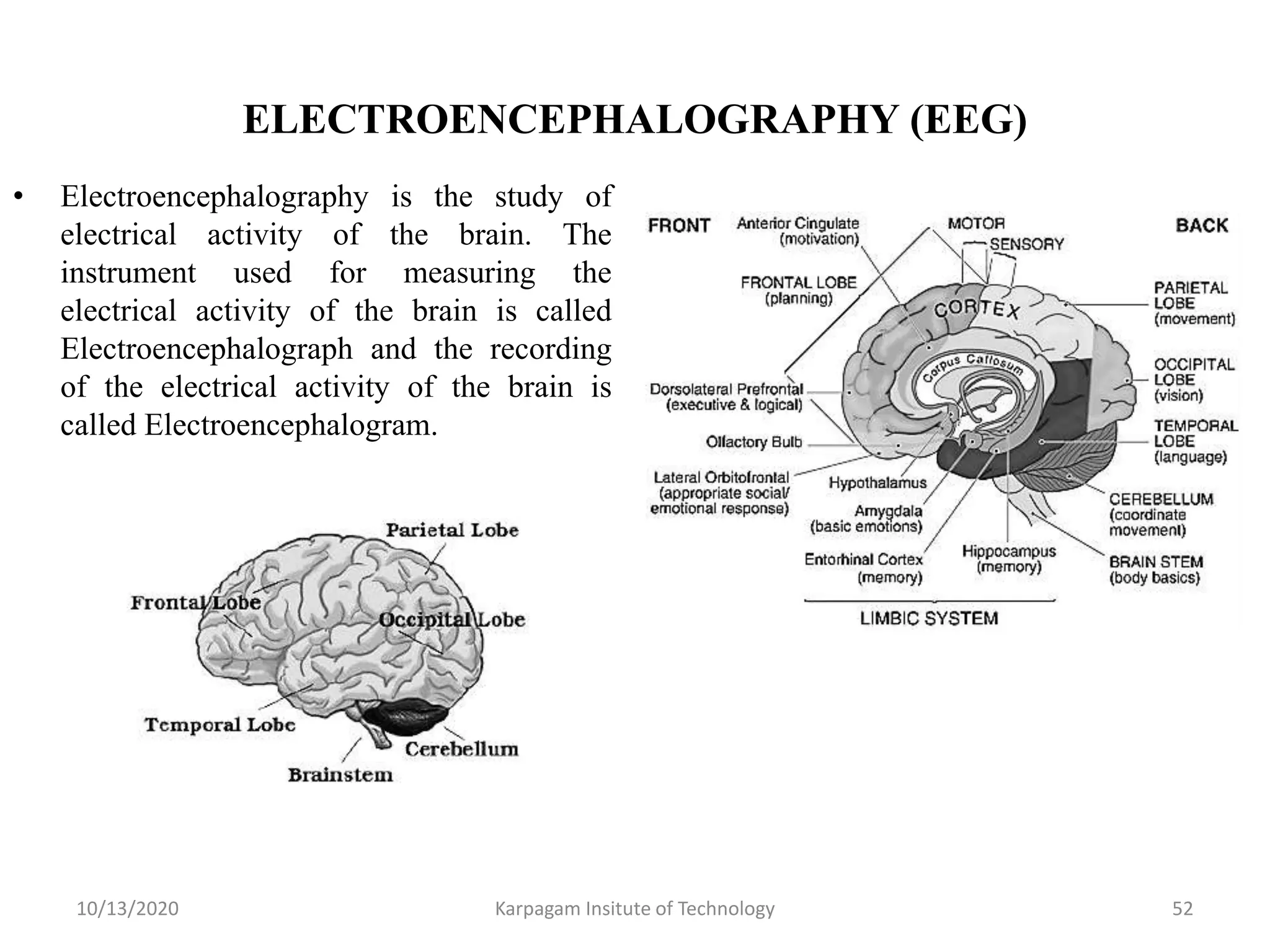
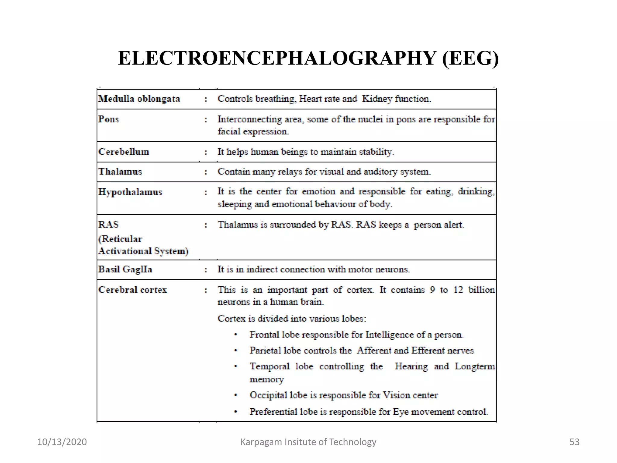
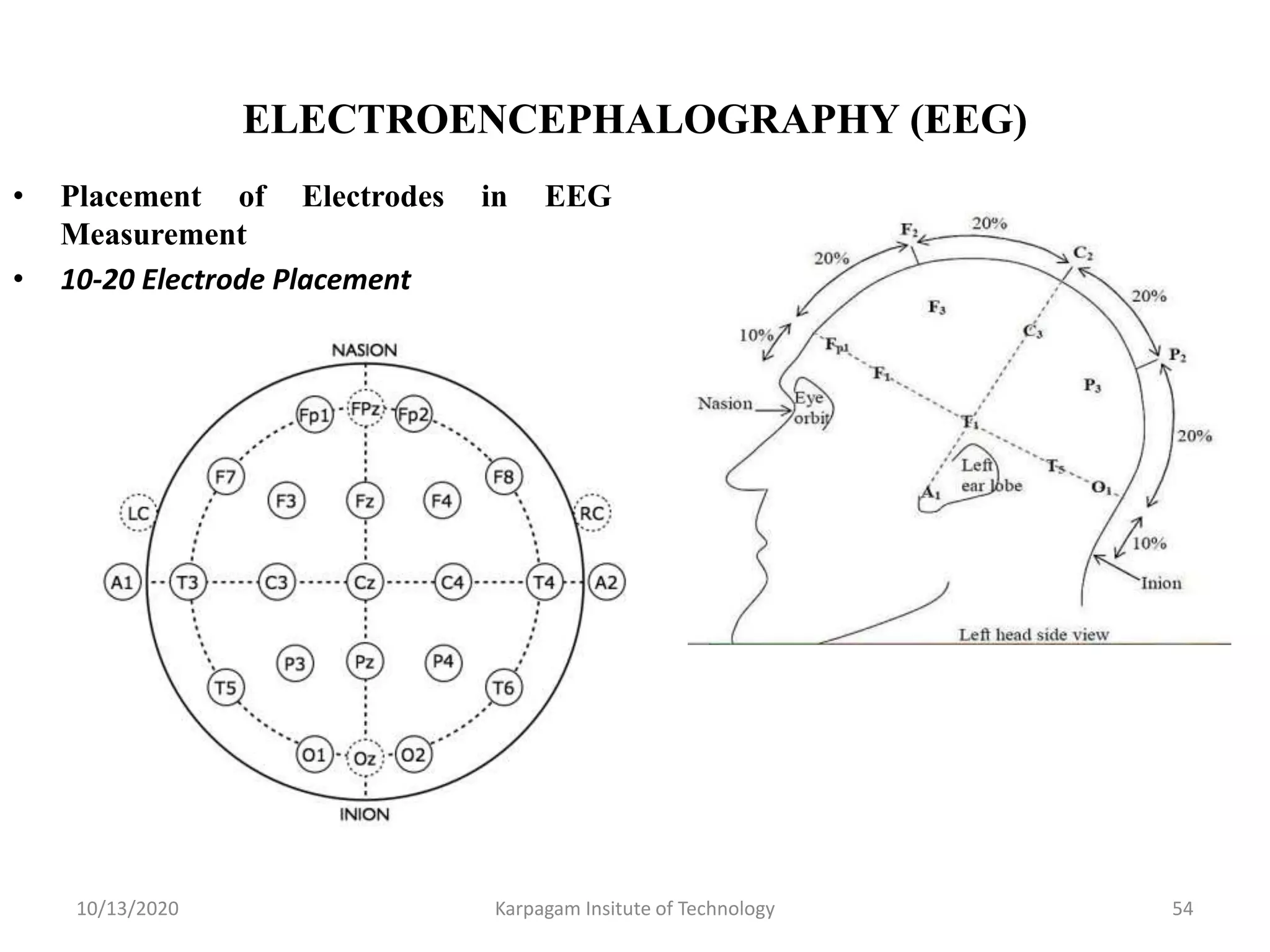
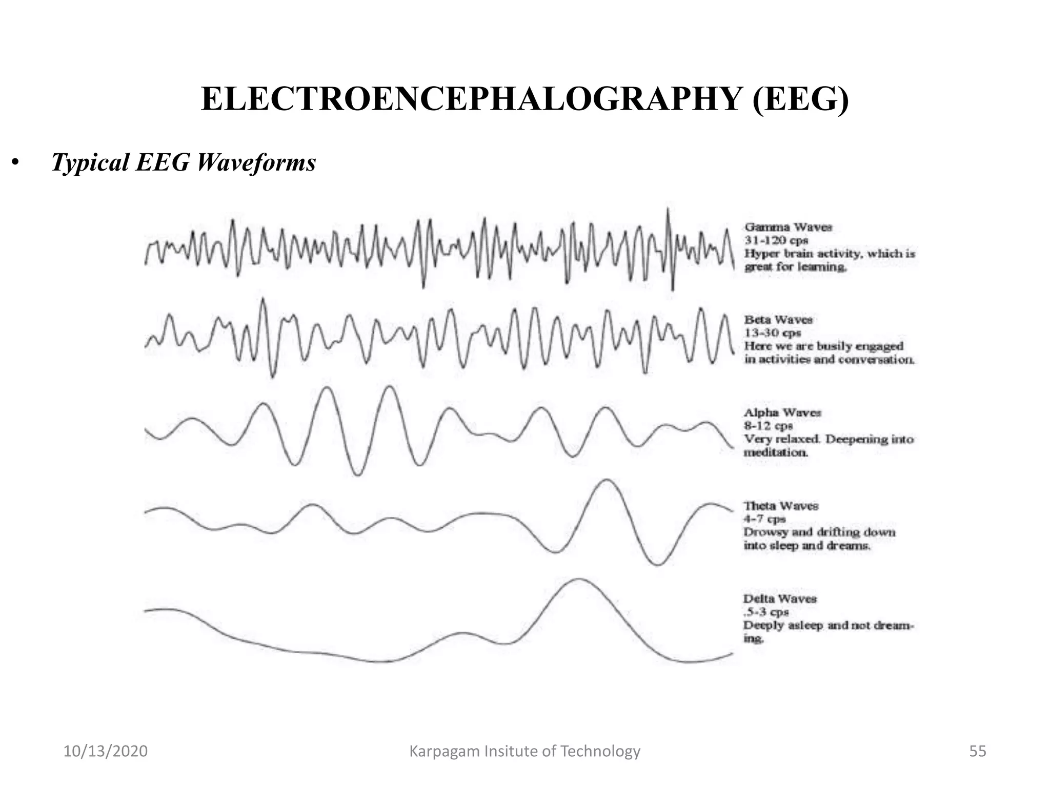
1. KARPAGAM INSTITUTE OF TECHNOLOGY,
COIMBATORE - 105
Course Code with Name : EC8073 – Medical Electronics
Staff Name / Designation : Dr. S. Gopinath/Associate Professor
Department : ECE
Year / Semester : III/VI
Department of Electronics and Communication Engineering
3. COURSE OBJECTIVES
• To gain knowledge about the various physiological parameters
both electrical and non electrical and the methods of recording
and also the method of transmitting these parameters.
• To study about the various assist devices used in the hospitals.
• To gain knowledge about equipment used for physical
medicine and the various recently developed diagnostic and
therapeutic techniques.
10/13/2020 3Karpagam Insitute of Technology
4. COURSE OUTCOMES
• Know the human body electro- physiological parameters and
recording of bio-potentials.
• Comprehend the non-electrical physiological parameters and
their measurement – body temperature, blood pressure, pulse,
blood cell count, blood flow meter etc.
• Interpret the various assist devices used in the hospitals viz.
pacemakers, defibrillators, dialyzers and ventilators.
• Comprehend physical medicine methods eg. ultrasonic,
shortwave, microwave surgical diathermies , and bio-telemetry
principles and methods.
• Know about recent trends in medical instrumentation.
10/13/2020 Karpagam Insitute of Technology 4
5. PROGRAM OUTCOMES
• Engineering Graduates will be able to:
1. Engineering knowledge: Apply the knowledge of
mathematics, science, engineering fundamentals, and an
engineering specialization to the solution of complex
engineering problems.
2. Problem analysis: Identify, formulate, review research
literature, and analyze complex engineering problems
reaching substantiated conclusions using first principles of
mathematics, natural sciences, and engineering sciences.
3. Design/development of solutions: Design solutions for
complex engineering problems and design system
components or processes that meet the specified needs with
appropriate consideration for the public health and safety, and
the cultural, societal, and environmental considerations.
10/13/2020 Karpagam Insitute of Technology 5
6. PROGRAM OUTCOMES
4. Conduct investigations of complex problems: Use research-
based knowledge and research methods including design of
experiments, analysis and interpretation of data, and synthesis
of the information to provide valid conclusions.
5. Modern tool usage: Create, select, and apply appropriate
techniques, resources, and modern engineering and IT tools
including prediction and modeling to complex engineering
activities with an understanding of the limitations.
6. The engineer and society: Apply reasoning informed by the
contextual knowledge to assess societal, health, safety, legal
and cultural issues and the consequent responsibilities relevant
to the professional engineering practice.
10/13/2020 Karpagam Insitute of Technology 6
7. PROGRAM OUTCOMES
7. Environment and sustainability: Understand the impact of the
professional engineering solutions in societal and environmental
contexts, and demonstrate the knowledge of, and need for
sustainable development.
8. Ethics: Apply ethical principles and commit to professional ethics
and responsibilities and norms of the engineering practice.
9. Individual and team work: Function effectively as an individual,
and as a member or leader in diverse teams, and in
multidisciplinary settings.
10. Communication: Communicate effectively on complex
engineering activities with the engineering community and with
society at large, such as, being able to comprehend and write
effective reports and design documentation, make effective
presentations, and give and receive clear instructions.
10/13/2020 Karpagam Insitute of Technology 7
8. PROGRAM OUTCOMES
11. Project management and finance: Demonstrate knowledge and
understanding of the engineering and management principles and
apply these to one‘s own work, as a member and leader in a
team, to manage projects and in multidisciplinary environments.
12. Life-long learning: Recognize the need for, and have the
preparation and ability to engage in independent and life-long
learning in the broadest context of technological change.
10/13/2020 Karpagam Insitute of Technology 8
9. PROGRAM SPECIFIC OUTCOMES
1. To analyze, design and develop solutions by applying
foundational concepts of electronics and communication
engineering.
2. To apply design principles and best practices for developing
quality products for scientific and business applications.
3. To adapt to emerging information and communication
technologies (ICT) to innovate ideas and solutions to
existing/novel problems.
10/13/2020 Karpagam Insitute of Technology 9
10. COURSE OUTCOME Vs. PROGRAM OUTCOMES AND
PROGRAM SPECIFIC OUTCOMES MAPPING
10/13/2020 Karpagam Institute of Technology 10
11. UNIT I : ELECTRO-PHYSIOLOGY AND BIO-
POTENTIAL RECORDING
• Sources of bio medical signals, Bio-potentials, Biopotential
electrodes, biological amplifiers, ECG, EEG, EMG, PCG, typical
waveforms and signal characteristics
10/13/2020 Karpagam Insitute of Technology 11
12. INTRODUCTION AND BIO MEDICAL
INSTRUMENTATION SYSTEM
• Medical Electronics is the application of engineering
principles and design concepts to medicine and biology for
healthcare purposes.
• Biomedical Instrument System
10/13/2020 Karpagam Insitute of Technology 12
13. SOURCES OF BIOMEDICAL SIGNALS
• Biomedical signals are those signals which are used primarily
for extracting information on a biological system under
investigation.
• Classification of Biosignals
• Classification according to source
• Classification according to biomedical application
• Classification according to signal characteristics
• Bio-Electric Signals, Bio-Acoustic Signals
• Bio-Mechanical Signal, Bio-Chemical Signals
• Bio-Magnetic Signal, Bio-Optical Signals
• Bio-Impedance Signals
10/13/2020 Karpagam Insitute of Technology 13
14. PHYSIOLOGICAL SYSTEMS OF HUMAN BODY
1) The Skeletal System - Bones & joints
2) Muscular System - Skeletal muscle
3) Nervous System - Brain, spinal cord & nerves
4) Endocrine System - Hormone-producing cells & glands
5) Cardiovascular System - Blood, heart & blood vessels
6) Respiratory System - Lungs & airways
7) Digestive System - Organs of the gastrointestinal tract
8) Urinary System - Kidneys, bladder and ureters
9) Reproductive System - Male and Female reproductive organs
10) The Integumentary System - The skin & derived structures
11) Lymphatic & Immune System - Lymphatic vessels & fluid
10/13/2020 Karpagam Insitute of Technology 14
15. ORIGIN OF BIO POTENTIALS
Structure of the cell
10/13/2020 15
The cell (from Latin cella, meaning "small room") is the basic
structural, functional, and biological unit of all known living
organisms. When a cell is excited, it generates an action potential
and hence an ionic current flows.
16. ESSENTIALS IN THE HUMAN CELL
WATER - Principal fluid in the cell (70% to 80%)
ELECTROLYTE – Consists of large & small quantities
PROTEIN – Concentration of the protein in the cell 10-20%
• Structural Protein – Provide Contractile mechanism of all muscles
• Globular Protein – Provide energy for cell function
LIPIDS – Soluble in fats & Solvents
CARBOHYDRATES – Supply cell’s energy through glycogen
CYTOPLASM – Filled with cytosol
RIBOSOMES –granual particles in cytosol and composed with RNA
LYSOSOMES –Used in digestive system
MITOCHONDRIA – Power house of the cell. Extracts energy from nucleus and oxygen.
10/13/2020 16
17. TRANSPORT OF IONS THROUGH CELL MEMBRANE
TWO FLUIDS
1. ICF- Intra Cellular Fluid – fluid lies inside the cell membrane.
2. ECF- Extra Cellular Fluid - fluid lies outside the cell membrane.
Active transport – obtained by concentration gradient
Passive transport – Transport of substances through the cell membrane.
Primary active transport
Energy is derived from the breakdown of “Adenosine Triphosphate (ATP)”
Eg. Sodium potassium pump and Calcium pump
Secondary active transport
Energy derived through ionic concentration gradient.
10/13/2020 17
18. RESTING AND ACTION POTENTIAL – Contd..
• Due to the change in the permeability, Sodium ions will get inside and produce
positive potential due to imbalance of potassium ions.
• The positive potential during excitation is called “Action potential” ( 20mV).
• Depolarisation (Discharging of cell)
• Repolarisation (Stopping
Sodium ions & Recharging of cell)
• Absolute Refractory Period
• Relative Refractory Period
• Conduction Velocity
• All or Nothing Law
10/13/2020 18
20. BIO-ELECTRIC POTENTIALS
• Potential generated at cellular level which is minute voltage generator.
• Bio electric signals in our body as follows
• ECG – Electro CardioGram
• EEG – Electro EncephaloGram
• EMG-Electro MyoGram
• ERG-Electro RectinoGram
• EGG-Electro GastroGram
• Sodium Potassium Pump
10/13/2020 20
21. BIO-POTENTIAL ELECTRODES
• Electrodes are used to pickup the electrical signals of the body.
• Types
• Surface electrode – Measure potential from the skin surface, heart, brain and nerves.
• Micro Electrode – Measure bioelectric potential near or within a single cell.
• Depth Electrode & Needle electrode – Penetrate the skin to record EEG potentials
& also measure the bioelectric potential from specific group of muscles.
• Half Cell Potential or Electrode Potential
• The Voltage dropped at an Electrode - Electrolyte interface is termed as half cell
potential.
• Perfectly polarised electrode – No net transfer of charge across metal electrolyte
interface.
• Perfectly nonpolarisable electrode – Unhindered transfer of charge across metal
electrolyte interface.
10/13/2020 21
23. TYPES OF BIO-POTENTIAL ELECTRODES
• Micro Electrodes - Measure the potential within a cell.
• Small in diameter which does not damage the cell during penetration.
• Micro electrodes – within a cell located
• Reference electrode – outside the cell located.
• Two types - Metallic electrode & Non Metallic electrode (Micropipet)
• Metallic – formed by electrolytically etching the tip of the fine tungsten filameter or
stainless steel wire.
• Micropipet – measure the potential through non metallic material.
• Depth Electrode - used to study the electrical activities of neuron of superficial
layers of the brain. It is also used to measure oxygen tension.
• Needle Electrode - used to record the peripheral nerve action potential (ERG).
10/13/2020 23
24. SURFACE ELECTRODE & OTHER ELECTRODES
• Measure the surface of the skin & sense the potential from heart, brain & nerves.
• Smaller area surface - measure EEG, EMG potentials.
• Large area surface – measure ECG potential.
• Suction cup Electrode - used for black surface of the body where the human tissue
is very soft.
• Multipoint Electrode - Frequently used for ECG measurement.
• Floating surface electrode - contact to electrolytic bridge to reduce artifact.
• Chemical electrode – measure oxygen, co2, pH and pO2 in blood
• {Hydrogen Electrode, Practive Reference Electrode, pH electrode, pO2 electrode,
pCO2 electrode}
10/13/2020 24
25. TYPES OF CHEMICAL ELECTRODES
• Hydrogen Electrode - Used as standard reference electrode with zero potential.
• Measure the pH of the body fluid. Hydrogen gas will be supplied during
measurement.
• Practical Reference Electrode – silver – silver chloride is used as reference electrode.
• pH electrode – chemical balance of the human body is identified by measurement
of pH content of the blood.
• pCO2 electrode – consists of standard glass pH electrode with rubber membrane
permeable to CO2.
• pO2 electrode – consists of piece of platinum wire embedded in an insulating glass
holder.
10/13/2020 25
28. BIO-AMPLIFIERS
• Bioamplifier is meant to gather and increase the signal integrity of
human neurophysiological electrical activity for further processing.
• Requirements
• Bio-Amplifiers must have high input impedance; the physiological
process that is being monitored should not be influenced in any way
by the amplifier.
• They must have isolation and protection circuits; the amplifier has to
offer protection to the patient from any hazard of electrical shock
• Voltage gain of bio-amplifiers should be more than 100db.
• Constant gain should be maintained throughout the required
bandwidth.
• CMRR of bio-amplifiers should be more than 80db.
10/13/2020 Karpagam Insitute of Technology 28
29. TYPES OF BIO-AMPLIFIERS
• Differential amplifier : It is one which will reject any common
mode signal that appears simultaneously at both amplifier input
terminals and amplifiers only the voltage difference that appears
across its input terminals.
• AC coupled amplifiers have a limited frequency response and are,
therefore, used only for special medical applications such as
electrocardiograph machine.
• Carrier amplifiers: It used with transducers which require an
external source of excitation.
• DC amplifiers: It is generally of the negative feedback type and are
used for medium gain applications down to about 1 mV signal levels
for full scale.
10/13/2020 Karpagam Insitute of Technology 29
30. TYPES OF BIO-AMPLIFIERS
• Chopper input dc amplifiers: It is preferred for low level
inputs to instrumentation systems because of their high
sensitivity, negligible drift and excellent common mode
rejection capability.
• Chopper-stabilized dc amplifiers: It is used for low level but
preferably wideband applications such as oscilloscopes, tape
recorders and light beam oscilloscope recorders.
• DC bridge amplifiers: It is employed with resistive
transducers which require an external source of excitation.
10/13/2020 Karpagam Insitute of Technology 30
31. DIFFERENTIAL AMPLIFIER WITH TRANSISTORS
• A differential amplifier with
two transistors is designed to
give the difference between
two input signals. The circuit
is shown in fig.
• There are two inputs, I/P1
and I/P2 and two outputs
V1OUT and V2OUT. I/P1 is
applied to the base of the
transistor T1 and I/P2 is
applied to the base of the
transistor T2.
10/13/2020 Karpagam Insitute of Technology 31
32. DIFFERENTIAL AMPLIFIER USING OP-AMP
• The circuit diagram of a
differential amplifier using
one op-amp is shown in fig.
• R1 and R2 are the input
resistors, Rf is the feedback
resistor, R3 is the input
resistor connected to the non-
inverting terminal and RL is
the load resistor.
10/13/2020 Karpagam Insitute of Technology 32
33. DIFFERENTIAL AMPLIFIER USING AN
INSTRUMENTATION AMPLIFIER
An instrumentation amplifier
is the front end component of
every measuring instrument
which improves the signal to
noise ratio of the input
electrical signal from the
transducer.
10/13/2020 Karpagam Insitute of Technology 33
34. DIFFERENTIAL AMPLIFIER USING AN
INSTRUMENTATION AMPLIFIER
Advantages of instrumentation amplifier
1) Typical gain values are inn the range of 100dB.
2) High CMMR ( ≈ 100dB).
3) High input impedance ( > 10MΩ ).
4) Low output resistance (<100Ω).
Applications of instrumentation amplifier
1) In Data acquisition from low output transducers such as strain gauges,
Thermocouples, Wheatstone bridge measurements e.t.c
2) In Medical instrumentation, Navigation, Radar instrumentation e.t.c
3) In Audio applications involving low amplitude audio signals in noisy
environments to improve the signal to noise ratio.
4) High-speed signal conditioning for video data acquisition and imaging
5) High frequency signal amplification in cable RF systems.
10/13/2020 Karpagam Insitute of Technology 34
35. OPTO ISOLATOR BASED AMPLIFIER
• Optically coupled isolation
amplifiers can realized using
only a single LED and
photodiode combination.
• While useful for a wide range
of digital applications, this
design has fundamental
limitations as to its linearity
and stability as a function of
time and temperature.
• Biopotential amplifiers have
to provide sufficient
protection from electrical
shock to both medical staff
and patient.
10/13/2020 Karpagam Insitute of Technology 35
36. CHOPPER AMPLIFIER
• Chopper amplifiers are normally preferred for converting the steady state,
time invariant biosignals into pulsated signals for the amplification and
signal processing purpose.
10/13/2020 Karpagam Insitute of Technology 36
37. ELECTROCARDIOGRAPHY (ECG)
• Electrocardiography is the study of electrical
activity of heart muscle and Electrocardiogram is
its recorded wave pattern. It reflects rhythmic
electrical depolarization and repolarization of
myocardium associated with contraction of atria
and ventricles.
• Human Heart has four chambers:
• • Left atrium (LA),
• • Right atrium (RA),
• • Left ventricle (LV),
• • Right ventricle(RV).
• Heart has four valves :
• • Tricuspid valve,
• • Mitral valve,
• • Pulmonary valve,
• • Aortic valve.
10/13/2020 Karpagam Insitute of Technology 37
38. ELECTROCARDIOGRAPHY (ECG)
• Functional diagram of Cardio Vascular System
(CVS)
• Bipolar Lead System - It is also known as
Einthoven leads. The signal is recorded using two
electrodes and final output is the difference in
potential between the two electrodes.
• Einthovan Triangle - Einthoven triangle is an
imaginary formation of three limb leads in a
triangle used in electrocardiography, formed by the
two shoulders and the pubis.
• Augmented Unipolar Limb Leads System
10/13/2020 Karpagam Insitute of Technology 38
39. ELECTROCARDIOGRAPHY (ECG) LEAD SYSTEMS
• LEAD aVL: The potential at LA is measured with respect to
augmented RA and augmented LL. RL is the ground (reference).
• Lead aVF: The potential at LL is measured with respect to augmented
LA and augmented RA. RL is the ground (reference).
• Unipolar Chest Leads - In unipolar chest lead system, explanatory
electrode is kept as the chest electrode.
10/13/2020 Karpagam Insitute of Technology 39
43. PHONOCARDIOGRAPHY ( PCG )
• The phonocardiogram is an instrument used for recording the sounds connected
with the pumping action of the heart. These sounds provide an indication of the
heart rate and its rhythmicity.
• The sounds are produced by the mechanical events that occur during the heart
cycle. These sounds are from the movement of the heart wall, closure of walls and
turbulence and leakage of blood flow.
10/13/2020 Karpagam Insitute of Technology 43
44. PHONOCARDIOGRAPHY ( PCG )
• Classification of Heart Sound
• Valve closure sound : These sounds occur at the beginning of systole and
the beginning of diastole.
• Valve opening sound: These sounds occur at the time of opening of atrio
ventricular valve and semilunar valve.
• Ventricular filling sound: These sounds occur at the times of filling (of
blood) of the ventricles.
• Extra cardiac sound: This sound occurs in mid or late systole or early
diastole.
• Systole : Contraction of heart muscles. The systolic pressure is 120 mm of
Hg
• Diastole : Relaxation of heart muscles. The diastolic pressure is 80 mm of
Hg.
10/13/2020 Karpagam Insitute of Technology 44
45. PHONOCARDIOGRAPHY ( PCG )
• PCG Signal Characteristics and Typical Waveforms
10/13/2020 Karpagam Insitute of Technology 45
46. PHONOCARDIOGRAPHY ( PCG )
• Aortic Stenosis: Aortic Stenosis occurs when blood flows from left
ventricle through aortic valve to other organs of the body.
• Mitral stenosis: This murmur is produced when the blood is passed from
left atrium to left ventricle. Usually this sound is very weak.
• Mitral regurgitation murmur: This murmur sound is produced when
blood flows in backward direction through mitral valve during systole.
• Aortic Regurgitation murmur: This murmur sound is produced during
diastole, when blood flows in backward direction from aorta to left
ventricle when the valves are damaged.
• PCG LEAD SYSTEM
10/13/2020 Karpagam Insitute of Technology 46
47. PHONOCARDIOGRAPHY ( PCG )
• PCG RECORDING SYSTEM
10/13/2020 Karpagam Insitute of Technology 47
48. PHONOCARDIOGRAPHY ( PCG )
• Applications
• Fetal PCG - PCG of a fetus
• Esophageal PCG - PCG of the food pipe that runs between the throat and
the stomach
• Tracheal PCG - PCG of the windpipe ( a tube that connects the throat to
the lungs)
10/13/2020 Karpagam Insitute of Technology 48
49. ELECTROMYOGRAPHY ( EMG )
• Electromyography is the study of electrical activity of the muscles.
• Electrodes Used For Measurement
• Surface Electrode: Usually, this electrode is used for EMG. But by using
this electrode it is not possible to take deeper potential, so needle
electrodes are used.
• Needle electrode: These are inserted into the tissues that are closer to the
muscle. EMG Recording System
10/13/2020 Karpagam Insitute of Technology 49
50. ELECTROMYOGRAPHY ( EMG )
• In Modern EMG systems, Nerve Conduction Time and Nerve Conduction
Velocity are measured. For this purpose, the nerve is initially stimulated.
• This conduction velocity is used to indicate location and type of nerve
lesion (A lesion is an infected or disessed or injured region in an organ).
10/13/2020 Karpagam Insitute of Technology 50
51. ELECTROMYOGRAPHY ( EMG )
• The electrical source is the muscle membrane potential of about –90 mV.
Measured EMG potentials range between less than 50 μV and up to 20 to
30 mV, depending on the muscle under observation.
10/13/2020 Karpagam Insitute of Technology 51
• Applications
• Neurology
• Psychiatry
• Clinical Neurophysiology
52. ELECTROENCEPHALOGRAPHY (EEG)
• Electroencephalography is the study of
electrical activity of the brain. The
instrument used for measuring the
electrical activity of the brain is called
Electroencephalograph and the recording
of the electrical activity of the brain is
called Electroencephalogram.
10/13/2020 Karpagam Insitute of Technology 52
56. Summary of Unit I
• Discussed about bio potentials.
• Learned about usage of bio potential electrodes and amplifiers.
• Studied the working principle of the ECG, EEG, EMG, PCG
and typical waveforms and characteristics.
10/13/2020 Karpagam Insitute of Technology 56
57. VIDEO TUTORIALS
S.No. Topic Link
1. Bio Amplifiers https://www.youtube.com/
watch?v=98lRv_8rQmo
2. Bio potential electrodes https://www.youtube.com/
watch?v=q_irkkOuXbY
3. Cardiac Conduction System and
Understanding ECG,
Animation.
https://www.youtube.com/
watch?v=RYZ4daFwMa8
4. EEG https://www.youtube.com/
watch?v=tZcKT4l_JZk
10/13/2020 Karpagam Insitute of Technology 57
58. ASSIGNMENT QUESTIONS
S.No. Assignment Topic Knowledge
Level
1. Demonstrate the generation of half cell
potential across electrode–electrolyte
interface with equivalent.
K3 level
2. Design a circuit that uses one op-amp plus
the other passive components that will detect
QRS complexes of the ECG even when the
amplitude of T exceeds that of the QRS
complex and provides output signals.
K3 level
3. Analyze the measurement of
Phonocardiography and how it is used to
identify the cardiac murmurs.
K4 level
10/13/2020 Karpagam Insitute of Technology 58
59. QUIZ QUESTIONS
1. The concentration of the protein in the cell ---------------
a.30-40%
b.70-80%
c.10-20%
d.50-60%
2. Lipids are
a.energy storage for cell function
b.Consists of large & small quantities
c.Filled with cytosol
d.Soluble in fats & Solvents
3. Cytoplasamconsists of ---
a.Cytosol
b.Cytosoland RNA
c.Glycogen
d.Fluid in the cell
10/13/2020 Karpagam Insitute of Technology 59
60. QUIZ QUESTIONS
4. Mitochondria is
a.Used in digestive system
b.Energy system
c.Contractile mechanism
d.Power house of the cell.
5. The positive potential during excitation is ---------------
a.Permeability
b.Depolarisation
c.Action Potential
d.Repolarisation
6. ___________ is the time period during which another stimulus given to
the neuron will not lead to a second action potential.
a.Relative Refractory Period
b.Absolute Refractory Period
c.Blanking Period
d.Ejection Period10/13/2020 Karpagam Insitute of Technology 60
61. QUIZ QUESTIONS
7) Which electrode is used for black surface of the body where the human
tissue is very soft?
a) Multipoint Electrode
b) Floating surface electrode
c) Suction cup Electrode
d) Chemical electrode
8) _________ means the two circuits are electrically isolated but coupled
by other means.
9) ____________ is the study of electrical activity of heart muscle and its
recorded wave pattern.
10) How many chambers present in the Human heart?
a) 6
b) 8
c) 4
d) 3
10/13/2020 Karpagam Insitute of Technology 61
62. QUIZ QUESTIONS
11) _________ are normally preferred for converting the steady state, time
invariant bio-signals into pulsated signals for the amplification and
signal processing purpose.
a) Differential Amplifier
b) Chopper Amplifier
c) Isolation Amplifier
d) Instrumentation Amplifier
12) Which valve prevents blood to come back to left ventricle?
a) Aortic valve
b) Tricuspid valve
c) Mitral valve
d) Pulmonary valve
10/13/2020 Karpagam Insitute of Technology 62
63. QUIZ QUESTIONS
13) Which vessels carry pure oxygenated blood from heart to various
organs of the body?
a) Cappillaries
b) Veins
c) Arteries
d) None of the above
14) Pulmonary circulation means _________________
15) Bipolar leads are also known as _________________
16) _______________an augmented unipolar limb lead in which the
positive electrode is on the left arm.
a) aVF lead
b) aVL lead
c) aVR lead
d) Lead aVR
10/13/2020 Karpagam Insitute of Technology 63
64. QUIZ QUESTIONS
17) Systole means _________ and its pressure is ______________
a) Relaxation and 30 mmHg.
b) Contraction and 120 mmHg.
c) Relaxation and 80 mmHg.
d) Contraction and 50 mmHg.
18) ______ means relaxation of heart muscles.
19) Coronary insufficiency means
a) ST segment is elevated
b) Train pulse instead of PQRST
c) QRS complex is widened
d) ST segment is suppressed
10/13/2020 Karpagam Insitute of Technology 64
65. QUIZ QUESTIONS
20) Which corresponds to the R wave of the ECG, is longer in duration?
a) Second sound
b) First sound
c) Third sound
d) Fourth sound
21) ________ sounds occur at the time of opening of atrio ventricular
valve and semilunar valve.
a) Valve Closure sound
b) Extra cardiac sound
c) Ventricular filling sound
d) Valve opening sound
22) ______ is an infected or injured region in an organ.
10/13/2020 Karpagam Insitute of Technology 65
66. QUIZ QUESTIONS
23) ________ helps human beings to maintain stability.
a) Pons
b) Medulla Oblongata
c) Thalamus
d) Cerebellum
24) _________ waves are seen in premature babies with a frequency range
of 0.5-4 Hz.
a) Beta
b) Delta
c) Alpha
d) Gamma
25) Alpha waves are normally obtained from _____________.
10/13/2020 Karpagam Insitute of Technology 66
67. QUIZ QUESTIONS
26) Which microphone is used for the movement of chest is transferred
through air cushion.
a) Air coupled microphone
b) Contact microphone
c) Condensor microphone
d) None of the above
27)How many cappillaries are present in the human body?
a) 5000
b) 8000
c) 4000
d) 2000
28) Evoked Potential means _______________________
29) The amplitude of EMG signals range from ___________________
30) ___________ means the technique of listening to sound produced by
the organs and vessels of the body.
10/13/2020 Karpagam Insitute of Technology 67
68. UNIT II : BIO-CHEMICALAND NON ELECTRICAL
PARAMETER MEASUREMENT
• pH, PO2, PCO2, Colorimeter, Blood flow meter, Cardiac output,
respiratory, blood pressure, temperature and pulse measurement,
Blood Cell Counters.
10/13/2020 Karpagam Insitute of Technology 68
69. pH MEASUREMENT
• The chemical balance in the body can be determined by the pH
value of blood and other body fluids. pH is defined as the
hydrogen ion concentration of a fluid.
• It is the logarithm of the reciprocal value of H+ ion
concentration. pH is the measure of acid-base balance in a
fluid.
10/13/2020 Karpagam Insitute of Technology 69
70. pH MEASUREMENT
• A pH meter is made up two parts : 1)
The electrodes and 2) A meter for
reading the potential generated by the
electrodes. The electrodes consist of a
thin glass membrane which allows only
the hydrogen ions to pass through it.
• The glass tube consists of a silver-silver
chloride (Ag /AgCl) electrode. A
reference electrode which is made up of
either calomel or silver-silver chloride
(Ag/AgCl) is placed in the solution in
which pH is being measured.
10/13/2020 Karpagam Insitute of Technology 70
71. pH MEASUREMENT
• The electrochemical measurement,
which is obtained by each of the
electrodes, is called Half-cell. The
electrode potential is called half-cell
potential.
10/13/2020 Karpagam Insitute of Technology 71
72. PO2 MEASUREMENT
• It is defined as the measurement of
partial pressure of oxygen dissolvent in
a liquid. The partial pressure of a gas is
proportional to the quantity of that gas
present in the blood.
• The effective functioning of both
respiratory and cardiovascular system
can be analyzed by PO2.
10/13/2020 Karpagam Insitute of Technology 72
73. PO2 MEASUREMENT
• Disadvantages
• The reduction process in the platinum electrode removes a finite amount of
the oxygen from the cathode and hence there is a gradual reduction of
current with respect to time.
• However, by using carefully designed modern PO2 electrodes and proper
procedures, the errors can be reduced.
10/13/2020 Karpagam Insitute of Technology 73
74. PCO2 MEASUREMENT
• It is defined as the measurement of partial pressure of carbon dioxide.
• The partial pressure of a gas is proportional to the quantity of that gas present
in the blood.
• The effective functioning of both respiratory and cardiovascular system can
be analyzed by PCO2 .
10/13/2020 Karpagam Insitute of Technology 74
75. PCO2 MEASUREMENT
• The partial pressure of carbon dioxide can
be measured with the help of PCO2
electrodes. There is a linear relationship
between the logarithm of PCO2 and pH of
a solution.
• In this electrode the membrane permeable
to CO2 is made up of silicone rubber
which is not permeable to other ions
which affect pH value.
10/13/2020 Karpagam Insitute of Technology 75
76. COLORIMETER
• The nature of these biological substances can be determined by
analyzing their absorbance and transmittance characteristics.
• The protein and iron levels in blood can be measured with the help of
colorimeters and photometers.
• Electrophoresis is the principle used in colorimeters. Electrophoresis
is the movement of solid phase with respect to a liquid (liquid means
but buffer solution).
• Applications
• To measure the quantity of protein in plasma, urine etc.
• To separate enzymes into their components isoenzymes.
• To identify antibodies.
10/13/2020 Karpagam Insitute of Technology 76
77. COLORIMETER
• Working of Colorimeter
• Colorimetric analysis is the study of absorbance and transmittance
properties of chemical substances.
10/13/2020 Karpagam Insitute of Technology 77
78. MAGNETIC BLOOD FLOW MEASUREMENTS
• The circulatory system of human being helps the flow of blood
throughout the body. During this process, adequate amount of
blood should be supplied for the organs to perform their functions.
• The blood flow rate can be determined with the help of blood flow
meters.
1) Magnetic Blood Flow Meters
2) Ultrasonic Blood Flow Meters
3) Thermal Convection Method
4) Radiographic Method
5) Indicator Dilution Method
10/13/2020 Karpagam Insitute of Technology 78
79. MAGENETIC BLOOD FLOW MEASUREMENTS
• The Magnetic blood flow meters
(also known as Electromagnetic
Blood Flow Meters) are based on
the principle of electromagnetic
induction.
• This principle states that when an
electrical conductor is moved
through a magnetic field, a
voltage is induced in the
conductor proportional to the
velocity of motion.
10/13/2020 Karpagam Insitute of Technology 79
80. ULTRASONIC BLOOD FLOW MEASUREMENTS
• In ultra sonic blood flow meter,
the velocity of the flowing blood
is determined with a beam of
ultrasonic energy.
1) Transit time ultrasonic flow meter
2) Doppler type ultrasonic flow meter
10/13/2020 Karpagam Insitute of Technology 80
81. THERMAL CONVECTION BLOOD FLOW
MEASUREMENTS
• When a hot object is placed in a colder
flowing medium, it gets cooled by thermal
convection.
• This principle is used in this method. Here
the rate of cooling is proportional to the rate
of flow of the medium.
• A thermistor placed on the blood stream is
kept at a constant temperature by a servo
system.
• The blood velocity can be determined by the
difference between the upstream and
downstream temperature indicated by the
sensor.
10/13/2020 Karpagam Insitute of Technology 81
82. RADIOGRAPHIC METHOD BLOOD FLOW
MEASUREMENTS
• The flow of blood cannot be made
visible with the help of X-rays due
to their same density as the
surrounding tissues.
• With the help of X-rays, the
progress of the contrast medium, the
obstructions and the blood flow in
the vessels can be estimated.
10/13/2020 Karpagam Insitute of Technology 82
83. INDICATOR DILUTION METHOD BLOOD FLOW
MEASUREMENTS
• The indicator dilution method helps
in the determination of rate of blood
flow and not the velocity of blood.
• Following are two types of
measurements:
• 1) Open circulation method
• 2) Closed circulation method
10/13/2020 Karpagam Insitute of Technology 83
84. CARDIAC OUTPUT MEASUREMENT
• Cardiac output is the amount of
blood delivered by the heart to the
aorta in one minute.
• For a normal adult, the cardiac
output is 4 - 6 liter/min.
• The decrease in cardiac output is
due to low pressure, reduced tissue
oxygenation, poor renal function,
shock and acidosis.
• Methods
• 1) Fick’s method
• 2) Indicator dilution method
• 3) Thermo dilution method
• 4) Impedance change method
10/13/2020 Karpagam Insitute of Technology 84
85. INDICATOR DILUTION METHOD
• In this method, a known amount of dye or radioisotope is used as an
indicator in the blood circulation.
• Measuring the concentration of the indicator with respect to time, we can
estimate the volume flow of blood.
10/13/2020 Karpagam Insitute of Technology 85
86. THERMO DILUTION METHOD
• 10 ml of 5% dextrose in water at room
temperature is injected as a thermal
indicator into the right atrium.
• After mixing, it is detected in the
pulmonary artery by a thermistor, placed at
the tip of a miniature catheter probe.
• The difference between the indicator
temperature and the circulating blood
temperature in the pulmonary artery is
measured.
10/13/2020 Karpagam Insitute of Technology 86
87. RESPIRATORY RATE MEASUREMENT
• Respiratory system provides a means of acquiring oxygen and eliminating
CO2. Various gas laws are involved in the understanding of respiratory
functions.
• Boyle’s Law states that at constant temperature, the volume of gas varies
inversely with the pressure.
• Charle’s Law states that, at constant pressure, the volume of gas is directly
proportional to the absolute temperature.
• Henry’s Law states that, if the temperature is constant, the quantity of the
gases going into a solution is directly proportional to the partial pressure of the
individual gas.
• Dalton’s Law: It states that, the total pressure exerted by a mixture of gases is
equal to the sum of the partial pressures of individual gases
10/13/2020 Karpagam Insitute of Technology 87
88. RESPIRATORY RATE MEASUREMENT
• Internal respiration: It is the exchange of gases between the blood stream
and nearby cells.
• External respiration: It is the exchange of gases between the lungs and blood
stream.
• Lung Volumes and Capacities (Respiration Parameters)
• Dead Air
• Tidal Volume (TV)
• Inspiratory Reserve Volume (IRV), Expiratory Reserve Volume (ERV)
• Residual Volume (RV), Minute Volume (MV)
• Airway Resistance, Lung Compliance, Lung Elasticity, Intra Thoracic
Pressure
• Total Lung Capacity (TLC), Vital Capacity (VC), Inspiratory Capacity(IC),
Functional Residual Capacity (FRC)
10/13/2020 Karpagam Insitute of Technology 88
89. TYPES OF RESPIRATORY RATE MEASUREMENT
• 1) Displacement method
• 2) Thermistor method
• 3) Impedance pneumography
• 4) CO2 method
• 5) Apnea detectors
• Displacement Method
• In this method, the transducer is held by an elastic band which goes around the
chest. The respiratory movements results in a corresponding resistance
changes of the strain gauge.
• Thermistor Method
• Generally, there is a temperature difference between inspired and expired air.
This temperature is sensed by placing thermistor in front of the nostrils.
Thermistor is placed by using suitable stand.
10/13/2020 Karpagam Insitute of Technology 89
90. TYPES OF RESPIRATORY RATE MEASUREMENT
• Impedance Pneumography
• CO2 Method
10/13/2020 Karpagam Insitute of Technology 90
91. TYPES OF RESPIRATORY RATE MEASUREMENT
• Apnoea Detectors
• Apnoea is the temporary
stoppage of breath
during sleeping in
between inspiration and
expiration.
• It leads to the arrest of
the circulation. It can
occur in conditions like
head injury, drug
overdose etc.
10/13/2020 Karpagam Insitute of Technology 91
92. BLOOD PRESSURE MEASUREMENT
• Blood pressure is considered as a good indicator of the status of the
cardiovascular system.
• In routine clinical tests, blood pressure is usually measured by means of an
indirect method using a sphygmomanometer .
• This method is easy to use and can be automated.
• Hydrostatic Pressure:
• If the force in a system under pressure is not varied, then the pressure is
known as Hydrostatic pressure.
• Hydrodynamic Pressure:
• If the force in a system under pressure is varied, then the pressure is
known as hydrodynamic pressure. Generally the physiological pressure is
hydrodynamic pressure.
10/13/2020 Karpagam Insitute of Technology 92
93. BLOOD PRESSURE MEASUREMENT
• Blood Pressure Measurement Methods
• 1) Indirect method using Sphygmomanometer – Auscultatory method
• 2) Indirect method using Sphygmomanometer – palpatory method
• 3) Indirect method using Sphygmomanometer – Oscillometric method
• 4) Direct method.
• Indirect BP measurement
10/13/2020 Karpagam Insitute of Technology 93
94. BLOOD PRESSURE MEASUREMENT
• Oscillometric method
• The oscillometric method involves the observation of oscillations in the
sphygmomanometer cuff pressure which are caused by the oscillations of
blood flow, i.e., the pulse.
10/13/2020 Karpagam Insitute of Technology 94
95. BLOOD PRESSURE MEASUREMENT
• Direct Method of BP Measurement
• Direct method of blood pressure is used when accurate blood pressure
reading is necessary.
10/13/2020 Karpagam Insitute of Technology 95
96. PULSE MEASUREMENT
• When heart muscle contracts, blood
is ejected from the ventricles and a
pulse of pressure is transmitted
through the circulatory system. This
pulse can be measured at various
points.
• Types of Pulse Measurement
• 1) Transmittance Method
• 2) Reflectance method.
10/13/2020 Karpagam Insitute of Technology 96
97. TEMPERATURE MEASUREMENT
• Temperature is one of the indicators
of the general well-being. Two types
of temperature measurements can be
obtained from the body. There are
• 1) Systemic temperature. - Systemic
temperature is a temperature of the
internal regions of the body.
• 2) Surface temperature - Skin
temperature is not constant
throughout the body. For a normal
person, it varies from 30˚C to 35˚C.
10/13/2020 Karpagam Insitute of Technology 97
98. BLOOD CELL COUNTERS
• The blood circulation is a special transport system which helps in the
exchange of chemical products between the specialized cells of various
organs.
• The circulatory system of normal adult human is 5 to 6 litres of blood per
minute, based on a heart rate of 72 beats a minute and a stroke volume of
70 ml.
• The different types of blood cells are:
• 1) Red Blood Cells (RBC)
• 2) White blood Cells (WBC)
• 3) Blood platelets (Thrombocytes)
10/13/2020 Karpagam Insitute of Technology 98
99. BLOOD CELL COUNTER
MEASUREMENT METHODS
• Hematocrit Determination
• To determine the relative proportion of
blood cells in a given volume of blood,
hematocrit or packed cell volume is used.
• Manual Method
• Blood cell counts by manual method are
performed using a microscope. At first the
blood is diluted in the ratio of 1:100 or
1:200 for counting RBCs and in the ratio
of 1:10 or 1:20 for WBCs.
• Conductivity Method (Coulter Method)
• The Coulter method of sizing and
counting particles is based on measurable
changes in electrical impedance produced
by nonconductive particles suspended in
an electrolyte.
10/13/2020 Karpagam Insitute of Technology 99
100. BLOOD CELL COUNTER MEASUREMENT METHODS
• LASER Based Cell Counting
• This modern technique is to determine the
number of RBCs, WBCs and platelets.
• The cell volume of the red blood cells and
the haemoglobin concentration can also be
obtained by this method.
• The principle used in this laser based
blood cell counting is ―the angle of
scattered light is different for different
sized blood cells‖.
• The hemoglobin concentration in the
RBCs also can be measured by this
method.
10/13/2020 Karpagam Insitute of Technology 100
101. Summary of Unit II
• Discussed about bio potential gases and its measurement.
• Studied the working principle of Colorimeter, Blood flow
meter, Cardiac output, respiratory, blood pressure, temperature
and pulse measurement, Blood Cell Counters.
10/13/2020 Karpagam Insitute of Technology 101
102. VIDEO TUTORIALS
S.No. Topic Link
1. Colorimeter https://www.youtube.com/
watch?v=QdufRwbkeKo
2. Measuring the respiratory rate https://www.youtube.com/
watch?v=35rHkV5KmkA
3. Blood Pressure Measurement https://www.youtube.com/
watch?v=f6HtqolhKqo
4. Haematology WBC https://www.youtube.com/
watch?v=q6rfJQVSals
5 BOHUI T-168 handheld non-
contact human body forehead
electronic thermometer
temperature
https://www.youtube.com/
watch?v=3RvsDDJvI3c
10/13/2020 Karpagam Insitute of Technology 102
103. ASSIGNMENT QUESTIONS
S.No. Assignment Topic Knowledge
Level
1. Calculate the stroke volume in milli litres if
the cardiac output is 5.25 litres/min and the
heart rate is 76 beats/min
K3 level
2. Analyze the measurement of pH value. K4 level
3. Elucidate how velocity of blood flow can be
measured in ultrasonic blood flow meter
K3 level
10/13/2020 Karpagam Insitute of Technology 103
104. QUIZ QUESTIONS
1. The normal pH value of human blood is ---------------
a.6.2
b. 5.8
c.7.4
d. 2.1
2. If the PH value exceeds 7.45 means, the body is considered to be in a
state of ________________
3. __________ electrode is used as reference electrode.
a.Na-Nacl
b.Ag-Agcl
c. K-Kcl
d. Mg-Mgcl
4. Electrophoresis means _______________________
10/13/2020 Karpagam Insitute of Technology 104
105. QUIZ QUESTIONS
5. ___________ is used to measure blood proteins and iron levels.
a. Blood flow meter
b. Blood pressure meter
c. Colorimeter
d. Pulse meter
6. Inspired gas contains _______ % oxygen and pressure is
_______mmHg.
a.35, 189
b.21, 760
c. 52, 284
d. 74,521
7._________ involves the cutting of blood vessels.
a.Ultrasonic blood flow meter
b.Turbine flow meter
c.Rotameter
d. b&c
10/13/2020 Karpagam Insitute of Technology 105
106. QUIZ QUESTIONS
8) _________ beam is used to measure the velocity of flowing blood.
9) _________ type blood flow meter is used to produce scattering due to
the result of moving blood cells.
10) In which method, the blood velocity can be determined between the
difference between upstream and downstream temperature.
a) Radiographic method
b) Thermal convection method
c) Doppler type
d) Dilution method
11) For a normal adult, the cardiac output is
a) 4-6 litre/min
b) 14-21 litre/min
c) 8-19 litre/min
d) 24-34 litre/min
10/13/2020 Karpagam Insitute of Technology 106
107. QUIZ QUESTIONS
12) Cardiac output is determined by ____________ method based on the
anlaysis of gas keeping property.
13) Hematocrit is normally ______ for men and _______ for women.
a) 21%, 12%
b) 10%, 43%
c) 11%, 32%
d) 45%, 40%
14) Boyles law states that
a) the volume of gas varies inversely with the pressure.
b) the volume of gas is directly proportional to the absolute temperature.
c) total pressure exerted by a mixture of gases is equal to the sum of the partial
pressures of individual gases
d) None of the above
15) ______ law states that, at constant pressure, the volume of gas is directly
proportional to the absolute temperature.
10/13/2020 Karpagam Insitute of Technology 107
108. QUIZ QUESTIONS
16) _____ is depth of breathing or the volume of gas inspired or expired
during each respiratory cycle
a) Minute volume
b) Tidal volume
c) Residual volume
d) Vital capacity
17) ___________ is the ability of the alveoli and lung tissue to expand on
inspiration.
18) __________ is the temporary stoppage of breath during sleeping in between
inspiration and expiration.
19) In Blood pressure measurement,
a) Hydrostatic pressure can be varied
b) Hydrodynamic pressure can be varied
c) Both can be varied
d) Non of the above
10/13/2020 Karpagam Insitute of Technology 108
109. QUIZ QUESTIONS
20) ___________ sound appears when systolic pressure exceeds the cuff
pressure.
21) ______ means relaxation of heart muscles.
22) The Pulse travels at the speed of _______________
a) 5 to 15 meters
b) 20 to 23 meters
c) 40 to 56 meters
d) 67 to 78 meters
23) The circulatory system of normal adult human is
a) 15 to 25 litres
b) 5 to 6 litres
c) 13 to 16 litres
d) 1 to 2 litres
10/13/2020 Karpagam Insitute of Technology 109
110. QUIZ QUESTIONS
24) In a day heart pumps,
a) 5600 litre of blood
b) 6100 litre of blood
c) 7200 litre of blood
d) 3100 litre of blood
25) A fluid plasma contains ______% of blood and blood cell contains
________ % of blood
a) 35, 45
b) 55, 45
c) 12, 24
d) 10, 20
26) ______ are known as erythrocytes and __________ are known as
leucocytes.
27) The rate of RBC generation per day is ___________
10/13/2020 Karpagam Insitute of Technology 110
111. QUIZ QUESTIONS
28) The average diameter of RBC and WBC is
a) 8 and 10 micrometer
b) 6 and 7 micrometer
c) 5 and 9 micrometer
d) 12 and 22 micrometer
29) The plasma in which the fibrinogen has been removed by the process
of precipitation is called ___________________
30) __________ is used to determine the number of RBCs, WBCs and
platelets.
a) Manual method
b) Conductivity method
c) Laser based cell counting
d) None of the above
10/13/2020 Karpagam Insitute of Technology 111
112. UNIT III : ASSIST DEVICES
• Cardiac pacemakers, DC Defibrillator, Dialyser, Ventilators,
Magnetic Resonance Imaging Systems, Ultrasonic Imaging
Systems
10/13/2020 Karpagam Insitute of Technology 112
113. Pacemaker
• A pacemaker is a small device that's placed in the chest or
abdomen to help control abnormal heart rhythms. This device
uses electrical pulses to prompt the heart to beat at a normal
rate.
• Fast Heartbeat – Tachycardia
• Slow Heartbet – Bradycardia
• To treat arrhythmias ( Problems with rate or rhythm of
heartbeat)
114. Understanding the Heart's Electrical System
• Heart has its own internal electrical
system that controls the rate and
rhythm of heartbeat.
• Each electrical signal normally begins
in a group of cells called the sinus
node or sinoatrial (SA) node.
• As the signal spreads from the top of
the heart to the bottom, it coordinates
the timing of heart cell activity.
115. Use of Pacemakers
• Pacemakers can:
• Speed up a slow heart rhythm.
• Help control an abnormal or fast heart rhythm.
• Make sure the ventricles contract normally
• Coordinate electrical signaling between the upper and lower
chambers of the heart.
• Coordinate electrical signaling between the ventricles.
• Cardiac resynchronization therapy (CRT) devices are used to
treat heart failure.
• Prevent dangerous arrhythmias caused by a disorder
called long QT syndrome (disorder of heart electrical activity).
116. CARDIAC PACEMAKERS
• Pace maker is an electrical pulse generator for initiating and maintaining
normal heart beat.
• The output signal of pacemaker is either applied external to chest or internal
to heart muscles.
• 1) Ventricular rate is too low, 2) Arrhythmia occurs , 3) Heart block occurs
• Simulation
• External Simulation , Internal Simulation
10/13/2020 Karpagam Insitute of Technology 116
118. CARDIAC PACEMAKERS
• Types of Pacing modes
Based on modes of operation, pacemakers are classified into 5 types
1) Ventricular asynchronous pacemaker.
2) Ventricular synchronous pacemaker.
3) Ventricular inhibited pacemaker.
4) Atrial synchronous pacemaker.
5) Atrial sequential ventricular inhibited pacemaker.
10/13/2020 Karpagam Insitute of Technology 118
119. CARDIAC PACEMAKERS
Ventricular asynchronous pacemaker
• This pacemaker is implemented in atria or ventricle. It is suitable for patients
who suffers by total AV block or atrial arrhythmia. It consists of a square
wave generator and a monostable multivibrator circuit.
10/13/2020 Karpagam Insitute of Technology 119
120. CARDIAC PACEMAKERS
Ventricular Synchronous Pacemaker
• It is suitable for patients who suffer by short period of AV block. Electrode is
placed in right ventricle of heart and it senses R wave. If the amplitude of the
sensed R wave is low, then Ventricular Synchronous Pacemaker is used.
• Advantage: This system avoids ventricular fibrillation.
• Disadvantage: It is very sensitive to Electromagnetic Interference (EMI)
generated by microwave oven, metal detector etc.
10/13/2020 Karpagam Insitute of Technology 120
121. CARDIAC PACEMAKERS
Ventricular Inhibited Pacemaker
It is also known as demand pacemaker as it turns ON only when R wave is
missing. The heart signal is sensed for missing R wave using a Missing Pulse
Detector circuit. The Pacing Pulses are generated and controlled accordingly.
10/13/2020 Karpagam Insitute of Technology 121
122. CARDIAC PACEMAKERS
Atrial Synchronous Pacemaker
• It is suitable for young patients who suffer from stable block. Electrode is
placed in atrium and it senses the P waves. If sensed p waves is lees in
amplitude, then the pacing circuit is turned ON. The amplifier unit will
amplify the P wave signals from heart and sends it to delay circuit.
• Atrial Sequential Ventricular Inhibited Pacemaker
• It is used to stimulate both atria and ventricle. In modern pacemakers, a
magnet is placed over the pacemaker location, on the skin area of the patient.
10/13/2020 Karpagam Insitute of Technology 122
123. CARDIAC PACEMAKERS
Pacemaker Batteries
• Mercury cells have a life time of 2-3 years.
• Lithium (Li) cells have a life time of 10 years.
• Rechargeable battery are not reliable- hence they are not preferred.
• Nuclear cells (NC) are smaller in size and categorised as High voltage NC
(28 years life time) and Low voltage NC.
• CARDIAC ARRHYTHMIA OR HEART ARRHYTHMIA
• Heart arrhythmia (also known as arrhythmia, dysrhythmia or irregular
heartbeat) is a group of conditions in which the heartbeat is irregular, too fast
or too slow.
• • A heart rate that is too fast – above 100 beats per minute in adults – is
• called tachycardia
• • A heart rate that is too slow – below 60 beats per minute – is called
bradycardia.
10/13/2020 Karpagam Insitute of Technology 123
125. DEFIBRILLATORS
• FIBRILLATION is defined as loss of synchronization
during which the normal rhythmic contraction of either
atria or ventricles are replaced by rapid irregular switching
of muscular wall. The equipment used for providing
electrical shock in order to restore normal pumping action
of heart is called Defibrillator.
• Anterior- anterior or anterior-posterior paddles.
• Types of Fibrillation
• Atrial fibrillation - During the atrial fibrillation, the
ventricles can function normally, but atrial is in fibrillation
condition, so, ventricles functions are also affected.
• Ventricular fibrillation - Ventricular fibrillation is very
dangerous. In this condition , the ventricles are unable to
pump the blood.
10/13/2020 Karpagam Insitute of Technology
125
126. DC DEFIBRILLATORS
• DC defibrillator was designed to overcome the disadvantage of ac
defibrillation method.
• In this method a capacitor is charged to high DC voltage and then rapidly
discharged through electrodes across the chest of the patient.
• This type of DC defibrillation method is capable of correcting both atrial
fibrillation and ventricular fibrillation.
10/13/2020 Karpagam Insitute of Technology
126
128. DC DEFIBRILLATORS WITH SYNCHRONIZER
• (i) Synchronization means, the defibrillator is connected in synchronous with
the pacemaker.
• (ii) Synchronized DC defibrillator allows the electric shock at the right point
on the ECG of the patient.
• (iii) During the T wave, the electric shock should not be applied to the
patient. The electric shock is delivered approximately 20-30ms after the peak
of R wave of patients ECG.
10/13/2020 Karpagam Insitute of Technology
128
129. DC DEFIBRILLATORS
• Electrodes Used For Defibrillation
• 1) For internal use, smaller paddles are used on infants and children.
• 2) For external use, a pair of electrodes is firmly pressed against the patient‘s
chest.
• 3) Conductive jelly or saline is applied between each paddle surface and the
skin.
10/13/2020 Karpagam Insitute of Technology
129
130. DC DEFIBRILLATORS
• Charging of Defibrillators
• 1) Various methods are adopted to recharge the defibrillator after use. In some
defibrillators charging is done by means of a charge switch located in the front
panel of the unit.
• 2) In recent models, the charge switch is located in the handle of one of its
paddles.
• 3) In few defibrillators, the charging process begins automatically after
discharge.
• Need For Insulated Handles
• 1) To prevent the accidental electric shock to the person applying the electrodes
to the patient, specially insulated handles are provided in the paddles.
• 2) A thumb switch is present in one or both of the handles and it is generally used
to discharge the defibrillator.
• 3) When the paddles are properly positioned, this prevents the operator from
receiving a shock.
10/13/2020 Karpagam Insitute of Technology
130
131. DIALYSER
• Dialysis s the process of removing excess water,
solutes, and toxins from the blood in people whose
kidneys can no longer perform these functions
naturally.
• Dialysis is used in patients with rapidly developing
loss of kidney function, called acute kidney injury or
slowly worsening kidney function and end-stage
kidney disease.
• A dialyser is an artificial kidney designed to provide
controllable transfer of solutes and water across a
semi permeable membrane separating flowing blood
and dialysate streams.
• Ultra filtration: All excess fluid must be removed
from the bloodstream as the patient's blood flows
through the dialyzer.
10/13/2020 Karpagam Insitute of Technology
131
133. TYPES OF DIALYSER
• Parallel Dialyser
• Coil dialyser
• Hollow fibre dialyser
10/13/2020 Karpagam Insitute of Technology
133
134. DIALYSER
• Risk of Dialysis
• Blood Loss
• Decrease of blood pressure which can lead to the loss of consciousness and
other complications Allergic reaction to the dialyzer material.
• Air entering the bloodstream (air embolism)
• Muscle cramping
• Pain, bruising, and swelling at the places where the needles are placed
(infiltration)
• Clotting of the vascular access
• Infection
10/13/2020 Karpagam Insitute of Technology
134
135. 10/13/2020 135
• Ventilation is a ―Supplemental Oxygen Therapy‖.
• Ventilators are used:
• • To get oxygen into the lungs and body
• • To help the body to get rid of carbon dioxide through the lungs
• • To ease the work of breathing—Some people can breath on their own, but it
is very hard. They feel short of breath and uncomfortable.
• • To breathe for a person who is not breathing because of injury to the nervous
system, like the brain or spinal cord, or who has very weak muscles.
10/13/2020
13
5
VENTILLATORS
137. 10/13/2020 137
• The ventilator blows gas (air plus oxygen as needed) into a person‘s
lungs. It can help a person by doing all of the breathing or just assisting
the person‘s breathing.
• The ventilator can deliver higher levels of oxygen than delivered by a
mask or other devices. The ventilator can also provide what is called
positive end expiratory pressure (PEEP).
• A patient on a ventilator in an Intensive Care Unit (ICU) is connected to a
monitor that measures heart rate, respiratory rate, blood pressure and
oxygen saturation.
10/13/2020
13
7
VENTILLATORS
138. 10/13/2020 138
• Ventilator is a machine that helps people breathe when they are not able to
breathe enough on their own. The ventilator is also called a respirator or
breathing machine. Most patients who need support from a ventilator are treated
in an ICU.
• Problems that can develop from using a ventilator :
• Infections
• Collapsed Lung
• Lung Damage
• Side effects of medications
• Inability to discontinue medical support
10/13/2020
13
8
VENTILLATORS
139. 10/13/2020 139
• For reduced breathing or respiratory failure (insufficiency), mechanical
devices or respirators are used in hospitals. These devices provide artificial
ventilation, supply enough oxygen and eliminate the right amount of carbon
dioxide, maintain the desired arterial partial pressure of carbon dioxide
(PaCO2) and desired arterial oxygen tension (PaO2).
10/13/2020
13
9
ARTIFICIAL VENTILLATORS
141. 10/13/2020 141
• It is a spectroscopic imaging technique used in medical settings to produce images
of the inside of the human body. An MRI is based on the principles of nuclear
magnetic resonance (NMR), which is a spectroscopic technique used to obtain
microscopic chemical and physical data about molecules.
• The magnetic resonance imaging is accomplished through the absorption and
emission of energy of the radio frequency (RF) range of the electromagnetic
spectrum.
• MRI utilizes non ionizing radiation (unlike x-rays). It has an ability to image in
any plane (unlike CT scans) and very low incidents of side effects. MRI has the
ability to diagnose, visualize, and evaluate various illnesses.
10/13/2020
14
1
MAGNETIC RESONANCE IMAGING
142. 10/13/2020 142
• The most important component of the MRI scanner is the magnet: The
magnets currently used in scanners today are in the 0.5-tesla to 2.0-tesla range
(5,000 to 20,000 gauss). Higher values are used for research. (Note: Earth‘s
magnetic field: 0.5 gauss)
• There are three types of magnets used in MRI systems:
• 1) Resistive magnets
• 2) Permanent magnets
• 3) Super conducting magnets (the most commonly used type in MRI scanners).
• In addition to the main magnet, the MRI machine also contains three gradient
magnets. These magnets have a much lower magnetic field and are used to
create a variable field. 10/13/2020
14
2
MRI CONSTRUCTION AND WORKING
145. 10/13/2020 145
• Ultrasound is a form of energy which consists of mechanical vibrations and the
frequencies of which are so high that they are above the range of human hearing.
Most biomedical applications of ultrasound employ frequencies in the range 1 to
15 MHz.
• Ultrasonic diagnostic aids are based on the Doppler shift aspect.
• Ultrasonography (USG) is a technique by which ultrasonic energy is used to
detect the state of the internal body organs.
• Bursts of ultrasonic energy are transmitted from a piezoelectric or
magnetostrictive transducer through the skin and into the internal anatomy.
• Ultrasonography (Non invasive method) uses mechanical energy at a level
which is not harmful to human tissues.
10/13/2020
14
5
ULTRASONIC IMAGING SYSTEMS
148. 10/13/2020 148
• In the pulsed type of ultrasound scanner, which is currently used for imaging, the
• vibrations have to be controlled effectively. This is achieved by a process known
as ―damping‖.
• Dampling is done by a backing material which has to fulfil two conditions:
• 1) The impedance of the material and crystal must be the same. This will reduce
the reflection at the boundary, between the crystal and the material.
• 2) The sound waves going into the backing material must be totally absorbed.
This helps in transmission of short pulses of sound waves into the medium.
10/13/2020
14
8
USI : DAMPING
149. 10/13/2020 149
• Linear: The linear transducer has a number of elements arranged side by side
and the image produced has a rectangular format.
• Sector: In this transducer, 1 to 3 crystals are used. Sector transducers can be
mechanically or electrically switched.
• Convex Array: The convex array transducer has a number of elements like the
linear array but these elements are arranged in a curvilinear fashion.
• Reflection: The tissues of the human body vary in their density and stiffness.
The velocities of sound through various tissues are also different.
• Specular reflection occurs when the interface is larger than the sound beam.
• Non-specular reflections occur when the interface is smaller than be sound
beam
• Scattering: This occurs when the beam encounters an interface that is irregular
and smaller than the sound beam.
• Attenuation: Part of the sound wave which encounters an interface, is reflected
and the rest of wave is transmitted into the medium to get reflected from deeper
interfaces
10/13/2020
14
9
USI : TRANSDUCER TYPES AND REFLECTION
150. 10/13/2020 150
• RESOLUTION: It is the ability to show two closely spaced interfaces as
separate echoes on the screen. Resolution should be considered in two axes:
Axial and Lateral.
• • Axial Resolution - Longitudinal axis.
• • Lateral Resolution - Horizontal axis.
• DISPLAY:
• The reflected echoes are now displayed on the screen as a useful iamge. This
various modes of display are:
• 1) A – mode - In this mode, the reflected echoes are depicted as vertical spikes
along the horizontal baseline.
• 2) B – mode - B-mode means brightness modulation. The reflected echoes are
depicted as dots on the screen. Every reflection produces a single dot. The
brightness of the dot depends on the intensity of the reflected echo.
• 3) M – mode (or) T-M mode - T-M-Mode stands for Time-Motion Mode. In this
mode, we have an uni-dimensional graphic representation of two-dimensional
structure. It is used to study moving objects like the valves and the walls of the
heart.
10/13/2020
15
0
USI : RESOLUTION AND DISPLAY
151. 10/13/2020 151
• Doppler Mode: This mode is used for the study of blood flow in various vessels
and across the valves in the heart. It has found wide applications in echo-
cardiograph, peripheral vascular diseases and in obstetrics.
• Recording devices
• The Polaroid Camera - This gives instantaneous photographic pictures, but is
too expensive to use in our country.
• 35 mm Camera - This is simple to use and also cost- effective. Good
reproduction of the image can be obtained.
• Multiformat Camera - Two types are available, automatic and manual. These
cameras use a standard 10*8 special imaging film which is single side coated.
• Video printer - This is the most popular system nowadays. In this method,
inexpensive heat-sensitive paper is used for recording.
10/13/2020
15
1
USI : RESOLUTION AND DISPLAY
152. 10/13/2020 152
• Artefact is a structure in the image which does not represent the actual part of
the body. They are artificial echoes which appear on the screen as factual echoes.
• Artefacts cause the following errors:
• False Structures in the image which are not actually present.
• Real objects that are missing in the image.
• Misregistered or distorted images .
• Artefacts may be created due to :
• Instrument malfunction.
• Improper operator techniques
• Interaction of sound with the tissues
• Artifacts caused by the malfunctioning of the equipment
• Artifacts caused by improper operator technique
• Unavoidable artifacts
• Advances in Biomedical instrumentation
10/13/2020
15
2
USI : ARTIFACTS
153. Summary of Unit III
• Learned about the role of assistive devices in medical
applications.
• Studied the working principle of Cardiac pacemakers, DC
Defibrillator, Dialyser, Ventilators, Magnetic Resonance
Imaging Systems, Ultrasonic Imaging Systems.
10/13/2020 Karpagam Insitute of Technology 153
154. VIDEO TUTORIALS
S.No. Topic Link
1. Types of Pacemakers https://www.youtube.com/
watch?v=GIMOa0HvLE0
2. Ultrasound Imaging Basics https://www.youtube.com/
watch?v=Gv3IN19HxEo
3. Adult Ventilator Breathing
system setup
https://www.youtube.com/
watch?v=Kc0pD29bD_c
4. Dialysis https://www.youtube.com/
watch?v=EU2skU3bgS8
5. Magnetic Resonance Imaging hhttps://www.youtube.co
m/watch?v=bxt-DXsMKDc
10/13/2020 Karpagam Insitute of Technology 154
155. ASSIGNMENT QUESTIONS
S.No. Assignment Topic Knowledge
Level
1. Apply the principles applied in the ON
demand cardiac pacemaker and cardiovert.
K4 level
2. How is atrial defibrillation arrested? Explain
the setup used.
K3 level
3. Analyze the working of CT and MRI scan
and differentiate them with features.
K3 level
10/13/2020 Karpagam Insitute of Technology 155
156. QUIZ QUESTIONS
1. _________ electrical pulse generator for starting or maintaining the normal
heart beat.
a.ECG
b. Pacemaker
c.EEG
d. Defibrillator
2. _________ is used to restart the normal rhythm of the heart in case of
cardiac stand still.
3. Which type of electrode is applied in the case of external stimulation and
what is the current range?
a. Needle electrode
b. Surface electrode
c. Depth electrode
d. Paddle shaped electrode
4. __________ electrode is applied in case of internal simulation.
10/13/2020 Karpagam Insitute of Technology 156
157. QUIZ QUESTIONS
5. ___________ does not need the open chest surgery.
a. Internal pacemaker
b. External pacemaker
c. Defibrillator
d. DC defibrillator
6. The voltage range of external defibrillator is ___________
7._________ means applying counter shock to resynchronize the heart.
a.Fibrillation
b.Artricle fibrillation
c.Counter shock
d. None of the above.
8. If 6000 V is applied to heart through DC defibrillation, it may lead to
___________________________________
10/13/2020 Karpagam Insitute of Technology 157
158. QUIZ QUESTIONS
9) ____________is the process of removing excess water, solutes, and
toxins from the blood in people whose kidneys can no longer perform
these functions naturally.
a) Fibrillation
b) Dialysis
c) Filtration
d) None of the above
10) During dialysis, the following risks may happen
a) Blood Loss
b) Infection
c) Muscle cramping
d) Clotting of the vascular access
11) Ventillation is a __________________________
10/13/2020 Karpagam Insitute of Technology 158
159. QUIZ QUESTIONS
12) _________________ refers to the process of getting the patient off the
ventilator.
13) ________________ is the volume of gas expired from the lungs each
minute.
14) __________ generate the inspiratory flow by applying a positive
pressure greater than the atmospheric pressure to the airways.
a) Anaesthesia ventilators
b) Intensive care ventilators
c) Negative pressure ventilators
d) Positive pressure ventilators
15) ______ the depth of breathing or the volume of gas inspired or expired
during each respiratory cycle.
a) Tidal volume
b) Expiratory volume
c) Minute volume
d) Sign volume
10/13/2020 Karpagam Insitute of Technology 159
160. QUIZ QUESTIONS
16) _____ is the highest level of pressure reached over several breathes.
a) Lung compliance
b) Airway resistance
c) Peak airway pressure
d) Mean airway pressure
17) Calibrated tidal volume settings range from
a) 0.010 litre to 4.8 litres
b) 21.5 litre to 70 litres
c) 0.2 litre to 0.7 litre
d) None of the above
18) __________ is a ventilation mode in which the patient initiates and breaths from the
ventilator at will.
19) One sign breath is _________% of the set tidal volume.
a) 35
b) 150
c) 210
d) 100
10/13/2020 Karpagam Insitute of Technology 160
161. QUIZ QUESTIONS
20) ___________ is a therapist – selected pressure level for the patient airway
at the end of expiration.
21) ______ determines the maximum pressure that can be reached in the
patient circuit during spontaneous mechanical and manual ventilation.
22) ____________ is a spectroscopic imaging technique used in medical
settings to produce images of the inside of the human body.
a) MRI
b) CT scan
c) Ultrasonic
d) X Ray
23) The first MRI was performed on
a) 1955
b) 1977
c) 1968
d) 1988
10/13/2020 Karpagam Insitute of Technology 161
162. QUIZ QUESTIONS
24) The magnets currently used in scanners today are in the __________tesla
to __________tesla range.
a) 1.5, 3.5
b) 5.5, 6.5
c) 0.5, 2.5
d) 7.8, 9.1
25) These magnets are commonly used in MRI scanners
a) Super conducting magnets
b) Resistive magnets
c) Permanent magnets
d) None of the above
26) ______ uses strong magnetic fields and non-ionizing electromagnetic fields
in the radio frequency range.
27) Velocities of ultrasound in soft tissues and bones _________ m/s and
__________m/s respectively
a) 500, 700
b) 900, 1450
c) 1500, 2250
d) 1570, 3600
10/13/2020 Karpagam Insitute of Technology 162
163. QUIZ QUESTIONS
28) _________________ is a technique by which ultrasonic energy is used
to detect the state of the internal body organs.
29) Which camera provides instantaneous photographic pictures, but is
too expensive to use. in our country?
a) 35 mm camera
b) Multiformat camera
c) Both a & b
d) Polaroid camera
30) Damping is done by a backing material which has to fulfil two
conditions
a) The impedance of the material and crystal must be the same.
b) The sound waves going into the backing material must be totally
absorbed.
c) Both a & b
d) None of the above
10/13/2020 Karpagam Insitute of Technology 163
164. References
• REFERENCES:
• Leslie Cromwell, ―Biomedical Instrumentation and Measurement‖, Prentice Hall
of India, New Delhi, 2007. (UNIT I – V)
• Khandpur, R.S., ―Handbook of Biomedical Instrumentation‖ , TATA Mc Graw-
Hill, New Delhi, 2003.
• John G.Webster, ―Medical Instrumentation Application and Design‖ , 3rd Edition,
Wiley India Edition, 2007
• Joseph J.Carr and John M.Brown, ―Introduction to Biomedical Equipment
Technology, John Wiley and Sons, New York, 2004.
• Dr.K.R.Valluvan, B.S. Sharomena Aarthi and Dr.D.Ganeshkumar, “Medical
Electronics”, Charulatha Publications, 2019.
10/13/2020 164