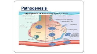
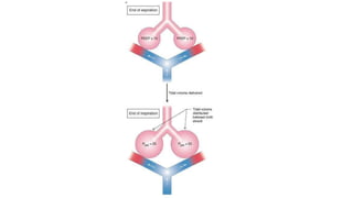
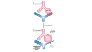
This document summarizes acute respiratory distress syndrome (ARDS) by defining it, describing its severity, pathophysiology, etiology, risk factors, goals of therapy, and ventilator strategies. ARDS is defined by timing, radiographic changes, and origin of edema not due to heart failure. Severity is based on oxygenation. The pathophysiology involves diffuse alveolar damage and endothelial injury. Common causes are sepsis, aspiration, and trauma. Treatment aims to allow lung healing while avoiding further injury through strategies like low tidal volumes and positive end-expiratory pressure to reduce volutrauma, barotrauma, and atelectrauma.
This document provides an overview of ARDS (acute respiratory distress syndrome) including its history, definition, pathophysiology, assessment, and treatment strategies. ARDS is characterized by acute hypoxemia, stiff lungs, and diffuse pulmonary infiltrates caused by inflammatory lung injury from direct or indirect insults. Key evidence-based treatment strategies discussed include lung protective ventilation with low tidal volumes, higher PEEP levels, targeting driving pressure, prone positioning, and rescue therapies like recruitment maneuvers which can improve oxygenation but their benefits are uncertain. The PROSEVA trial showed a significant reduction in 28-day mortality for prone positioning in severe ARDS patients.
This document summarizes the key points of a clinical review on acute lung injury and acute respiratory distress syndrome (ARDS). It defines ARDS and discusses its causes, histopathology, progression in stages from exudative to fibroproliferative, and treatment approaches including mechanical ventilation with low tidal volumes, use of positive end-expiratory pressure, prone positioning, corticosteroids, fluid management, and vasodilators. While some treatments like low tidal volume ventilation and conservative fluid management have shown benefits, corticosteroids and higher levels of PEEP have not conclusively improved clinical outcomes for patients with ARDS.
Acute respiratory distress syndrome (ARDS) is a condition where the lungs cannot provide enough oxygen to the blood due to damage to the tiny air sacs in the lungs called alveoli. It is caused by lung injury from infections, trauma, or other illnesses. Symptoms include rapid breathing and low blood oxygen. Diagnosis is based on chest x-ray findings and low blood oxygen levels. Treatment focuses on supportive care through mechanical ventilation and treating the underlying cause. Complications can include additional lung and organ damage if not properly managed.
This document provides a historical overview of acute respiratory distress syndrome (ARDS) and discusses early understandings and management approaches. It describes some of the first documented cases and autopsy findings of ARDS from the 1960s. Pioneering work in the 1970s established positive end-expiratory pressure (PEEP) as an important management strategy for improving oxygenation in ARDS patients. Studies throughout the 1970s explored how different levels of PEEP impact oxygenation and lung mechanics. The concept of "baby lung" emerged in the 1980s with the use of computed tomography showing that ARDS often spares some normal lung regions, challenging views of diffuse, symmetric involvement.
Acute respiratory distress syndrome (ARDS) is a clinical syndrome characterized by rapid onset of severe breathing difficulties, low oxygen levels in the blood, and diffuse lung inflammation and fluid buildup leading to respiratory failure. It has two phases - an initial exudative phase where lung inflammation causes fluid buildup in the lungs, and a later proliferative phase where the lungs try to heal but can develop pulmonary fibrosis. Treatment focuses on supportive care, minimizing additional lung injury from mechanical ventilation, maintaining low fluid levels to prevent further pulmonary edema, and managing the underlying cause of the ARDS when possible. The mainstay of ventilation treatment is using low tidal volumes and adequate positive end-expiratory pressure to safely recruit and maintain open lung volumes without over
Acute respiratory distress syndrome (ARDS) is defined as acute lung inflammation and increased permeability causing hypoxemia. It is characterized by bilateral infiltrates and low PaO2/FiO2 ratio with no cardiac cause. ARDS can be caused directly by lung injury or indirectly. It has three stages - exudative, proliferative, and recovery. Mechanical ventilation can worsen ARDS through barotrauma, atelectotrauma, and volutrauma. Lung protective ventilation aims to prevent further injury through small tidal volumes, high respiratory rate, low pressures and adequate PEEP. The prone position and ECMO may be rescue therapies for moderate-severe ARDS when conventional measures fail.
This document discusses strategies for mechanical ventilation in patients with acute respiratory distress syndrome (ARDS). It recommends using a protective ventilation strategy with low tidal volumes between 4-6 ml/kg and limiting airway plateau pressure to below 30 cmH2O to prevent ventilator-induced lung injury. Prone positioning is gaining acceptance as a standard of care for severe ARDS as it improves oxygenation and mortality outcomes. Early application of prone positioning for at least 16 hours per session is recommended for eligible ARDS patients meeting criteria. Recruitment maneuvers combined with high positive end-expiratory pressure are also advocated as part of an "open lung" approach to maximize aeration and oxygenation while minimizing pressures and volumes.
Acute Respiratory Distress Syndrome (ARDS) is a clinical syndrome characterized by hypoxemia, bilateral pulmonary infiltrates, and respiratory failure. The document defines ARDS and discusses its etiology, pathophysiology, clinical features, diagnosis, and evidence-based treatment recommendations. Key points include low tidal volume ventilation to minimize lung injury, conservative fluid management, use of PEEP to recruit alveoli while limiting pressures, and treating the underlying cause of ARDS. Outcomes remain poor with high mortality rates, though some patients fully recover lung function over time.
This document discusses ARDS (acute respiratory distress syndrome), including its history, definitions, pathophysiology, and evidence-based treatment strategies. ARDS is characterized by diffuse pulmonary inflammation and reduced lung compliance. Traditional ventilator strategies have been shown to cause ventilator-induced lung injury, so current recommendations focus on lung-protective ventilation with low tidal volumes and high PEEP. Additional rescue therapies for refractory hypoxemia include recruitment maneuvers, proning, and ECMO. Proper diagnosis requires consideration of alternative conditions and use of diagnostic tools like echocardiogram, bronchoscopy, and chest CT scan.
This document discusses various strategies for managing acute respiratory distress syndrome (ARDS). It begins by describing the pathophysiology of ARDS including pulmonary capillary leak, surfactant inactivation, and diffuse lung injury. It then discusses various ventilator strategies and adjunctive therapies for ARDS management such as recruitment maneuvers, prone positioning, high frequency oscillatory ventilation, liquid ventilation, and application of surfactant. The document emphasizes an open lung approach with low tidal volumes and higher positive end expiratory pressures to minimize ventilator-induced lung injury while maintaining adequate oxygenation and ventilation.
Acute Respiratory Distress Syndrome (ARDS) is an acute hypoxemic respiratory failure following a lung or systemic insult without heart failure. It involves diffuse bilateral lung infiltrates, normal heart functioning, and profound hypoxemia. Common causes include pneumonia, aspiration, and sepsis. Patients experience rapid onset of labored breathing and hypoxemia. Chest imaging shows bilateral infiltrates. Treatment focuses on supportive care, mechanical ventilation with low tidal volumes, and treating the underlying condition. While the mortality rate is high, especially with sepsis, outcomes have improved in recent decades.
Acute respiratory distress syndrome (ARDS) is a severe life-threatening lung condition that involves diffuse pulmonary inflammation and the formation of a hyaline membrane in the alveoli. It exists in two forms: neonatal ARDS, which affects premature infants and infants of diabetic mothers, and adult ARDS, which is usually caused by factors like shock, infection, inhalation of toxins, or trauma. Clinically, both forms cause sudden respiratory distress, tachypnea, hypoxia, and cyanosis. ARDS is diagnosed based on history, physical exam, chest x-ray, and blood tests. Treatment focuses on circulatory and respiratory support through oxygen therapy, ventilation, positioning, and medication. Complications can include
Acute Respiratory Distress Syndrome (ARDS) is a sudden, progressive form of respiratory failure characterized by severe dyspnea, hypoxemia, and decreased lung compliance. It develops from direct or indirect lung injuries and is thought to be caused by stimulation of the inflammatory and immune systems, resulting in leakage of fluid into the lungs. The clinical progression of ARDS involves exudative, proliferative, and fibrotic phases that can lead to respiratory failure if not promptly treated with oxygen supplementation, mechanical ventilation, and other supportive therapies.
- ARDS is an acute respiratory condition characterized by diffuse lung inflammation and fluid buildup in the lungs, causing hypoxemia. Common causes include sepsis, aspiration, and pneumonia.
- The document discusses the definition, pathogenesis, clinical presentation, diagnosis, and management of ARDS. The primary goals of management are treating the underlying cause, maintaining oxygenation levels through ventilation strategies like low tidal volumes, and preventing further lung injury.
- Low tidal volume ventilation, which aims to limit overexpansion of alveoli, is the best proven strategy to improve survival based on current evidence. Other adjuncts like prone positioning and PEEP may also help optimize oxygenation in some cases.
Acute respiratory distress syndrome (ARDS) is a condition that damages the lungs and prevents sufficient oxygen from entering the bloodstream. It is caused by injury to the lungs from direct or indirect insults. In ARDS, the alveolar-capillary membrane is compromised, allowing fluid to enter the lungs. Treatment involves intensive care, use of a ventilator to deliver oxygen under pressure, and addressing the underlying cause while preventing further lung injury. The goal is to support breathing and remove fluid from the damaged lungs.
Acute respiratory failure is defined as low blood oxygen and high blood carbon dioxide levels caused by impaired gas exchange in the lungs. It can be caused by conditions affecting breathing muscles/nerves, lung diseases, injuries, or drugs/alcohol. Symptoms include shortness of breath, fatigue, and confusion. Treatment involves oxygen therapy, ventilator support if needed, and treating any underlying conditions. Nursing care focuses on positioning, infection prevention, nutrition, psychological support, and close monitoring of the patient.
Acute respiratory distress syndrome (ARDS) is a clinical syndrome characterized by rapid onset of severe breathing difficulties, low oxygen levels in the blood, and diffuse pulmonary infiltrates leading to respiratory failure. It has three phases: exudative, proliferative, and fibrotic. ARDS can develop due to various causes like pneumonia, sepsis, trauma, inhalation of toxic substances. Diagnosis involves chest x-ray findings and low oxygen levels with normal pulmonary artery pressure. Treatment focuses on treating the underlying cause, providing oxygen supplementation, respiratory support through mechanical ventilation, and managing complications.
Updates on Acute respiratory distress syndromeHamdi Turkey
The document provides an overview of acute respiratory distress syndrome (ARDS). It begins with a case presentation of a patient exhibiting symptoms of ARDS and then outlines the learning objectives which include understanding the definition, pathology, ventilation strategies, and adjunct therapies for ARDS. It reviews the history and evolving definitions of ARDS from 1967 to the current Berlin Definition from 2012. Key aspects of the Berlin Definition are described. The document discusses the incidence, outcomes, risk factors, pathophysiology involving different phases, clinical features, investigations, management goals and therapies for ARDS. Images are included showing histology, chest x-rays and CT scans of ARDS patients.
This document discusses acute respiratory distress syndrome (ARDS). It begins with an introduction and definition of ARDS. ARDS is an acute respiratory failure where the alveolar capillary membrane becomes damaged and more permeable, resulting in hypoxemia. The document then covers the etiology and risk factors of ARDS, which can be direct lung injury from things like pneumonia or indirect injury from sepsis. The pathophysiology of ARDS is explained through a schematic. Clinical manifestations like dyspnea and hypoxemia are outlined. Diagnostic evaluations and potential complications of ARDS are also reviewed. The document concludes with discussions of the medical management of ARDS including mechanical ventilation support, settings, modes of ventilation and use of PEE
Acute Respiratory Distress Syndrome (ARDS) is an acute lung injury characterized by increased permeability and decreased compliance. The Berlin Definition from 2013 is the accepted clinical definition, requiring hypoxemia, bilateral opacities on CXR, and no heart failure. ARDS has multiple risk factors and a pathophysiology involving exudative, proliferative, and sometimes fibrotic stages. Diagnosis is based on criteria of hypoxemia, pulmonary edema, bilateral infiltrates, and low compliance. Treatment focuses on treating the underlying cause, supporting oxygenation, and preventing complications through ventilation support, nutrition, fluid balance, and infection control.
This document discusses acute respiratory distress syndrome (ARDS). It defines ARDS as diffuse inflammatory lung injury leading to impaired gas exchange. ARDS is not a primary disorder but occurs due to infectious or non-infectious conditions like pneumonia or sepsis. Treatment involves treating the underlying cause, mechanical ventilation with a protective strategy using low tidal volumes and high PEEP, fluid management to avoid positive balance, and possibly steroids in moderate to severe cases. Outcomes are improved by following evidence-based guidelines for ARDS therapies.
This document provides an overview of acute respiratory distress syndrome (ARDS). It defines ARDS and discusses its causes, pathophysiology, diagnosis, incidence, prognosis and long-term outcomes. Treatment focuses on supportive care including mechanical ventilation with low tidal volumes, conservative fluid management, prone positioning and other strategies to improve oxygenation. Corticosteroids are not recommended for treatment due to lack of proven benefit. With treatment, prognosis depends on the underlying cause, but many ARDS survivors can expect to return to normal lifestyles within a year.
This document discusses acute respiratory distress syndrome (ARDS), including its definition, causes, pathogenesis, management challenges, and treatment strategies. ARDS is characterized by acute lung injury leading to hypoxemia. Common causes include pneumonia, sepsis, and trauma. The lung injury progresses through exudative, proliferative, and fibrotic phases. Conservative fluid management, low tidal volume ventilation, and positive end-expiratory pressure are the primary treatment approaches.
This document provides an overview of acute respiratory distress syndrome (ARDS) including its definition, pathophysiology, clinical presentation, diagnosis, and management. Some key points:
- ARDS is characterized by acute hypoxemic respiratory failure due to widespread inflammation and fluid buildup in the lungs.
- Treatment involves supportive care with mechanical ventilation using low tidal volumes, maintaining adequate oxygen levels, treating the underlying cause, and considering rescue therapies for severe cases like prone positioning or extracorporeal membrane oxygenation.
- Mortality remains high at around 26-58% depending on severity, with the most common causes of death being complications of the initial insult or secondary infections like pneumonia. Ongoing research focuses on
1) ARDS is an acute lung condition caused by direct or indirect injury to the lungs. It is characterized by inflammation and fluid buildup in the lungs.
2) The pathophysiology involves activation of the innate immune system and release of cytokines, which damage the alveolar-capillary membrane leading to leakage of fluid into the lungs.
3) Treatment focuses on mechanical ventilation with low tidal volumes, application of PEEP, conservative fluid management, prone positioning in severe cases, and treating the underlying cause.
Respiratory failure is characterized by severe dysfunction of pulmonary ventilation and/or oxygenation caused by various diseases, resulting in hypoxia and retention of carbon dioxide. It is defined as a PaO2 of less than 8.0 kPa (60 mmHg), and/or a PaCO2 of greater than 6.67 kPa (50 mmHg). The main causes are ventilation dysfunction due to airway obstruction or limitation, and oxygenation dysfunction due to pulmonary edema, interstitial lung disease, or ARDS. The key pathophysiological changes are hypoxia, retention of carbon dioxide, and acidosis, which can affect multiple organ systems and lead to complications.
The document presents information about a seminar on Acute Respiratory Distress Syndrome (ARDS). The seminar aims to provide in-depth knowledge of ARDS including defining it, describing the pathophysiology and management. ARDS is a life-threatening condition that prevents enough oxygen from entering the blood. It occurs when the lungs become severely inflamed and fluid builds up in the tiny air sacs of the lungs. The seminar will discuss etiology, risk factors, clinical manifestations, diagnostic evaluation, complications, and the nurse's role in management.
This document provides an overview of ventilator settings and their clinical application. It begins with the objectives and provides background on pulmonary physiology including lung volumes, compliance, resistance, and time constants. It then covers types of respiratory failure and diseases that impact compliance and resistance. The remainder focuses on ventilator settings like FiO2, PIP, PEEP, rate, Ti/Te ratio, flow and their significance. Manipulations to optimize oxygenation and CO2 elimination are discussed along with the advantages and disadvantages of increasing various settings. The goal of assisted ventilation is to achieve adequate oxygenation and CO2 elimination while minimizing risks of barotrauma.
1) Recruitment maneuvers (RMs) aim to reopen collapsed alveoli in ARDS patients through temporarily increasing transpulmonary pressure. Common types include sighs, sustained inflations, and stepwise increases in pressure.
2) While RMs often improve short-term oxygenation, clinical trials have found no evidence of reduced mortality or improved outcomes. One large trial found RMs may actually increase mortality.
3) Not all ARDS patients respond equally to RMs due to factors like etiology, severity, and lung recruitability. RMs should only be considered for hypoxemic individuals based on an individual risk-benefit assessment.
The document discusses venting. In a few short sentences, it introduces the topic of venting without providing many details. The document does not have enough context or information to generate a multi-sentence summary while maintaining accuracy.
1) Respiratory failure occurs when the respiratory system fails in gas exchange, and can be hypoxaemic (low oxygen tension) or ventilatory (insufficient carbon dioxide elimination). Common causes include COPD, asthma, and neuromuscular fatigue.
2) Acute respiratory distress syndrome (ARDS) is the clinical manifestation of acute pulmonary inflammatory states that damage the alveolar-capillary membrane. Risk factors include sepsis, pneumonia, aspiration, trauma, and pancreatitis.
3) Treatment of respiratory failure focuses on correcting symptoms like hypoxemia, interrupting pathogenesis, and treating the underlying cause. Mechanical ventilation may be necessary for severe cases.
This document provides an overview of respiratory failure and acute respiratory distress syndrome (ARDS). It discusses the diagnosis of respiratory failure using blood gases and imaging tests. The pathophysiology of ARDS involves generalized lung inflammation that leads to pulmonary edema. Mechanical ventilation aims to improve gas exchange while avoiding lung damage, using low tidal volumes and optimizing positive end-expiratory pressure levels. The treatment of respiratory failure focuses on addressing the underlying cause, symptoms, and pathogenesis.
1) Acute respiratory distress syndrome (ARDS) is a life-threatening lung condition caused by injury to the lungs. It can result from direct lung injury, such as pneumonia, or indirect injury, like sepsis.
2) ARDS progresses through exudative and proliferative phases characterized by fluid accumulation and scarring in the lungs. This impairs gas exchange and causes respiratory failure.
3) Mechanical ventilation is used to treat respiratory failure but can further damage the lungs if not done carefully. The ARDSNet trial showed using low tidal volumes of 6 ml/kg improved survival compared to larger volumes.
The document discusses lung volumes, capacities, and pulmonary function tests. Key points include:
- Pulmonary function testing measures ventilation, diffusion, and blood flow to evaluate lung health. Spirometry is the cornerstone test and measures volumes inhaled and exhaled over time.
- Other tests include lung volume determination, diffusing capacity tests, and six-minute walk tests. Results are compared to predicted normal values.
- Spirometry evaluates the airways and lung parenchyma. It measures volumes like forced vital capacity and flows like FEV1. Pattern recognition from these values helps diagnose restrictive or obstructive lung diseases.
- Flow-volume loops provide additional information, showing concave loops in asthma and dog-
The document discusses the case of a 27-year-old postpartum woman presenting with worsening dyspnea and hypoxia. It then reviews the key considerations and management strategies for acute respiratory distress syndrome (ARDS), including low tidal volume ventilation, open lung strategies using recruitment maneuvers and high positive end-expiratory pressure, unconventional approaches like airway pressure release ventilation and high frequency oscillatory ventilation, and adjunctive therapies such as prone positioning. The optimal ventilator mode, settings, and adjunctive strategies depend on the individual patient's severity of lung injury and response to different interventions.
How to ventilate COPD and ARDS in Intensive care unit. safe lung ventilation. PEEP, Tidal volume, mode of ventilation. limits of ventilation. ventilator alarms
Acute respiratory distress syndrome (ARDS) is a clinical syndrome characterized by rapid onset of severe respiratory distress, hypoxemia, and diffuse pulmonary infiltrates. It is commonly caused by sepsis, pneumonia, trauma, or multiple transfusions. The pathophysiology involves injury to the alveolar-capillary barrier leading to accumulation of protein-rich edema fluid in the lungs. Treatment focuses on supportive care, mechanical ventilation with low tidal volumes and positive end-expiratory pressure to prevent alveolar collapse, and maintaining a low left atrial pressure to minimize pulmonary edema.
This document discusses mechanical ventilation. It begins by defining ventilation and the parts of a ventilator. It then covers the history of mechanical ventilation and describes various ventilator modes and settings such as PEEP, tidal volume, and inspiratory/expiratory ratio. The document outlines indications for ventilation and discusses monitoring ventilated patients. Key steps in ventilation like initiation, sedation, and weaning are summarized.
The document provides information about acute respiratory distress syndrome (ARDS). It begins with a brief history of ARDS and provides the clinical definition. It describes the diagnostic criteria and etiology, including that most cases are caused by sepsis, pneumonia, or trauma. It then discusses the normal lung physiology and pathophysiology of ARDS, which involves three phases: exudative, proliferative, and fibrotic. The management section outlines the principles of therapy to provide adequate gas exchange while avoiding secondary injury, including mechanical ventilation protocols, fluid management, and other strategies. It concludes with a discussion of prognosis and recent advances in ARDS management such as protective ventilation strategies.
The document provides information on indications for mechanical ventilation, criteria for instituting ventilation based on pulmonary function and blood gas parameters, settings for mechanical ventilation including tidal volume, respiratory rate, PEEP, and oxygen concentration. It also outlines the basics of various ventilator modes including assist-control, pressure support, and SIMV. Guidelines are provided for initiating mechanical ventilation and troubleshooting issues like high pressures, low volumes, and patient-ventilator dysynchrony. The nurse's key roles in monitoring the patient and equipment are also summarized.
Acute respiratory distress syndrome (ARDS) is a clinical syndrome characterized by rapid onset of bilateral pulmonary infiltrates and hypoxemia leading to respiratory failure. It is caused by acute diffuse inflammatory lung injury from a direct or indirect pulmonary insult. Diagnosis requires excluding left heart failure and evaluating for underlying causes. Pathologically, it involves diffuse alveolar damage and pulmonary edema. Treatment focuses on supportive care including mechanical ventilation, fluid management, and treatment of underlying conditions. While mortality has decreased in recent decades, ARDS still carries a significant risk of death.
This document discusses respiratory system mechanics in mechanically ventilated patients. It covers topics like compliance, resistance, work of breathing, modes of ventilation, and patient-ventilator asynchrony. It also describes the key phase variables in mechanical ventilation - trigger, which initiates inspiration, limit, which ends inspiratory flow, and cycle, which ends inspiration. Pressure, flow, and electrical activity can be used as triggers to initiate breaths based on patient effort. Initial settings for mechanical ventilation like tidal volume, PEEP, flow rate, and I:E ratio are also outlined.
This document provides an overview of Acute Respiratory Distress Syndrome (ARDS). It discusses the epidemiology, causes, clinical course and features, diagnostic criteria, investigations and management of ARDS. The key points are: ARDS affects approximately 10% of ICU patients annually and has a mortality rate of around 30%; it is caused by direct or indirect lung injury and progresses through exudative, proliferative and fibrotic phases; diagnosis is based on acute onset hypoxemia, bilateral opacities on chest imaging and low oxygen levels; management focuses on treating the underlying cause, lung protective ventilation with low tidal volumes, PEEP and prone positioning if severe. With treatment, most ARDS survivors recover lung function within 6-
The document provides information about a pulmonary function test (PFT), including the physiology, types of tests, purposes, indications, and contraindications. A PFT can evaluate lung volumes, capacities, flows, and gas exchange. It helps diagnose and classify pulmonary diseases as obstructive or restrictive based on abnormalities in flows versus volumes. Key tests described are closed-circuit helium dilution, open-circuit nitrogen washout, and body plethysmography. PFTs are used to identify lung impairment, determine its severity, and evaluate response to treatment.
Compliance Resistance & Work Of Breathing Zareer Tafadar
This document discusses the mechanics of respiration and resistance to breathing. It covers:
1. Elastic resistance makes up around 65% of total resistance and is due to the elastic recoil of lung tissue and surface tension forces. Lung compliance measures a lung's elastic resistance.
2. Non-elastic resistance accounts for the remaining 35% and includes airway resistance. Dynamic compliance is lower than static compliance due to factors like airway obstruction.
3. Several lung diseases can decrease compliance by increasing elastic or non-elastic resistance, requiring more work from respiratory muscles. Surfactant reduces surface tension forces and the work of breathing.
1. The document discusses respiratory failure, describing it as a failure to maintain adequate gas exchange resulting in hypoxemia and potentially hypercapnia.
2. Types of respiratory failure are classified as type 1 (hypoxemic) or type 2 (hypercapnic), and common causes of each type are provided.
3. Diagnostic testing and management approaches are outlined, focusing on arterial blood gas analysis, oxygen therapy using devices like nasal cannulas or Venturi masks, and treating the underlying cause.
This document provides definitions for various medical terminology related to cardiopulmonary and respiratory systems. It includes terms for different types of breathing difficulties, oxygen and carbon dioxide levels in blood, heart conditions like arrhythmias, heart attacks, and heart failure. Diagnostic tests are also defined such as ECG, CXR, ABG's, pulmonary function tests, bronchoscopy, echocardiograms, CT scans, and cardiac catheterization. Emergency procedures like CPR, intubation, chest tube insertion, and defibrillation are also listed.
This document discusses different ventilator waveforms and modes of ventilation. It describes basic modes like pressure and volume ventilation. It identifies different types of waveform abnormalities that can occur which indicate patient-ventilator desynchrony, such as auto-PEEP, trigger desynchrony, and cycle desynchrony. The document contains diagrams of normal and abnormal ventilator waveforms to help illustrate concepts like auto-PEEP and pressure ventilation flow patterns.
The Foundation of Medicine Curriculum includes topics like medical terminology, history of medicine, concepts of health and disease, and communication skills. Medical terminology is the professional language of healthcare and uses word roots from Latin and Greek to name body parts and diseases. There are four main elements to a medical term: word root, combining form, suffix, and prefix. By understanding the meanings of these elements like word roots for body parts and suffixes for processes, the meaning of complex medical terms can be deduced by analyzing the smaller word parts. Correct pronunciation of terms is aided by inserting combining vowels between word parts.
1. The document discusses acid-base imbalances and how to interpret arterial blood gases (ABGs).
2. It outlines the steps to analyze ABGs which include determining if there is acidemia or alkalemia, identifying if the disturbance is respiratory or metabolic, and assessing compensation.
3. Causes of different acid-base imbalances are provided such as respiratory acidosis resulting from airway obstruction or increased carbon dioxide production, and metabolic acidosis occurring in conditions like diabetic ketoacidosis or lactic acidosis.
This document discusses different types of patient-ventilator dyssynchrony. It begins with background information on how the main purpose of a ventilator is to decrease the work of breathing. Normally, respiratory muscles account for 1-3% of oxygen consumption, but this can increase to 20% for patients in acute respiratory failure undergoing CPR due to the increased work of breathing.
The document then discusses different factors that can contribute to different types of patient-ventilator dyssynchrony, including trigger-related dyssynchrony from a high trigger threshold, muscle weakness, leaks, auto-PEEP, or expiratory flow limitation. Target-related and cycle-related dyssynchrony are also mentioned
This document summarizes specific organ support considerations in the ICU, including the brain, heart, lungs, kidneys, liver, hematology/coagulation, skin/soft tissue, and endocrine system. Key points covered include oxygenation and ventilation strategies for respiratory support, indications for renal replacement therapy, transfusion thresholds for blood products, and general measures to prevent complications like bed sores.
The document discusses monitoring respiratory mechanics during mechanical ventilation. It describes the resistive and elastic components of the respiratory system and how peak pressure, plateau pressure, compliance, and elastance are measured and affected. Peak pressure is the maximum pressure achieved in a breath and is impacted by resistance, flow, volume, and elastance. Plateau pressure measures lung parenchymal properties at a given volume. Compliance is the change in volume per unit change in pressure and can be measured statically or dynamically.
Mechanical ventilation 6- in obstructive airway diseaseDr fakhir Raza
This document discusses ventilation challenges in patients with obstructive airway disease. Increased airway resistance from conditions like bronchospasm can lead to inefficient exhalation requiring prolonged expiratory time. This can result in breath stacking where the patient triggers another breath before fully exhaling, and gas trapping where extra air remains in the alveoli at expiration's end, increasing end-expiratory pressure known as auto-PEEP. Diagnosing these issues involves seeing if expiratory flow doesn't reach zero or wheezing persists to the next breath. Managing them includes increasing exhalation time, decreasing tidal volume, adjusting ventilator settings accordingly based on the mode used.
This document discusses mechanical ventilation and the mechanics of breathing. It explains that breathing can occur spontaneously through the work of respiratory muscles, or through positive pressure ventilation using devices like endotracheal tubes. The balance between the lungs' elastic recoil inward and the chest wall's recoil outward determines the lung volume. Transpulmonary pressure, the difference between alveolar and pleural pressures, controls lung volume and inflation. Boyle's law states that for a fixed amount of gas, pressure and volume are inversely proportional.
This document discusses mechanical ventilation and the mechanics of breathing. It explains that breathing can occur spontaneously through the work of respiratory muscles, or through positive pressure ventilation using devices like endotracheal tubes. The balance between the lungs' elastic recoil inward and the chest wall's recoil outward determines lung volume. Transpulmonary pressure, the difference between alveolar and pleural pressures, increases to inflate the lungs either through spontaneous breathing or positive pressure ventilation. Boyle's law states that for a fixed amount of gas, pressure and volume are inversely proportional.
The document discusses different modes of mechanical ventilation:
1) Volume-controlled ventilation (VCV) targets a set flow and cycles based on a set volume, with the pressure waveform determined by the respiratory system and patient effort.
2) Pressure-controlled ventilation (PCV) targets a set pressure and cycles based on a set time, with the flow waveform again determined by the respiratory system and patient effort.
3) Pressure support ventilation (PSV) is pressure-targeted and flow-cycled, with the inspiratory time varying and the flow waveform determined by set variables and the respiratory system.
Sepsis is a life-threatening organ dysfunction caused by a dysregulated response to infection. Septic shock, a subset of sepsis, involves circulatory and cellular abnormalities with high mortality. Sepsis signs include temperature changes, increased heart and breathing rates, and abnormal white blood cell counts. Diagnosis involves assessing these signs, blood pressure, lactate levels, and cultures of potential infection sources. Treatment focuses on rapidly restoring perfusion with IV fluids and antibiotics, controlling the infection source, and supporting organ functions.
This document discusses gastrointestinal support and nutrition in the ICU, including:
1. Problems such as starvation and stress metabolism that ICU patients face and the need to provide adequate nutrition while avoiding overfeeding.
2. Guidelines for assessing risk of malnutrition using the MUST tool and determining when and how to provide enteral or parenteral nutrition.
3. Managing risks like stress ulceration and gastrointestinal bleeding, including identifying patients at risk, treatment protocols, and rescue measures.
Management of liver failure in general intensive careDr fakhir Raza
The document provides guidance on the management of acute liver failure and cirrhotic patients in the intensive care unit. It recommends:
1) Treating extrahepatic organ failure early and preventing aggravating factors to reduce morbidity and mortality in acute liver failure patients.
2) Initiating N-acetylcysteine therapy for all acute liver failure patients regardless of etiology to improve outcomes.
3) Consulting a liver transplant center to discuss further investigations or transplant evaluation if initial tests are negative for acute liver failure patients.
Respiratory therapists have general responsibilities of being a team member, ensuring patient safety, and implementing the ABCDEF bundle for quality improvement. Their specific responsibilities include conducting SAT and SBT components of the ABCDEF bundle, having thorough knowledge of lung mechanics to apply for early weaning, assisting physicians with difficult patients, leading proning teams, and applying chest physiotherapy.
A – Assess, Prevent and Manage Pain
B – Both SATs and SBTs
C – Choice of Sedation
D – Delirium: Assess, Prevent and Manage
E – Early Mobility and Exercise
F – Family Engagement and Empowerment
*www.iculiberation.org
latest knowledge practical points short presentation
It will serve as guideline for Covid-19 corona virus
it will help in preparing ICU as well as policy making
institutions should device their own strategy
Integrating Ayurveda into Parkinson’s Management: A Holistic ApproachAyurveda ForAll
Explore the benefits of combining Ayurveda with conventional Parkinson's treatments. Learn how a holistic approach can manage symptoms, enhance well-being, and balance body energies. Discover the steps to safely integrate Ayurvedic practices into your Parkinson’s care plan, including expert guidance on diet, herbal remedies, and lifestyle modifications.
Clinic ^%[+27633867063*Abortion Pills For Sale In Tembisa Central19various
Clinic ^%[+27633867063*Abortion Pills For Sale In Tembisa Central Clinic ^%[+27633867063*Abortion Pills For Sale In Tembisa CentralClinic ^%[+27633867063*Abortion Pills For Sale In Tembisa CentralClinic ^%[+27633867063*Abortion Pills For Sale In Tembisa CentralClinic ^%[+27633867063*Abortion Pills For Sale In Tembisa Central
TEST BANK For Basic and Clinical Pharmacology, 14th Edition by Bertram G. Kat...rightmanforbloodline
TEST BANK For Basic and Clinical Pharmacology, 14th Edition by Bertram G. Katzung, Verified Chapters 1 - 66, Complete Newest Version.
Local Advanced Lung Cancer: Artificial Intelligence, Synergetics, Complex Sys...Oleg Kshivets
Overall life span (LS) was 1671.7±1721.6 days and cumulative 5YS reached 62.4%, 10 years – 50.4%, 20 years – 44.6%. 94 LCP lived more than 5 years without cancer (LS=2958.6±1723.6 days), 22 – more than 10 years (LS=5571±1841.8 days). 67 LCP died because of LC (LS=471.9±344 days). AT significantly improved 5YS (68% vs. 53.7%) (P=0.028 by log-rank test). Cox modeling displayed that 5YS of LCP significantly depended on: N0-N12, T3-4, blood cell circuit, cell ratio factors (ratio between cancer cells-CC and blood cells subpopulations), LC cell dynamics, recalcification time, heparin tolerance, prothrombin index, protein, AT, procedure type (P=0.000-0.031). Neural networks, genetic algorithm selection and bootstrap simulation revealed relationships between 5YS and N0-12 (rank=1), thrombocytes/CC (rank=2), segmented neutrophils/CC (3), eosinophils/CC (4), erythrocytes/CC (5), healthy cells/CC (6), lymphocytes/CC (7), stick neutrophils/CC (8), leucocytes/CC (9), monocytes/CC (10). Correct prediction of 5YS was 100% by neural networks computing (error=0.000; area under ROC curve=1.0).
Basavarajeeyam is a Sreshta Sangraha grantha (Compiled book ), written by Neelkanta kotturu Basavaraja Virachita. It contains 25 Prakaranas, First 24 Chapters related to Rogas& 25th to Rasadravyas.
Does Over-Masturbation Contribute to Chronic Prostatitis.pptxwalterHu5
In some case, your chronic prostatitis may be related to over-masturbation. Generally, natural medicine Diuretic and Anti-inflammatory Pill can help mee get a cure.
2. Berlin Definition
• Timing: within 1 wk of clinical insult (or onset of respiratory
symptoms)
• radiographic changes: (bilateral opacities not fully explained by
effusions, consolidation, or atelectasis)
• origin of edema: (not fully explained by cardiac failure or fluid
overload)
Acute respiratory distress syndrome: the Berlin Definition. JAMA. 2012; 307(23):2526-33 (ISSN: 1538-3598) ; Ranieri VM; Rubenfeld GD; Thompson BT; Ferguson ND;
Caldwell E; Fan E; Camporota L; Slutsky AS
3. Severity of ARDS
• Severity based on the PaO2/FiO2ratio on 5 cm of continuous positive
airway pressure (CPAP)
• Mild (PaO2/FiO2 200-300)
• Moderate (PaO2/FiO2 100-200)
• Severe (PaO2/FiO2≤100)
4. Pathophysiology
• ARDS is associated with diffuse alveolar damage (DAD) and lung
capillary endothelial injury. The early phase is described as being
exudative, whereas the later phase is fibroproliferative in character.
5.
6. Etiology
• ARDS risk factors include
• direct lung injury (most commonly, aspiration of gastric contents),
• systemic illnesses, and injuries.
• The most common risk factor for ARDS is sepsis.
7. Risk Factor
• 20% no risk factor
• Bacteremia
• Sepsis
• Trauma, with or without pulmonary contusion
• Fractures, particularly multiple fractures and long bone fractures
• Burns
• Massive transfusion
• Pneumonia
• Aspiration
• Drug overdose
• Near drowning
• Postperfusion injury after cardiopulmonary bypass
• Pancreatitis
• Fat embolism
8. Goal of therapy in ARDS
• Besides treating the underlying etiology, the treatment of ARDS is
predominantly supportive.
• The goal is to allow time for the injured lung parenchyma to properly
heal while avoiding damage to the uninjured lung
9. Types of injury in ARDS
• Three types of ventilator-induced lung injury have been described
• Volutrauma
• Barotrauma
• Atelectrauma
• Ventilator strategies employed in ARDS are primarily used to minimize
these types of lung injury.
10. Volutrauma
• High Tidal Volumes – increased stretch – further inflammation and
lung injury in already damaged lung
• Low TV volumes (6ml/kg)
• Ideal body weight is used for calculation of weight
11. Volume control vs Pressure control in ARDS
• Target is to limit the Tidal volume
• In volume control the Tidal volume is guaranteed
• In Pressure control the pressure should be adjusted to avoid high
Tidal volumes
12. Barotrauma
• Unlike ETT and large airways, alveoli can not bear large pressures and
pressure changes
• pneumothorax, pneumomediastinum, and subcutaneous emphysema
are complications of barotrauma
• Plateau pressure is the maximum pressure in the alveoli during
respiratory cycle
• In Volume control ventilation even lower tidal volumes then 6ml/kg
may be required
• Maximum Plateau pressure allowed in 30cm of H2O
13. Atelectrauma
• The lung injury that is perpetuated by repetitive opening and closing
of alveoli is known as atelectrauma
14. Alveolar Pressures
• Opening Pressure: minimum pressure required to open a closed
alveolus
• Closing Pressure: minimum pressure required within an open alveolus
to keep it open
15. PEEP (Peak End Expiratory Pressure)
• It provides positive alveolar pressure through out the respiratory cycle
• It should be set in between opening and closing pressures
• PEEP is used to keep the alveoli open and NOT to actually OPEN UP the
closed alveoli
• To open the closed alveoli, PEEP should be briefly increased above the
opening pressure
• Recruitment maneuvers, are transient increases in alveolar pressure
that are intended to exceed opening pressure, can be utilized to open
collapsed alveoli
16. PEEP can also have detrimental effects
• If not enough to keep alveoli open
• If too high to compromise perfusion
22. Permissive hypercapnia
• Permissive hypercapnia refers to hypercapnia despite the maximum
possible respiratory rate in the setting of low tidal volume ventilation
• It is “permitted” because of the known benefits of maintaining low
tidal volume in the setting of ARDS.
Editor's Notes
Both volume and pressure modes of ventilation can be
used for low tidal volume ventilation. As discussed in Chap.
3, with volume-controlled ventilation (VCV), flow (target)
and volume (cycle) are set; therefore, changes in the patient’s
lung compliance, airway resistance, and patient effort will
affect proximal airway pressure and not the delivered tidal
volume. For example, the development of strong inspiratory
efforts by the patient during VCV will not alter tidal volume
but will instead decrease proximal airway pressure.
With pressure-controlled ventilation (PCV), the target variable
is proximal airway pressure, and the cycle variable is time.
Tidal volume, not directly set in this mode of ventilation, is
determined by proximal airway pressure, inspiratory time, and
characteristics of the respiratory system (lung compliance, airway
resistance, and patient effort). Changes in the respiratory
system will not alter proximal airway pressure but will instead
affect flow and consequently tidal volume. For example, the
development of strong inspiratory efforts by the patient during
PCV will not alter proximal airway pressure and will instead
lead to increased inspiratory flow and consequently increased
tidal volume. In this case, tidal volume can be reduced by
decreasing proximal airway pressure. If PCV is used for
patients with ARDS, it is crucial that the resultant tidal volume
be closely monitored to ensure that lung overinflation does not
occur in the setting of changes in the respiratory system.
plateau pressure can be
viewed as the maximum pressure during the respiratory cycle
in the alveolus; preventing high plateau pressure is important
in the management of ARDS. To achieve lower plateau pressure
with VCV, tidal volume must be reduced. For patients
with very low lung compliance, tidal volume below 6 mL per
kg may be necessary to achieve an acceptable plateau pressure.
The goal plateau pressure is usually below 30 cm H2O.
With PCV, flow is administered to quickly achieve and
maintain the set proximal airway pressure. As air flows into
the patient, alveolar pressure rises, and less flow is needed to
achieve the set proximal airway pressure. If the inspiratory
time (cycle) is long enough for flow to cease altogether,
alveolar pressure will equal proximal airway pressure. Thus,
setting lower proximal airway pressure will ensure lower
alveolar pressure and help prevent barotrauma.
Alveolar collapse is a hallmark of ARDS. The repetitive
opening and closing of collapsed alveoli with mechanical
ventilation is detrimental, as high sheer stresses are generated
at the interface of collapsed and aerated tissue when a collapsed
alveolus is reopened. The lung injury that is perpetuated
by this repetitive opening and closing of alveoli is known
as atelectrauma. Surfactant is a substance that reduces alveolar
wall surface tension, preventing alveolar collapse. Because
of alveolar epithelial damage and inflammation, there are
both a decrease in surfactant production and also a decrease
in surfactant function, resulting in alveolar collapse and
atelectasis.
The use of increased PEEP can minimize atelectrauma. As
discussed in Chap. 1, PEEP is setting proximal airway pressure
to be positive during the expiratory phase. Opening
pressure, which is the minimum pressure required to open a
closed alveolus, is higher than closing pressure, which is the minimum pressure required within an open alveolus to keep
it open. That is, more force is needed to separate alveolar
walls that have become stuck to each other than is needed to
prevent alveolar walls from closing in the first place. Figure 5.1
demonstrates the relationship between alveolar pressure and
the state of the alveolus. An alveolus with alveolar pressure
below closing pressure will be closed (Point A). If alveolar
pressure is increased to above closing pressure, but still below
opening pressure, the alveolus remains closed as it has not
risen above opening pressure (Point B). The alveolus will
open only once alveolar pressure has increased to above
opening pressure (Point C). After the alveolus has opened, if
alveolar pressure is reduced to below opening pressure, yet
still above closing pressure, the alveolus remains open
(Point D). If alveolar pressure is then reduced below closing
pressure, the alveolus will close (Point E). If alveolar pressure
is again increased above closing pressure, but still below
opening pressure, the alveolus remains closed (Point F). Note that alveolar pressure at Point B, Point D, and Point F is the
same, yet the alveolus is closed at Point B and Point F and
open at Point D. Theoretically, setting PEEP above closing
pressure will prevent open alveoli from closing and will
reduce atelectrauma.
Not only would setting PEEP above closing pressure of the alveoli prevent atelectrauma, it may maintain alveoli in
an open state that would otherwise remain collapsed during
the entire respiratory cycle. Because these open alveoli can
now receive part of the delivered tidal volume, lung compliance
may improve. These open alveoli can now participate in
gas exchange, improving hypoxemia. It is important to note
that PEEP does not actually “open” closed alveoli, but if set
above closing pressure, it can prevent open alveoli from closing.
In order to actually open a closed alveolus, alveolar pressure
must exceed opening pressure. Recruitment maneuvers,
which are transient increases in alveolar pressure that are
intended to exceed opening pressure, can be utilized to open
collapsed alveoli. While recruitment maneuvers have been
shown to improve oxygenation in patients with ARDS, they
have not been shown to improve clinically important outcomes
like mortality or hospital length of stay.
PEEP can also have detrimental effects, particularly if it is
not effective in preventing alveolar collapse and atelectasis.
To illustrate this phenomenon, imagine that the administration
of PEEP for a given patient does not lead to more alveoli
remaining open. In this case, PEEP raises end-expiratory
pressure of the open alveoli and thus increases their end-expiratory
volume. This increased end-expiratory volume
PEEP can also have detrimental effects, particularly if it is
not effective in preventing alveolar collapse and atelectasis.
To illustrate this phenomenon, imagine that the administration
of PEEP for a given patient does not lead to more alveoli
remaining open. In this case, PEEP raises end-expiratory
pressure of the open alveoli and thus increases their end-expiratory
volume. This increased end-expiratory volume places these alveoli on a less compliant portion of the
pressure-volume
curve because lungs are less compliant at
higher volumes (Fig. 1.4). Given that the administration of
PEEP did not result in more alveoli remaining open for this
patient, the entire tidal volume will be delivered to the same
set of open alveoli. Since these alveoli are starting at an elevated
end-expiratory volume and pressure, they will end at an
elevated end-inspiratory volume and pressure. This alveolar
overdistension can cause barotrauma and lung injury.
Additionally, overdistended alveoli may compress their
associated
capillaries, shunting blood away from open, well-ventilated
alveoli to collapsed, poorly ventilated alveoli. This
shunted blood does not participate in gas exchange and, in
fact, worsens hypoxemia
(a) Without
increased PEEP, the alveolus on the right remains open, while the
alveolus on the left remains closed. Assuming the closed alveolus
has high opening pressure, the entire tidal volume with inspiration
will enter only the open alveolus, leading to high end-inspiratory
(plateau) pressure. The closed alveolus does not participate in gas
exchange with the capillary.
(b) If increased PEEP maintains the
collapsed alveolus open, both alveoli remain open at the end of
expiration. The entire tidal volume with inspiration will be distributed
between the two alveoli, improving overall lung compliance
and possibly reducing end-inspiratory (plateau) pressure. Both
alveoli participate in gas exchange with the capillaries.
If
increased PEEP does not result in keeping the closed alveolus open,
it may cause overdistension of the already open alveolus with resultant
compression of its associated capillary, leading to shunting of
blood to the collapsed, poorly ventilated alveolus.
PEEP positive end-expiratory pressure
The total amount of air that is inhaled and exhaled per minute,
minute ventilation VE ( ), is equal to tidal volume (VT)
multiplied by respiratory rate (RR):
Minute ventilation is comprised of air that participates in
gas exchange, known as alveolar ventilation VA ( ), and air
that does not participate in gas exchange, known as dead-space
ventilation VD ( )