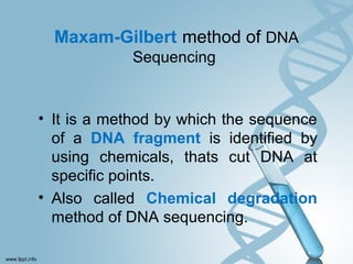
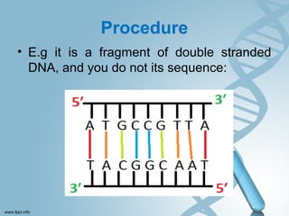
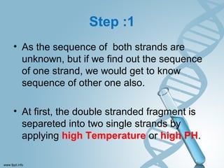
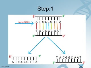
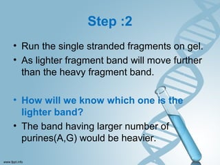
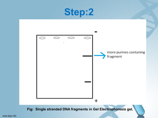
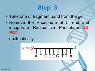
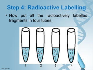
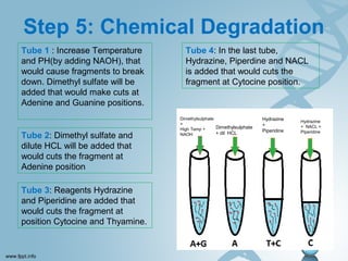
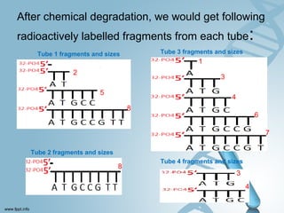
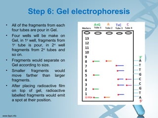
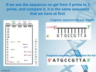
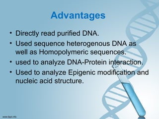
Maxam-Gilbert
method of
DNA Sequencing
Maxam-Gilbert method of DNA
Sequencing
• It is a method by which the sequence
of a DNA fragment is identified by
using chemicals, thats cut DNA at
specific points.
• Also called Chemical degradation
method of DNA sequencing.
Developed by developed
by
Allan Maxam and Walter
Gilbert
in
1976–1977
Procedure
Procedure
• E.g it is a fragment of double stranded
DNA, and you do not its sequence:
Step :1
• As the sequence of both strands are
unknown, but if we find out the sequence
of one strand, we would get to know
sequence of other one also.
• At first, the double stranded fragment is
separeted into two single strands by
applying high Temperature or high PH.
Step:1
Step :2
• Run the single stranded fragments on gel.
• As lighter fragment band will move further
than the heavy fragment band.
• How will we know which one is the
lighter band?
• The band having larger number of
purines(A,G) would be heavier.
Step:2
Fig: Single stranded DNA fragments in Gel Electrophoresis gel.
Step :3
• Take one of fragment band from the gel.
• Remove the Phosphate at 5′ end and
incoporate Radioactive Phosphate 32-
PO4
enztmatically.
Step 4: Radioactive Labelling
• Now put all the radioactively labelled
fragments in four tubes.
1 2 3 4
Step 5: Chemical Degradation
Tube 1 : Increase Temperature
and PH(by adding NAOH), that
would cause fragments to break
down. Dimethyl sulfate will be
added that would make cuts at
Adenine and Guanine positions.
Tube 2: Dimethyl sulfate and
dilute HCL will be added that
would cuts the fragment at
Adenine position
Tube 3: Reagents Hydrazine
and Piperidine are added that
would cuts the fragment at
position Cytocine and Thyamine.
Tube 4: In the last tube,
Hydrazine, Piperdine and NACL
is added that would cuts the
fragment at Cytocine position.
Dimethylsulphate
+
High Temp +
NAOH
Dimethylsulphate
+ dil HCL
Hydrazine
+
Piperidine
Hydrazine
+ NACL +
Piperidine
After chemical degradation, we would get following
radioactively labelled fragments from each tube:
Tube 1 fragments and sizes
Tube 2 fragments and sizes
Tube 3 fragments and sizes
Tube 4 fragments and sizes
2
5
8
8
1
3
4
6
7
3
4
Step 6: Gel electrophoresis
• All of the fragments from each
four tubes are pour in Gel.
• Four wells will be make on
Gel, in 1st
well, fragments from
1st
tube is pour, in 2nd
well
fragments from 2nd
tubes and
so on.
• Fragments would separate on
Gel according to size.
• Smaller fragments would
move farther than larger
fragments.
• After placing radioactive film
on top of gel, radioactive
labelled fragments would emit
a spot at their position.
If we see the sequence on gel from 5 prime to 3
prime, and compare it, it is the same sequence
that we have at first
:Fragment sequence that was selected
:Fragment sequence identified from the Gel
Advantages
• Directly read purified DNA.
• Used sequence heterogenous DNA as
well as Homopolymeric sequences.
• used to analyze DNA-Protein interaction.
• Used to analyze Epigenic modification and
nucleic acid structure.
Disadvantages
• Use of toxic chemicals and extensive use
of radioactive isotopes. highly poisonous
and unstable.
• Cannot read more than 500bp.
• Setup is quite complex.
• It is difficult to make Maxam-Gilbert DNA
sequencing kit.
• Read size decrease with incomplete
cleavage reactions.
Maxam-Gilbert method of DNA sequencing

Maxam-Gilbert method of DNA sequencing

  • 1.
  • 2.
    Maxam-Gilbert method ofDNA Sequencing • It is a method by which the sequence of a DNA fragment is identified by using chemicals, thats cut DNA at specific points. • Also called Chemical degradation method of DNA sequencing.
  • 3.
    Developed by developed by AllanMaxam and Walter Gilbert in 1976–1977
  • 4.
  • 5.
    Procedure • E.g itis a fragment of double stranded DNA, and you do not its sequence:
  • 6.
    Step :1 • Asthe sequence of both strands are unknown, but if we find out the sequence of one strand, we would get to know sequence of other one also. • At first, the double stranded fragment is separeted into two single strands by applying high Temperature or high PH.
  • 7.
  • 8.
    Step :2 • Runthe single stranded fragments on gel. • As lighter fragment band will move further than the heavy fragment band. • How will we know which one is the lighter band? • The band having larger number of purines(A,G) would be heavier.
  • 9.
    Step:2 Fig: Single strandedDNA fragments in Gel Electrophoresis gel.
  • 10.
    Step :3 • Takeone of fragment band from the gel. • Remove the Phosphate at 5′ end and incoporate Radioactive Phosphate 32- PO4 enztmatically.
  • 11.
    Step 4: RadioactiveLabelling • Now put all the radioactively labelled fragments in four tubes. 1 2 3 4
  • 12.
    Step 5: ChemicalDegradation Tube 1 : Increase Temperature and PH(by adding NAOH), that would cause fragments to break down. Dimethyl sulfate will be added that would make cuts at Adenine and Guanine positions. Tube 2: Dimethyl sulfate and dilute HCL will be added that would cuts the fragment at Adenine position Tube 3: Reagents Hydrazine and Piperidine are added that would cuts the fragment at position Cytocine and Thyamine. Tube 4: In the last tube, Hydrazine, Piperdine and NACL is added that would cuts the fragment at Cytocine position. Dimethylsulphate + High Temp + NAOH Dimethylsulphate + dil HCL Hydrazine + Piperidine Hydrazine + NACL + Piperidine
  • 13.
    After chemical degradation,we would get following radioactively labelled fragments from each tube: Tube 1 fragments and sizes Tube 2 fragments and sizes Tube 3 fragments and sizes Tube 4 fragments and sizes 2 5 8 8 1 3 4 6 7 3 4
  • 14.
    Step 6: Gelelectrophoresis • All of the fragments from each four tubes are pour in Gel. • Four wells will be make on Gel, in 1st well, fragments from 1st tube is pour, in 2nd well fragments from 2nd tubes and so on. • Fragments would separate on Gel according to size. • Smaller fragments would move farther than larger fragments. • After placing radioactive film on top of gel, radioactive labelled fragments would emit a spot at their position.
  • 15.
    If we seethe sequence on gel from 5 prime to 3 prime, and compare it, it is the same sequence that we have at first :Fragment sequence that was selected :Fragment sequence identified from the Gel
  • 16.
    Advantages • Directly readpurified DNA. • Used sequence heterogenous DNA as well as Homopolymeric sequences. • used to analyze DNA-Protein interaction. • Used to analyze Epigenic modification and nucleic acid structure.
  • 17.
    Disadvantages • Use oftoxic chemicals and extensive use of radioactive isotopes. highly poisonous and unstable. • Cannot read more than 500bp. • Setup is quite complex. • It is difficult to make Maxam-Gilbert DNA sequencing kit. • Read size decrease with incomplete cleavage reactions.