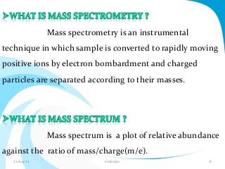
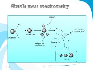
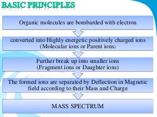
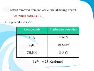
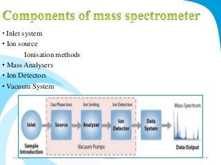
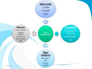
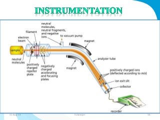
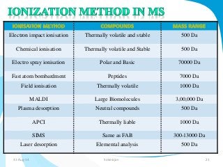
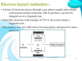
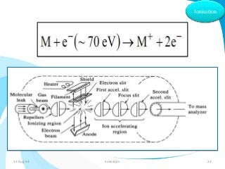
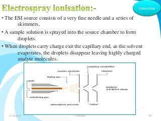
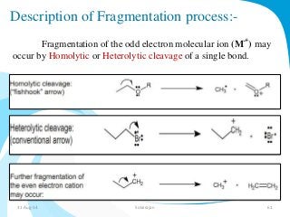
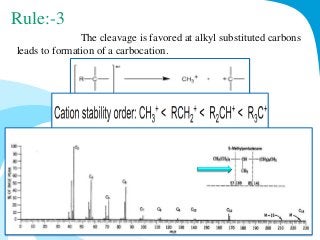
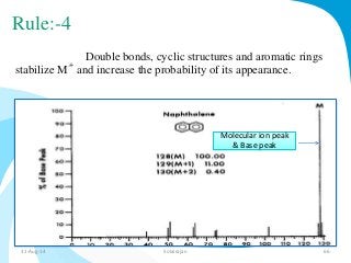
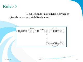
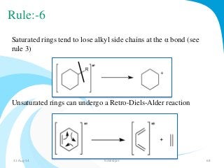
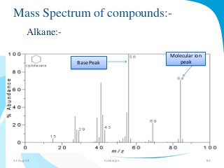
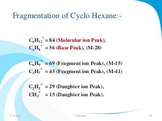
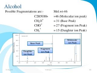
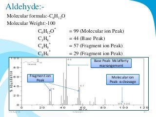
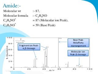
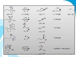
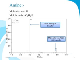
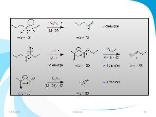
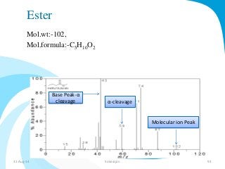
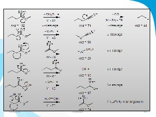
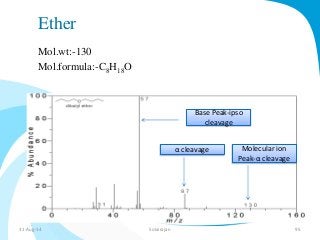
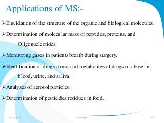
MASS SPECTROMETRY 
A.Solairajan, 
M.Pharm,1st year, 
S.B.C.P. 
31-Aug-14 Solairajan 1
What is Mass 
Number? 
Zoo Zoo 
31-Aug-14 Solairajan 2
Mass number:- 
The Mass number (A), also called atomic mass number or nucleon 
number is the total number of protons and neutrons in an atomic 
nucleus. 
No.of protons 
(Z) 
No.of 
neutrons (N) 
Mass Number (A) 
31-Aug-14 Solairajan 3
Example:- 
Carbon atom have 6 protons and 6 neutrons in the centre of 
the nucleus, 2 electrons situated in the inner orbital where as 
other 4 electrons are outside the orbital. 
We can represent carbon atom like 
Mass 
Number(A) 
No. of protons(Z) 
Carbon Atom:- 
31-Aug-14 Solairajan 4
31-Aug-14 Solairajan 5
31-Aug-14 Solairajan 6
31-Aug-14 Solairajan 7
Mass spectrometry is an instrumental 
technique in which sample is converted to rapidly moving 
positive ions by electron bombardment and charged 
particles are separated according to their masses. 
Mass spectrum is a plot of relative abundance 
against the ratio of mass/charge(m/e). 
31-Aug-14 Solairajan 8
31-Aug-14 Solairajan 9
•To measure relative molecular masses. 
• To know the fragmentation of the molecules. 
• Comparison of mass spectra with known 
compounds. 
31-Aug-14 Solairajan 10
Organic molecules are bombarded with electron 
converted into Highly energetic positively charged ions 
(Molecular ions or Parent ions) 
Further break up into smaller ions 
(Fragment ions or Daughter ions) 
The formed ions are separated by Deflection in Magnetic 
field according to their Mass and Charge 
MASS SPECTRUM 
31-Aug-14 Solairajan 11
Loss of electron from a molecule leads to radical cation. 
e- 
Molecular ion 
15 eV 
70 eV 
31-Aug-14 Solairajan 12
 Electron removed from molecule orbital having lowest 
ionization potential (IP). 
 In general n < π < σ 
Compounds Ionization potential 
CH4 12.6 eV 
C2H4 10.52 eV 
CH3NH2 10.3 eV 
1 eV = 23 Kcal/mol 
31-Aug-14 Solairajan 13
• Inlet system 
• Ion source 
Ionisation methods 
• Mass Analysers 
• Ion Detectors 
• Vacuum System 
31-Aug-14 Solairajan 14
Inlet system 
1.Solid 
2.Liquid 
3.Gas 
Mass 
spectrometer 
Ionisation 
ESI,EI,FAB, 
CI,FI,FD, 
MALDI,PD, 
TI 
Mass 
Analyser 
Quadrupole, 
TOF, 
Ion trap, 
FT-ICR 
Detector 
Photo multiplier, 
Electron 
multiplier, 
Faraday cup, 
Microchannel 
plate 
31-Aug-14 Solairajan 15
31-Aug-14 Solairajan 16
• SOLIDS SAMPLES with lower vapour pressure 
Inlet 
system 
directly inserted into the ionization chamber and volatilization 
is controlled by heating the probe. 
• LIQUIDS are handled by hypodermic needles 
injection through a silicon rubber dam. 
• GASES SAMPLES are leaked into the ionisation chamber 
directly by the help of mercury manometer. 
31-Aug-14 Solairajan 17
Ionisation 
 The ion source is the part of the mass spectrometer that ionizes the 
material under analysis (the analyte). 
 The ions are then transported by magnetic or electric fields to the 
mass analyzer. 
 Molecular ions are formed when energy of the electron beam 
reaches to 10-15 eV. 
 Fragmentation of the ion reaches only at higher bombardment 
energies at 70 eV. 
31-Aug-14 Solairajan 18
. 
Samples are ionised outside the ion source. 
This technique include, 
1.Electron impact ionization (EIS) 
2.Chemical ionization.(CI) 
3.Field ionisation.(FI) 
Samples are ionised inside the ion source. 
This technique include, 
1. Field desorption.(FD) 
2. Fast atom bombardment.(FAB) 
Ionisation 
31-Aug-314. Laser desorption.(LD) Solairajan 19
Ionisation 
31-Aug-14 Solairajan 20
Electron impact ionisation Thermally volatile and stable 500 Da 
Chemical ionisation Thermally volatile and Stable 500 Da 
Electro spray ionisation Polar and Basic 70000 Da 
Fast atom bombardment Peptides 7000 Da 
Field ionisation Thermally volatile 1000 Da 
MALDI Large Biomolecules 3,00,000 Da 
Plasma desorption Neutral compounds 500 Da 
APCI Thermally liable 1000 Da 
SIMS Same as FAB 300-13000 Da 
Laser desorption Elemental analysis 500 Da 
31-Aug-14 Solairajan 21
Ionisation 
• A beam of electrons passes through a gas-phase sample and collides 
with neutral analyte molecules (M) to produce a positively 
charged ion or a fragment ion. 
• Generally electrons with energies of 70 eV are used to form a 
fragment ions. 
• The positive ions are collected in focusing plates and passed to mass 
analyzer. 
31-Aug-14 Solairajan 22
Ionisation 
31-Aug-14 Solairajan 23
• The ESI source consists of a very fine needle and a series of 
skimmers. 
• A sample solution is sprayed into the source chamber to form 
droplets. 
Ionisation 
•When droplets carry charge exit the capillary end, as the solvent 
evaporates, the droplets disappear leaving highly charged 
analyte molecules. 
31-Aug-14 Solairajan 24
Ionisation 
• Chemical Impact Ionisation between interactions of sample 
with large amount of reagent gas. 
• Commonly used reagent gases include 
methane,ammonia,isobutane. 
• Oxygen and hydrogens are used in Negative ion chemical 
ionisation in MS. 
31-Aug-14 Solairajan 25
Ionisation 
The vaporised sample is introduced into the mass spectrometer with 
an excess of a reagent gas (methane) at pressure of about 1 torr. 
The excess carrier gas is ionized by electron impact to the primary 
.+ 
+ 
ions CHand CH. 
4 
3 
These may react with the excess methane to give secondary ions. 
31-Aug-14 Solairajan 26
• In this method the molecule pass through sharp metal anode 
carrying an electric field of 1010 v m-1 
• Electrons are analysed in primary focusing cathode slit. 
• ADV :- abundance of molecular ions. 
• DISADV :- lower resolution. 
Ionisation 
31-Aug-14 Solairajan 27
Ionisation 
• Useful for nonvolatile and thermolabile compounds. 
• Sample is applied to field ion emitter and the solvent allowed to 
evaporate. 
• Evaporated sample that leads to chemical ionisation or EIS. 
• Example:-Nucleotides & Quarternary ammonium compounds. 
31-Aug-14 Solairajan 28
• Argon gas ionised by hot filament and focused beam that 
bombards the sample. 
Ionisation 
• Beam impinges the sample, a series of molecular reactions occur 
and analyse in MS analyser. 
• Ex:-Insulin,Amino glycosides,Phospholipids. 
31-Aug-14 Solairajan 29
Ionisation 
• Sample is coated with a high energetic fragment Californium 252. 
• This fission fragment desorbs positive,negative, and neutral molecules. 
• 252cf generates 1012 power at 10,000k, this may ionise the target 
molecule. 
• This method involves the interaction of laser beam with sample to 
produce both vaporisation and ionisation. 
• The vaporised sample passed to mass spectrometers for analysis. 
• Appl:-used for elemental analysis. 31-Aug-14 Solairajan 30
Ionisation 
 MALDI is a LIMS method of vaporizing and ionizing and sample 
molecules are dispersed in a solid matrix such as nicotinic acid. 
 A UV laser pulse ablates the matrix which carries some of the 
large molecules into the gas phase in an ionized form so they can be 
extracted into a mass spectrometer. 
MALDI 
31-Aug-14 Solairajan 31
Atmospheric pressure chemical ionisation (APCI) is an 
analogous ionisation method to chemical ionisation(CI). 
Corona discharge is used to ionize the analyte in the 
atmospheric pressure region. 
Ionisation 
31-Aug-14 Solairajan 32
Ionisation 
 Secondary ion mass spectrometry (SIMS) is based on the 
observation that charged particles (Secondary Ions) are ejected 
from a sample surface when bombarded by a primary beam of 
heavy particles. 
 Primary beam species useful in SIMS include Cs+, O2+, O , 
Ar+, and Ga+ 
31-Aug-14 Solairajan 33
An ion, after leaving ion source, the ions are separated 
according to their m/e ratio. 
Mass 
Analyzer 
In this area, the ions are accelerated by both electrostatic 
and magnetically 
Types:- 
Magnetic sector mass analysers 
Double focussing analysers 
Quadrupole mass analysers 
Time of Flight analysers (TOF) 
Ion trap analyser 
Ion cyclotron analyser 
31-Aug-14 Solairajan 34
m/z= H2r2/2V 
H Magnetic field 
R Radius of the curvature 
Mass 
Analyzer 
31-Aug-1V4 AppliedSo lavirajoan ltage 35
It contains two analysers namely 
•Electrostatic analyser 
•Magnetic sector analyser. 
Mass 
Analyzer 
31-Aug-14 Solairajan 36
Mass 
Analyzer 
The quadrupole consists of two pairs of parallel rods with applied 
DC and RF voltages. 
Ions are scanned by varying the DC/Rf quadrupole voltages. 
31-Aug-14 Solairajan 37
Mass 
Analyzer 
31-Aug-14 Solairajan 38
Mass 
Analyzer 
TOF analyzer – ions are accelerated through a flight tube and 
the time of fight to the detector is measured. 
Typical flight times are 1 to 50μs. 
31-Aug-14 Solairajan 39
The quadrupole ion trap typically consists of a ring 
electrode and two hyperbolic end cap electrodes. 
Mass 
Analyzer 
As the radio frequency voltage is increased, the orbits of 
heavier ions become stabilised,and passed into the detector. 
31-Aug-14 Solairajan 40
Mass 
Analyzer 
 Fourier transform-ICR mass spectrometry, is a type of mass 
analyzer for determining the mass-to-charge ratio (m/z) of ions 
based on the cyclotron frequency of the ions in a fixed magnetic 
field. 
The ions are trapped in a Penning trap(a magnetic field with 
electric trapping plates) where they are excited to a larger 
cyclotron radius by an oscillating electric field perpendicular to the 
magnetic field. 
31-Aug-14 Solairajan 41
The excitation also results in the ions moving in phase (in a packet). 
The signal is detected as an image current on a pair of plates which the 
packet of ions passes close to as they cyclotron. The resulting signal is 
called a free induction decay (FID), transient or interferogram that 
converts signal. 
The useful signal is extracted from this data by performing a Fourier 
transform to give a mass spectrum. 
31-Aug-14 Solairajan 42
31-Aug-14 Solairajan 43
Faraday cup 
Electron Multiplier 
photomultiplier 
Micro Channel Plate 
Detector 
31-Aug-14 Solairajan 44
Detector 
 The basic principle is that the incident ion strikes the dynode 
surface which emits electrons and induces a current which is 
amplified and recorded. 
 The dynode electrode is made of a secondary emitting 
material like CsSb, GaP or BeO. 
 It is ideally suited to isotope analysis. 
Faradaycup 
31-Aug-14 Solairajan 45
Detector 
 Electron multipliers are the most common especially when 
positive and negative ions need to be detected on the same 
instrument. 
 Dynodes made up of copper-beryllium which transduces the 
initial ion current ,and electron emitted by first dynode are focused 
magnetically from dynode to the next. 
 Final cascade current is amplified more than million times. 
Electron 
multipliers 
31-Aug-14 Solairajan 46
Detector 
 The dynode consists of a substance( a scintillator) which emits 
photons(light). 
 The emitted light is detected by photo multiplier tube and is 
converted into electric current. 
 These detectors are useful in studies on metastable ions 
31-Aug-14 Solairajan 47
Detector 
31-Aug-14 Solairajan 48
All mass spectrometers need a vacuum to allow ions to 
reach the detector without colliding with other gaseous molecules 
or atoms. If such collisions did occur, the instrument would suffer 
from reduced resolution and sensitivity. 
31-Aug-14 Solairajan 49
TYPES OF PEAKS IN MS 
•Molecular ion peak 
•Fragment ions peak 
•Rearrangement ions peak 
•Metastable ion peaks 
•Multicharged ions 
•Base peak 
•Negative ion peak 
Molecular ion Peak:- 
When a sample is bombarded with electrons of 9 to 15 eV 
energy, the molecular ion is produced, by loss of single electron. 
M e- 
M 
+ 
+ 2 e 
- 
31-Aug-14 Solairajan 50
Fragment ions Peak:- 
when an energy is given further more upto 70 eV, 
fragment ions produced, it have lower mass number. 
Rearrangement ion Peak:- 
Recombination of fragment ion is known as 
Rearrangement Peaks. 
Metastable ion Peak:- 
The ions resulting from the decomposition between the 
source region and magnetic analyser are called as Meta stable 
ions.These appear as broad peaks called Metastable ion Peaks. 
Multicharged ions:- 
Ions may exist with 2 or 3 charges instead of usual 
single charge.The peaks due to these charged ions are known as 
Multicharged ion peaks. 
31-Aug-14 Solairajan 51
Base Peak:- 
The largest peak in the mass spectrum corresponding 
to the most abundant ion or most intense peak in the spectrum is 
called as Base Peak. 
Negative ion Peak:- 
Negative ions are formed from electron bombardment 
of sample. These results due to the capture of electron by a molecule 
during collision of molecules 
Fragment ion 
peak 
31-Aug-14 Solairajan 52
FRAGMENTATION 
Fragmentation is a type of chemical dissociation. 
Fragmentation takes place by a process of heterolysis or homolysis. 
Types of Fragmentation:- 
• Collision induced dissociation(CID) 
• Electron capture dissociation(ECD) 
• Electron transfer dissociation(ETD) 
• Electron detachment dissociation(EDD) 
• Photo dissociation 
Infrared multiphoton dissociation(IRMPD) 
Blackbody infrared radiative dissociation(BIRD) 
• Surface induced dissociation(SID) 
• Charge remote fragmentation 
• Higher energy C-trap dissociation(HCD) 
31-Aug-14 Solairajan 53
Collision Induced Dissociation 
 Molecular ions are accelerated by electrical potential to high 
kinetic energy and then allowed to collide with neutral molecules like 
He,N or Ar. 
 Collision between these molecules leads to bond breakage and 
formation of fragment ions. 
 These fragment ions are analysed by mass spectrometer. 
 Example:- Triple quadrupole spectrometer produces CID 
fragments. 
31-Aug-14 Solairajan 54
SORI-CID:-(Sustained Off-Resonance Irradiation Collision-Induced 
Dissociation) 
 It is one of CID technique used in Fourier transform ion 
cyclotron resonance mass spectrometry. 
 In this method accelerating ions in cyclotron motion and 
increasing the pressure resulting collisions produce CID fragments. 
31-Aug-14 Solairajan 55
Electron Capture Dissociation:- 
 It is a method of fragmenting gas phase ions for tandem mass 
spectrometric analysis (structural elucidation). 
 ECD involves the direct introduction of low energy electrons to 
trapped gas phase ions. 
 Electron-capture dissociation typically involves a multiply 
protonated molecule M interacting with a free electron to form an odd-electron 
ion. 
31-Aug-14 Solairajan 56
Electron Transfer Dissociation:- 
ETD induces fragmentation of cations by transferring electrons to them. 
Example:-peptides or proteins. 
Electron Detachment Dissociation:- 
EDD is a method for fragmenting anionic species in mass spectrometry. 
31-Aug-14 Solairajan 57
Photo Dissociation:- 
Photodissociation is a chemical reaction in which a chemical compound is 
broken down by photons. 
IRMPD:- 
Absorption of multiple infra red photons by a molecule and leads to 
dissociation. 
BIRD:- 
Long interaction of molecule with radiation field like carbon dioxide laser. 
Surface-induced dissociation:-(SID) 
It is a technique used in mass spectrometry to fragment molecular ions in the 
gas phase by collision of an ion with a surface under high vacuum. 
31-Aug-14 Solairajan 58
Charge Remote Fragmentation:- 
It is a type of covalent bond breaking that occurs in a gas 
phase ion in which the cleaved bond is not adjacent to the location of the 
charge. 
This fragmentation can be observed using tandem mass 
spectrometry. 
Higher-energy C-trap dissociation:-(HCD) 
It is a fragmentation technique, used 
for peptide modification analysis. 
Immonium ions generated via HCD pinpoint modifications such 
as phospho tyrosine. 
An added octopole collision cell facilitates de novo sequencing. 
31-Aug-14 Solairajan 59
Fragmentation of the Molecular ion 
Fragmentation of a molecular ion, M, produces a 
radical and a cation. 
-Only the cation is detected by MS. 
A-B 
A 
Radical Cation 
+ 
A 
B 
B 
• + 
Molecular ion 
(a radical cation) 
+ • 
+ • 
+ 
Cation Radical 
31-Aug-14 Solairajan 60
Description of Fragmentation process:- 
Fragmentation of the odd electron molecular ion (M 
.+ 
) may 
occur by Homolytic or Heterolytic cleavage of a single bond. 
31-Aug-14 Solairajan 61
Mass interpretation 
Fragmentation rules 
Mclafferty rearrangement 
Alpha cleavage 
Beta cleavage 
Nitrogen rule 
Retro diels alder reaction 
IHD 
31-Aug-14 Solairajan 62
Fragmentation rules:- (9 rules) 
Rule:-1 
The height of the M 
.+ 
peak decreases with increasing degree 
of branching. 
31-Aug-14 Solairajan 63
Rule:-2 
The height of the M 
.+ 
Peak decreases with 
increasing molecular weight. 
Example:- Fatty molecules, steroids. 
31-Aug-14 Solairajan 64
Rule:-3 
The cleavage is favored at alkyl substituted carbons 
leads to formation of a carbocation. 
31-Aug-14 Solairajan 65
Rule:-4 
Double bonds, cyclic structures and aromatic rings 
stabilize M 
.+ 
and increase the probability of its appearance. 
Molecular ion peak 
& Base peak 
31-Aug-14 Solairajan 66
Rule:-5 
Double bonds favor allylic cleavage to 
give the resonance stabilized cation. 
31-Aug-14 Solairajan 67
Rule:-6 
Saturated rings tend to lose alkyl side chains at the α bond (see 
rule 3) 
Unsaturated rings can undergo a Retro-Diels-Alder reaction 
31-Aug-14 Solairajan 68
Rule:-7 
Alkyl substituted aromatic compounds are cleaved 
preferably at the β bond to the ring, giving the resonance stabilized 
benzyl ion (or) tropyllium ion. 
31-Aug-14 Solairajan 69
Rule:-8 
C-C bonds next to hetero atom are frequently 
cleaved, leaving the charge on the hetero atom (resonance 
stabilization). 
31-Aug-14 Solairajan 70
Rule:-9 
Cleavage is often associated with elimination of small 
stable, neutral molecules, such as 
CO,olefins,water,ammonia,H2S,HCN,ketene or alcohols (often with 
rearrangements) 
Ex:-Mclafferty rearrangement 
31-Aug-14 Solairajan 71
Mclafferty Rearrangement:- 
 Mclafferty arrangement can occur in 
ketones,aldehydes,carboxylic acids, and esters. 
 In this rearrangement a radical center in molecular ion derived 
from a lone pair or pi bond, removes hydrogen from the Gamma 
position(γ), a pi bond is formed between the β and γ position, and 
the bond between the α and β positions is broken. 
31-Aug-14 Solairajan 72
α cleavage 
 Alpha cleavage in mass spectrometry is a 
characteristic fragmentation of the molecular ion derived 
from carbonyl compounds, in which the bond linking the carbonyl 
carbon to the atom occupying an alpha position breaks. 
It is an expected pathway for carbonyl 
compounds,ethers,halides,alcohols,and amines. 
31-Aug-14 Solairajan 73
31-Aug-14 Solairajan 74
β cleavage 
Beta cleavage in mass spectrometry is 
a characteristic fragmentation of the molecular ion derived 
from some organic compounds, most 
notably alcohols, ethers, and amines, in which the bond 
connecting alpha- and beta-carbons break. 
31-Aug-14 Solairajan 75
31-Aug-14 Solairajan 76
Retro-Diels-Alder reactions:- 
 Retro Diels-Alder fragmentation occurs in 3-cyano-cyclohexene, 
lets first look at the fragmentation of cyclohexene. 
 First ionization occurs and electrons from the double bond 
transfer to an adjacent carbon and an electron from the bond 
between the 3 and 4 carbons transfers to form a second double bond 
that is conjugate with the first one. 
These rearrangements cleave the molecule between the 3 and 4 
carbon and 5 and 6 (where another electron is transferred to form a 
double bond between the 4 and 5 carbons). This leaves an olefin and 
a diene. 
31-Aug-14 Solairajan 77
3 
6 
2 
1 
4 
5 
4 
5 
Diene Olefin 
31-Aug-14 Solairajan 78
Nitrogen rule:- 
The nitrogen rule states, that a molecule that has no or even 
number of nitrogen atoms has an even nominal mass, whereas a 
molecule that has an odd number of nitrogen atoms has an odd 
nominal mass. 
Example:-1 Example:-2 
31-Aug-14 Solairajan 79
Contd…. 
The molecular ion appears at m/z 121, indicating an odd number 
of nitrogen atoms in the structure. 
Odd number of 
molecular ion 
31-Aug-14 Solairajan 80
IHD:- 
 In a hydrocarbon where all carbon atoms have only single bonds 
and no rings are involved, the compound would have maximum 
number of H atoms. 
 If any of the bonds are replaced with double or triple bonds, there 
would be deficiency of H atoms. By calculating the index of 
hydrogen deficiency(IHD), we can calculate molecular formula and 
how many multiple bonds and rings are involved. IHD is also called 
the Degree of Unsaturation. 
 A double bond and ring each counts as one IHD. 
 A triple bond counts as two IHD. 
31-Aug-14 Solairajan 81
Example:-1 
Example:-2 
CH2=CH2 
31-Aug-14 Solairajan 82
31-Aug-14 Solairajan 83
Mass Spectrum of compounds:- 
Alkane:- 
Base Peak 
Molecular ion 
peak 
31-Aug-14 Solairajan 84
Fragmentation of Cyclo Hexane:- 
C6H12 
+ 
= 84 (Molecular ion Peak), 
C4H8 
+ 
= 56 (Base Peak), (M-28) 
C6H9 
+ 
= 69 (Fragment ion Peak), (M-15) 
C3H7 
+ 
= 43 (Fragment ion Peak), (M-41) 
C2H5 
+ 
= 29 (Daughter ion Peak), 
CH3 
+ 
= 15 (Daughter ion Peak). 
31-Aug-14 Solairajan 85
Alcohol 
Possible Fragmentations are:- Mol.wt-46 
C2H5OH+ =46 (Molecular ion peak) 
+ 
CHO 
= 31 (Base Peak) 
3+ 
CHO 
= 27 (Fragment ion Peak) 
CH3 
+ 
= 15 (Daughter ion Peak) 
Base Peak 
Molecular 
ion Peak 
Fragment 
Daughter ion Peak 
ion Peak 
31-Aug-14 Solairajan 86
Aldehyde:- 
Molecular formula:-C6H12O 
Molecular Weight:-100 
+ 
C6H12O 
= 99 (Molecular ion Peak) 
C3H8 
+ 
= 44 (Base Peak) 
C4H9 
+ 
= 57 (Fragment ion Peak) 
C2H5 
+ 
= 29 (Fragment ion Peak) 
Fragment ion 
Peak 
Base Peak- Mclafferty 
rearrangement 
Molecular ion 
Peak- α cleavage 
31-Aug-14 Solairajan 87
31-Aug-14 Solairajan 88
Amide:- 
Molecular wt :- 87, 
Molecular formula :- CHNO 
49+ 
CHNO 
49= 87 (Molecular ion Peak), 
+ 
C2H5NO 
= 59 (Base Peak) 
Fragment ion Peak- 
α,β cleavage 
Base Peak- 
Mclafferty 
rearrangement 
Molecular ion 
Peak-β cleavage 
31-Aug-14 Solairajan 89
31-Aug-14 Solairajan 90
Amine:- 
Molecular wt:-59 
Mol.formula :-C3H9N 
Base Peak-β-H 
transfer 
Molecular ion Peak- 
β-H transfer 
31-Aug-14 Solairajan 91
31-Aug-14 Solairajan 92
Ester 
Mol.wt:-102, 
Mol.formula:-C5H10O2 
Base Peak-α 
cleavage 
Molecular ion Peak 
α-cleavage 
31-Aug-14 Solairajan 93
31-Aug-14 Solairajan 94
Ether 
Mol.wt:-130 
Mol.formula:-C8H18O 
Base Peak-ipso 
cleavage 
α cleavage Molecular ion 
Peak-α cleavage 
31-Aug-14 Solairajan 95
31-Aug-14 Solairajan 96
GC-MS 
Gas chromatography–mass spectrometry (GC-MS) is a 
method that combines the features of gas-liquid chromatography 
and mass spectrometry to identify different substances within a test 
sample. 
Applications of GC-MS include :- 
Drug detection, 
Fire investigation, 
Environmental analysis, 
Explosives investigation, and 
Identification of unknown samples. 
31-Aug-14 Solairajan 97
GC-MS 
31-Aug-14 Solairajan 98
Tandem MS:- 
What is Tandem MS:- 
-Uses 2 (or more) mass analyzers in a single instrument. 
-One purifies the analyte ion from a mixture using a 
magnetic field. 
-The other analyzes fragments of the analyte ion for 
identification and quantification. 
31-Aug-14 Solairajan 99
31-Aug-14 Solairajan 100
Tandem mass spectrometry, also known 
as MS/MS or MS2, involves multiple steps of mass spectrometry 
selection, with some form of fragmentation occurring in between 
the stages. 
31-Aug-14 Solairajan 101
Components of Tandem Mass 
Spectrometer 
MS-1 Collision cell MS-2 
31-Aug-14 Solairajan 102
Applications of Tandem MS 
Biotechnology & Pharmaceutical 
 To determine chemical structure of drugs and drug metabolites. 
 Detection/quantification of impurities, drugs and their metabolites 
in biological fluids and tissues. 
 Analysis of liquid mixtures 
 Fingerprinting 
Nutraceuticals/herbal drugs/tracing source of natural products 
or drugs 
Clinical testing & Toxicology 
 Inborn errors of metabolism, cancer, diabetes, various poisons, 
drugs of abuse, etc. 
31-Aug-14 Solairajan 103
MALDI-MS 
Matrix-assisted laser desorption/ionization (MALDI) is a 
soft ionization technique used in mass spectrometry allowing the 
analysis of biomolecules (biopolymers such 
as DNA, proteins, peptides and sugars) and 
large organic molecules (such as polymers, dendrimers and 
other macromolecules). 
31-Aug-14 Solairajan 104
 MALDI is based on the bombardment of sample molecules 
with a laser light to bring about sample ionisation. 
 The sample is pre-mixed with a highly 
absorbing matrix compound for the most consistent and reliable 
results. 
 The matrix transforms the laser energy into excitation 
energy for the sample, which leads to sputtering of analyte and 
matrix ions from the surface of the mixture. 
 Most commercially available MALDI mass spectrometers 
now have a pulsed nitrogen laser of wavelength 337 nm. 
31-Aug-14 Solairajan 105
Common matrix in MALDI 
Matrix Solvent Applications 
2,5-dihydroxy benzoic 
acid 
Acetonitrile,water,metha 
nol,acetone,CHcl3 
Peptides,Nucleotides, 
oligo nucleotides 
3,5-dimethoxy-4- 
hydroxycinnamic acid 
Acetonitrile, 
water,acetone, CHcl3 
Peptides,proteins,lipids 
4-hydroxy-3- 
methoxycinnamic acid 
Acetonitrile, water, 
propanol 
Proteins 
Picolinic acid Ethanol Oligo nucleotides 
31-Aug-14 Solairajan 106
31-Aug-14 Solairajan 107
31-Aug-14 Solairajan 108
Applications of MS:- 
Elucidation of the structure of the organic and biological molecules. 
Determination of molecular mass of peptides, proteins, and 
Oligonucleotides. 
Monitoring gases in patients breath during surgery. 
Identification of drugs abuse and metabolites of drugs of abuse in 
blood, urine, and saliva. 
Analyses of aerosol particles. 
Determination of pesticides residues in food. 
31-Aug-14 Solairajan 109
References:- 
Instrumental methods of chemical analysis by willard 
Organic spectroscopy by William kemp 
Spectroscopic identification of organic compounds by Silverstein 
Instrumental analysis by skoog 
Wikipedia 
ww2.chemistry.gatech.edu/class/4341-6371/fahrni/set02.pdf 
31-Aug-14 Solairajan 110
By 
A.Solairajan 
31-Aug-14 Solairajan 111

Mass spectrometry

  • 1.
    MASS SPECTROMETRY A.Solairajan, M.Pharm,1st year, S.B.C.P. 31-Aug-14 Solairajan 1
  • 2.
    What is Mass Number? Zoo Zoo 31-Aug-14 Solairajan 2
  • 3.
    Mass number:- TheMass number (A), also called atomic mass number or nucleon number is the total number of protons and neutrons in an atomic nucleus. No.of protons (Z) No.of neutrons (N) Mass Number (A) 31-Aug-14 Solairajan 3
  • 4.
    Example:- Carbon atomhave 6 protons and 6 neutrons in the centre of the nucleus, 2 electrons situated in the inner orbital where as other 4 electrons are outside the orbital. We can represent carbon atom like Mass Number(A) No. of protons(Z) Carbon Atom:- 31-Aug-14 Solairajan 4
  • 5.
  • 6.
  • 7.
  • 8.
    Mass spectrometry isan instrumental technique in which sample is converted to rapidly moving positive ions by electron bombardment and charged particles are separated according to their masses. Mass spectrum is a plot of relative abundance against the ratio of mass/charge(m/e). 31-Aug-14 Solairajan 8
  • 9.
  • 10.
    •To measure relativemolecular masses. • To know the fragmentation of the molecules. • Comparison of mass spectra with known compounds. 31-Aug-14 Solairajan 10
  • 11.
    Organic molecules arebombarded with electron converted into Highly energetic positively charged ions (Molecular ions or Parent ions) Further break up into smaller ions (Fragment ions or Daughter ions) The formed ions are separated by Deflection in Magnetic field according to their Mass and Charge MASS SPECTRUM 31-Aug-14 Solairajan 11
  • 12.
    Loss of electronfrom a molecule leads to radical cation. e- Molecular ion 15 eV 70 eV 31-Aug-14 Solairajan 12
  • 13.
     Electron removedfrom molecule orbital having lowest ionization potential (IP).  In general n < π < σ Compounds Ionization potential CH4 12.6 eV C2H4 10.52 eV CH3NH2 10.3 eV 1 eV = 23 Kcal/mol 31-Aug-14 Solairajan 13
  • 14.
    • Inlet system • Ion source Ionisation methods • Mass Analysers • Ion Detectors • Vacuum System 31-Aug-14 Solairajan 14
  • 15.
    Inlet system 1.Solid 2.Liquid 3.Gas Mass spectrometer Ionisation ESI,EI,FAB, CI,FI,FD, MALDI,PD, TI Mass Analyser Quadrupole, TOF, Ion trap, FT-ICR Detector Photo multiplier, Electron multiplier, Faraday cup, Microchannel plate 31-Aug-14 Solairajan 15
  • 16.
  • 17.
    • SOLIDS SAMPLESwith lower vapour pressure Inlet system directly inserted into the ionization chamber and volatilization is controlled by heating the probe. • LIQUIDS are handled by hypodermic needles injection through a silicon rubber dam. • GASES SAMPLES are leaked into the ionisation chamber directly by the help of mercury manometer. 31-Aug-14 Solairajan 17
  • 18.
    Ionisation  Theion source is the part of the mass spectrometer that ionizes the material under analysis (the analyte).  The ions are then transported by magnetic or electric fields to the mass analyzer.  Molecular ions are formed when energy of the electron beam reaches to 10-15 eV.  Fragmentation of the ion reaches only at higher bombardment energies at 70 eV. 31-Aug-14 Solairajan 18
  • 19.
    . Samples areionised outside the ion source. This technique include, 1.Electron impact ionization (EIS) 2.Chemical ionization.(CI) 3.Field ionisation.(FI) Samples are ionised inside the ion source. This technique include, 1. Field desorption.(FD) 2. Fast atom bombardment.(FAB) Ionisation 31-Aug-314. Laser desorption.(LD) Solairajan 19
  • 20.
  • 21.
    Electron impact ionisationThermally volatile and stable 500 Da Chemical ionisation Thermally volatile and Stable 500 Da Electro spray ionisation Polar and Basic 70000 Da Fast atom bombardment Peptides 7000 Da Field ionisation Thermally volatile 1000 Da MALDI Large Biomolecules 3,00,000 Da Plasma desorption Neutral compounds 500 Da APCI Thermally liable 1000 Da SIMS Same as FAB 300-13000 Da Laser desorption Elemental analysis 500 Da 31-Aug-14 Solairajan 21
  • 22.
    Ionisation • Abeam of electrons passes through a gas-phase sample and collides with neutral analyte molecules (M) to produce a positively charged ion or a fragment ion. • Generally electrons with energies of 70 eV are used to form a fragment ions. • The positive ions are collected in focusing plates and passed to mass analyzer. 31-Aug-14 Solairajan 22
  • 23.
  • 24.
    • The ESIsource consists of a very fine needle and a series of skimmers. • A sample solution is sprayed into the source chamber to form droplets. Ionisation •When droplets carry charge exit the capillary end, as the solvent evaporates, the droplets disappear leaving highly charged analyte molecules. 31-Aug-14 Solairajan 24
  • 25.
    Ionisation • ChemicalImpact Ionisation between interactions of sample with large amount of reagent gas. • Commonly used reagent gases include methane,ammonia,isobutane. • Oxygen and hydrogens are used in Negative ion chemical ionisation in MS. 31-Aug-14 Solairajan 25
  • 26.
    Ionisation The vaporisedsample is introduced into the mass spectrometer with an excess of a reagent gas (methane) at pressure of about 1 torr. The excess carrier gas is ionized by electron impact to the primary .+ + ions CHand CH. 4 3 These may react with the excess methane to give secondary ions. 31-Aug-14 Solairajan 26
  • 27.
    • In thismethod the molecule pass through sharp metal anode carrying an electric field of 1010 v m-1 • Electrons are analysed in primary focusing cathode slit. • ADV :- abundance of molecular ions. • DISADV :- lower resolution. Ionisation 31-Aug-14 Solairajan 27
  • 28.
    Ionisation • Usefulfor nonvolatile and thermolabile compounds. • Sample is applied to field ion emitter and the solvent allowed to evaporate. • Evaporated sample that leads to chemical ionisation or EIS. • Example:-Nucleotides & Quarternary ammonium compounds. 31-Aug-14 Solairajan 28
  • 29.
    • Argon gasionised by hot filament and focused beam that bombards the sample. Ionisation • Beam impinges the sample, a series of molecular reactions occur and analyse in MS analyser. • Ex:-Insulin,Amino glycosides,Phospholipids. 31-Aug-14 Solairajan 29
  • 30.
    Ionisation • Sampleis coated with a high energetic fragment Californium 252. • This fission fragment desorbs positive,negative, and neutral molecules. • 252cf generates 1012 power at 10,000k, this may ionise the target molecule. • This method involves the interaction of laser beam with sample to produce both vaporisation and ionisation. • The vaporised sample passed to mass spectrometers for analysis. • Appl:-used for elemental analysis. 31-Aug-14 Solairajan 30
  • 31.
    Ionisation  MALDIis a LIMS method of vaporizing and ionizing and sample molecules are dispersed in a solid matrix such as nicotinic acid.  A UV laser pulse ablates the matrix which carries some of the large molecules into the gas phase in an ionized form so they can be extracted into a mass spectrometer. MALDI 31-Aug-14 Solairajan 31
  • 32.
    Atmospheric pressure chemicalionisation (APCI) is an analogous ionisation method to chemical ionisation(CI). Corona discharge is used to ionize the analyte in the atmospheric pressure region. Ionisation 31-Aug-14 Solairajan 32
  • 33.
    Ionisation  Secondaryion mass spectrometry (SIMS) is based on the observation that charged particles (Secondary Ions) are ejected from a sample surface when bombarded by a primary beam of heavy particles.  Primary beam species useful in SIMS include Cs+, O2+, O , Ar+, and Ga+ 31-Aug-14 Solairajan 33
  • 34.
    An ion, afterleaving ion source, the ions are separated according to their m/e ratio. Mass Analyzer In this area, the ions are accelerated by both electrostatic and magnetically Types:- Magnetic sector mass analysers Double focussing analysers Quadrupole mass analysers Time of Flight analysers (TOF) Ion trap analyser Ion cyclotron analyser 31-Aug-14 Solairajan 34
  • 35.
    m/z= H2r2/2V HMagnetic field R Radius of the curvature Mass Analyzer 31-Aug-1V4 AppliedSo lavirajoan ltage 35
  • 36.
    It contains twoanalysers namely •Electrostatic analyser •Magnetic sector analyser. Mass Analyzer 31-Aug-14 Solairajan 36
  • 37.
    Mass Analyzer Thequadrupole consists of two pairs of parallel rods with applied DC and RF voltages. Ions are scanned by varying the DC/Rf quadrupole voltages. 31-Aug-14 Solairajan 37
  • 38.
  • 39.
    Mass Analyzer TOFanalyzer – ions are accelerated through a flight tube and the time of fight to the detector is measured. Typical flight times are 1 to 50μs. 31-Aug-14 Solairajan 39
  • 40.
    The quadrupole iontrap typically consists of a ring electrode and two hyperbolic end cap electrodes. Mass Analyzer As the radio frequency voltage is increased, the orbits of heavier ions become stabilised,and passed into the detector. 31-Aug-14 Solairajan 40
  • 41.
    Mass Analyzer Fourier transform-ICR mass spectrometry, is a type of mass analyzer for determining the mass-to-charge ratio (m/z) of ions based on the cyclotron frequency of the ions in a fixed magnetic field. The ions are trapped in a Penning trap(a magnetic field with electric trapping plates) where they are excited to a larger cyclotron radius by an oscillating electric field perpendicular to the magnetic field. 31-Aug-14 Solairajan 41
  • 42.
    The excitation alsoresults in the ions moving in phase (in a packet). The signal is detected as an image current on a pair of plates which the packet of ions passes close to as they cyclotron. The resulting signal is called a free induction decay (FID), transient or interferogram that converts signal. The useful signal is extracted from this data by performing a Fourier transform to give a mass spectrum. 31-Aug-14 Solairajan 42
  • 43.
  • 44.
    Faraday cup ElectronMultiplier photomultiplier Micro Channel Plate Detector 31-Aug-14 Solairajan 44
  • 45.
    Detector  Thebasic principle is that the incident ion strikes the dynode surface which emits electrons and induces a current which is amplified and recorded.  The dynode electrode is made of a secondary emitting material like CsSb, GaP or BeO.  It is ideally suited to isotope analysis. Faradaycup 31-Aug-14 Solairajan 45
  • 46.
    Detector  Electronmultipliers are the most common especially when positive and negative ions need to be detected on the same instrument.  Dynodes made up of copper-beryllium which transduces the initial ion current ,and electron emitted by first dynode are focused magnetically from dynode to the next.  Final cascade current is amplified more than million times. Electron multipliers 31-Aug-14 Solairajan 46
  • 47.
    Detector  Thedynode consists of a substance( a scintillator) which emits photons(light).  The emitted light is detected by photo multiplier tube and is converted into electric current.  These detectors are useful in studies on metastable ions 31-Aug-14 Solairajan 47
  • 48.
  • 49.
    All mass spectrometersneed a vacuum to allow ions to reach the detector without colliding with other gaseous molecules or atoms. If such collisions did occur, the instrument would suffer from reduced resolution and sensitivity. 31-Aug-14 Solairajan 49
  • 50.
    TYPES OF PEAKSIN MS •Molecular ion peak •Fragment ions peak •Rearrangement ions peak •Metastable ion peaks •Multicharged ions •Base peak •Negative ion peak Molecular ion Peak:- When a sample is bombarded with electrons of 9 to 15 eV energy, the molecular ion is produced, by loss of single electron. M e- M + + 2 e - 31-Aug-14 Solairajan 50
  • 51.
    Fragment ions Peak:- when an energy is given further more upto 70 eV, fragment ions produced, it have lower mass number. Rearrangement ion Peak:- Recombination of fragment ion is known as Rearrangement Peaks. Metastable ion Peak:- The ions resulting from the decomposition between the source region and magnetic analyser are called as Meta stable ions.These appear as broad peaks called Metastable ion Peaks. Multicharged ions:- Ions may exist with 2 or 3 charges instead of usual single charge.The peaks due to these charged ions are known as Multicharged ion peaks. 31-Aug-14 Solairajan 51
  • 52.
    Base Peak:- Thelargest peak in the mass spectrum corresponding to the most abundant ion or most intense peak in the spectrum is called as Base Peak. Negative ion Peak:- Negative ions are formed from electron bombardment of sample. These results due to the capture of electron by a molecule during collision of molecules Fragment ion peak 31-Aug-14 Solairajan 52
  • 53.
    FRAGMENTATION Fragmentation isa type of chemical dissociation. Fragmentation takes place by a process of heterolysis or homolysis. Types of Fragmentation:- • Collision induced dissociation(CID) • Electron capture dissociation(ECD) • Electron transfer dissociation(ETD) • Electron detachment dissociation(EDD) • Photo dissociation Infrared multiphoton dissociation(IRMPD) Blackbody infrared radiative dissociation(BIRD) • Surface induced dissociation(SID) • Charge remote fragmentation • Higher energy C-trap dissociation(HCD) 31-Aug-14 Solairajan 53
  • 54.
    Collision Induced Dissociation  Molecular ions are accelerated by electrical potential to high kinetic energy and then allowed to collide with neutral molecules like He,N or Ar.  Collision between these molecules leads to bond breakage and formation of fragment ions.  These fragment ions are analysed by mass spectrometer.  Example:- Triple quadrupole spectrometer produces CID fragments. 31-Aug-14 Solairajan 54
  • 55.
    SORI-CID:-(Sustained Off-Resonance IrradiationCollision-Induced Dissociation)  It is one of CID technique used in Fourier transform ion cyclotron resonance mass spectrometry.  In this method accelerating ions in cyclotron motion and increasing the pressure resulting collisions produce CID fragments. 31-Aug-14 Solairajan 55
  • 56.
    Electron Capture Dissociation:-  It is a method of fragmenting gas phase ions for tandem mass spectrometric analysis (structural elucidation).  ECD involves the direct introduction of low energy electrons to trapped gas phase ions.  Electron-capture dissociation typically involves a multiply protonated molecule M interacting with a free electron to form an odd-electron ion. 31-Aug-14 Solairajan 56
  • 57.
    Electron Transfer Dissociation:- ETD induces fragmentation of cations by transferring electrons to them. Example:-peptides or proteins. Electron Detachment Dissociation:- EDD is a method for fragmenting anionic species in mass spectrometry. 31-Aug-14 Solairajan 57
  • 58.
    Photo Dissociation:- Photodissociationis a chemical reaction in which a chemical compound is broken down by photons. IRMPD:- Absorption of multiple infra red photons by a molecule and leads to dissociation. BIRD:- Long interaction of molecule with radiation field like carbon dioxide laser. Surface-induced dissociation:-(SID) It is a technique used in mass spectrometry to fragment molecular ions in the gas phase by collision of an ion with a surface under high vacuum. 31-Aug-14 Solairajan 58
  • 59.
    Charge Remote Fragmentation:- It is a type of covalent bond breaking that occurs in a gas phase ion in which the cleaved bond is not adjacent to the location of the charge. This fragmentation can be observed using tandem mass spectrometry. Higher-energy C-trap dissociation:-(HCD) It is a fragmentation technique, used for peptide modification analysis. Immonium ions generated via HCD pinpoint modifications such as phospho tyrosine. An added octopole collision cell facilitates de novo sequencing. 31-Aug-14 Solairajan 59
  • 60.
    Fragmentation of theMolecular ion Fragmentation of a molecular ion, M, produces a radical and a cation. -Only the cation is detected by MS. A-B A Radical Cation + A B B • + Molecular ion (a radical cation) + • + • + Cation Radical 31-Aug-14 Solairajan 60
  • 61.
    Description of Fragmentationprocess:- Fragmentation of the odd electron molecular ion (M .+ ) may occur by Homolytic or Heterolytic cleavage of a single bond. 31-Aug-14 Solairajan 61
  • 62.
    Mass interpretation Fragmentationrules Mclafferty rearrangement Alpha cleavage Beta cleavage Nitrogen rule Retro diels alder reaction IHD 31-Aug-14 Solairajan 62
  • 63.
    Fragmentation rules:- (9rules) Rule:-1 The height of the M .+ peak decreases with increasing degree of branching. 31-Aug-14 Solairajan 63
  • 64.
    Rule:-2 The heightof the M .+ Peak decreases with increasing molecular weight. Example:- Fatty molecules, steroids. 31-Aug-14 Solairajan 64
  • 65.
    Rule:-3 The cleavageis favored at alkyl substituted carbons leads to formation of a carbocation. 31-Aug-14 Solairajan 65
  • 66.
    Rule:-4 Double bonds,cyclic structures and aromatic rings stabilize M .+ and increase the probability of its appearance. Molecular ion peak & Base peak 31-Aug-14 Solairajan 66
  • 67.
    Rule:-5 Double bondsfavor allylic cleavage to give the resonance stabilized cation. 31-Aug-14 Solairajan 67
  • 68.
    Rule:-6 Saturated ringstend to lose alkyl side chains at the α bond (see rule 3) Unsaturated rings can undergo a Retro-Diels-Alder reaction 31-Aug-14 Solairajan 68
  • 69.
    Rule:-7 Alkyl substitutedaromatic compounds are cleaved preferably at the β bond to the ring, giving the resonance stabilized benzyl ion (or) tropyllium ion. 31-Aug-14 Solairajan 69
  • 70.
    Rule:-8 C-C bondsnext to hetero atom are frequently cleaved, leaving the charge on the hetero atom (resonance stabilization). 31-Aug-14 Solairajan 70
  • 71.
    Rule:-9 Cleavage isoften associated with elimination of small stable, neutral molecules, such as CO,olefins,water,ammonia,H2S,HCN,ketene or alcohols (often with rearrangements) Ex:-Mclafferty rearrangement 31-Aug-14 Solairajan 71
  • 72.
    Mclafferty Rearrangement:- Mclafferty arrangement can occur in ketones,aldehydes,carboxylic acids, and esters.  In this rearrangement a radical center in molecular ion derived from a lone pair or pi bond, removes hydrogen from the Gamma position(γ), a pi bond is formed between the β and γ position, and the bond between the α and β positions is broken. 31-Aug-14 Solairajan 72
  • 73.
    α cleavage Alpha cleavage in mass spectrometry is a characteristic fragmentation of the molecular ion derived from carbonyl compounds, in which the bond linking the carbonyl carbon to the atom occupying an alpha position breaks. It is an expected pathway for carbonyl compounds,ethers,halides,alcohols,and amines. 31-Aug-14 Solairajan 73
  • 74.
  • 75.
    β cleavage Betacleavage in mass spectrometry is a characteristic fragmentation of the molecular ion derived from some organic compounds, most notably alcohols, ethers, and amines, in which the bond connecting alpha- and beta-carbons break. 31-Aug-14 Solairajan 75
  • 76.
  • 77.
    Retro-Diels-Alder reactions:- Retro Diels-Alder fragmentation occurs in 3-cyano-cyclohexene, lets first look at the fragmentation of cyclohexene.  First ionization occurs and electrons from the double bond transfer to an adjacent carbon and an electron from the bond between the 3 and 4 carbons transfers to form a second double bond that is conjugate with the first one. These rearrangements cleave the molecule between the 3 and 4 carbon and 5 and 6 (where another electron is transferred to form a double bond between the 4 and 5 carbons). This leaves an olefin and a diene. 31-Aug-14 Solairajan 77
  • 78.
    3 6 2 1 4 5 4 5 Diene Olefin 31-Aug-14 Solairajan 78
  • 79.
    Nitrogen rule:- Thenitrogen rule states, that a molecule that has no or even number of nitrogen atoms has an even nominal mass, whereas a molecule that has an odd number of nitrogen atoms has an odd nominal mass. Example:-1 Example:-2 31-Aug-14 Solairajan 79
  • 80.
    Contd…. The molecularion appears at m/z 121, indicating an odd number of nitrogen atoms in the structure. Odd number of molecular ion 31-Aug-14 Solairajan 80
  • 81.
    IHD:-  Ina hydrocarbon where all carbon atoms have only single bonds and no rings are involved, the compound would have maximum number of H atoms.  If any of the bonds are replaced with double or triple bonds, there would be deficiency of H atoms. By calculating the index of hydrogen deficiency(IHD), we can calculate molecular formula and how many multiple bonds and rings are involved. IHD is also called the Degree of Unsaturation.  A double bond and ring each counts as one IHD.  A triple bond counts as two IHD. 31-Aug-14 Solairajan 81
  • 82.
    Example:-1 Example:-2 CH2=CH2 31-Aug-14 Solairajan 82
  • 83.
  • 84.
    Mass Spectrum ofcompounds:- Alkane:- Base Peak Molecular ion peak 31-Aug-14 Solairajan 84
  • 85.
    Fragmentation of CycloHexane:- C6H12 + = 84 (Molecular ion Peak), C4H8 + = 56 (Base Peak), (M-28) C6H9 + = 69 (Fragment ion Peak), (M-15) C3H7 + = 43 (Fragment ion Peak), (M-41) C2H5 + = 29 (Daughter ion Peak), CH3 + = 15 (Daughter ion Peak). 31-Aug-14 Solairajan 85
  • 86.
    Alcohol Possible Fragmentationsare:- Mol.wt-46 C2H5OH+ =46 (Molecular ion peak) + CHO = 31 (Base Peak) 3+ CHO = 27 (Fragment ion Peak) CH3 + = 15 (Daughter ion Peak) Base Peak Molecular ion Peak Fragment Daughter ion Peak ion Peak 31-Aug-14 Solairajan 86
  • 87.
    Aldehyde:- Molecular formula:-C6H12O Molecular Weight:-100 + C6H12O = 99 (Molecular ion Peak) C3H8 + = 44 (Base Peak) C4H9 + = 57 (Fragment ion Peak) C2H5 + = 29 (Fragment ion Peak) Fragment ion Peak Base Peak- Mclafferty rearrangement Molecular ion Peak- α cleavage 31-Aug-14 Solairajan 87
  • 88.
  • 89.
    Amide:- Molecular wt:- 87, Molecular formula :- CHNO 49+ CHNO 49= 87 (Molecular ion Peak), + C2H5NO = 59 (Base Peak) Fragment ion Peak- α,β cleavage Base Peak- Mclafferty rearrangement Molecular ion Peak-β cleavage 31-Aug-14 Solairajan 89
  • 90.
  • 91.
    Amine:- Molecular wt:-59 Mol.formula :-C3H9N Base Peak-β-H transfer Molecular ion Peak- β-H transfer 31-Aug-14 Solairajan 91
  • 92.
  • 93.
    Ester Mol.wt:-102, Mol.formula:-C5H10O2 Base Peak-α cleavage Molecular ion Peak α-cleavage 31-Aug-14 Solairajan 93
  • 94.
  • 95.
    Ether Mol.wt:-130 Mol.formula:-C8H18O Base Peak-ipso cleavage α cleavage Molecular ion Peak-α cleavage 31-Aug-14 Solairajan 95
  • 96.
  • 97.
    GC-MS Gas chromatography–massspectrometry (GC-MS) is a method that combines the features of gas-liquid chromatography and mass spectrometry to identify different substances within a test sample. Applications of GC-MS include :- Drug detection, Fire investigation, Environmental analysis, Explosives investigation, and Identification of unknown samples. 31-Aug-14 Solairajan 97
  • 98.
  • 99.
    Tandem MS:- Whatis Tandem MS:- -Uses 2 (or more) mass analyzers in a single instrument. -One purifies the analyte ion from a mixture using a magnetic field. -The other analyzes fragments of the analyte ion for identification and quantification. 31-Aug-14 Solairajan 99
  • 100.
  • 101.
    Tandem mass spectrometry,also known as MS/MS or MS2, involves multiple steps of mass spectrometry selection, with some form of fragmentation occurring in between the stages. 31-Aug-14 Solairajan 101
  • 102.
    Components of TandemMass Spectrometer MS-1 Collision cell MS-2 31-Aug-14 Solairajan 102
  • 103.
    Applications of TandemMS Biotechnology & Pharmaceutical  To determine chemical structure of drugs and drug metabolites.  Detection/quantification of impurities, drugs and their metabolites in biological fluids and tissues.  Analysis of liquid mixtures  Fingerprinting Nutraceuticals/herbal drugs/tracing source of natural products or drugs Clinical testing & Toxicology  Inborn errors of metabolism, cancer, diabetes, various poisons, drugs of abuse, etc. 31-Aug-14 Solairajan 103
  • 104.
    MALDI-MS Matrix-assisted laserdesorption/ionization (MALDI) is a soft ionization technique used in mass spectrometry allowing the analysis of biomolecules (biopolymers such as DNA, proteins, peptides and sugars) and large organic molecules (such as polymers, dendrimers and other macromolecules). 31-Aug-14 Solairajan 104
  • 105.
     MALDI isbased on the bombardment of sample molecules with a laser light to bring about sample ionisation.  The sample is pre-mixed with a highly absorbing matrix compound for the most consistent and reliable results.  The matrix transforms the laser energy into excitation energy for the sample, which leads to sputtering of analyte and matrix ions from the surface of the mixture.  Most commercially available MALDI mass spectrometers now have a pulsed nitrogen laser of wavelength 337 nm. 31-Aug-14 Solairajan 105
  • 106.
    Common matrix inMALDI Matrix Solvent Applications 2,5-dihydroxy benzoic acid Acetonitrile,water,metha nol,acetone,CHcl3 Peptides,Nucleotides, oligo nucleotides 3,5-dimethoxy-4- hydroxycinnamic acid Acetonitrile, water,acetone, CHcl3 Peptides,proteins,lipids 4-hydroxy-3- methoxycinnamic acid Acetonitrile, water, propanol Proteins Picolinic acid Ethanol Oligo nucleotides 31-Aug-14 Solairajan 106
  • 107.
  • 108.
  • 109.
    Applications of MS:- Elucidation of the structure of the organic and biological molecules. Determination of molecular mass of peptides, proteins, and Oligonucleotides. Monitoring gases in patients breath during surgery. Identification of drugs abuse and metabolites of drugs of abuse in blood, urine, and saliva. Analyses of aerosol particles. Determination of pesticides residues in food. 31-Aug-14 Solairajan 109
  • 110.
    References:- Instrumental methodsof chemical analysis by willard Organic spectroscopy by William kemp Spectroscopic identification of organic compounds by Silverstein Instrumental analysis by skoog Wikipedia ww2.chemistry.gatech.edu/class/4341-6371/fahrni/set02.pdf 31-Aug-14 Solairajan 110
  • 111.