This document provides a comprehensive overview of mass spectrometry, detailing its history, major discoveries, techniques, and components, including ionization methods and mass analyzers. It outlines the evolution of mass spectrometry from the late 19th century to modern advancements and its applications in various fields. Key information about the theoretical and practical aspects of mass spectrometry, including sample introduction and ion detection, is also discussed.
History of MassSpectrometry
2
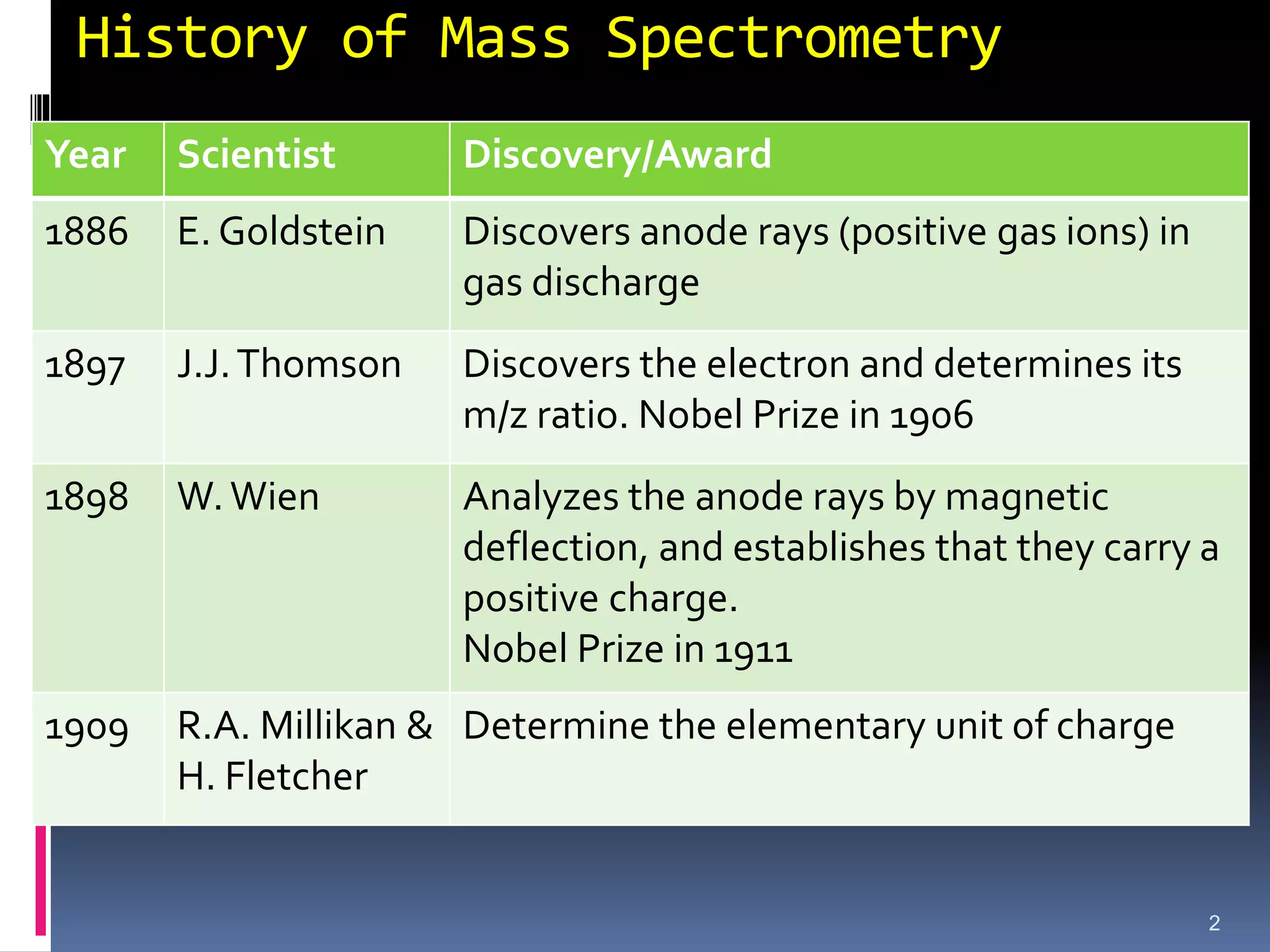
Year Scientist Discovery/Award
1886 E. Goldstein Discovers anode rays (positive gas ions) in
gas discharge
1897 J.J.Thomson Discovers the electron and determines its
m/z ratio. Nobel Prize in 1906
1898 W.Wien Analyzes the anode rays by magnetic
deflection, and establishes that they carry a
positive charge.
Nobel Prize in 1911
1909 R.A. Millikan &
H. Fletcher
Determine the elementary unit of charge
3.
Mass Spectrometry
3
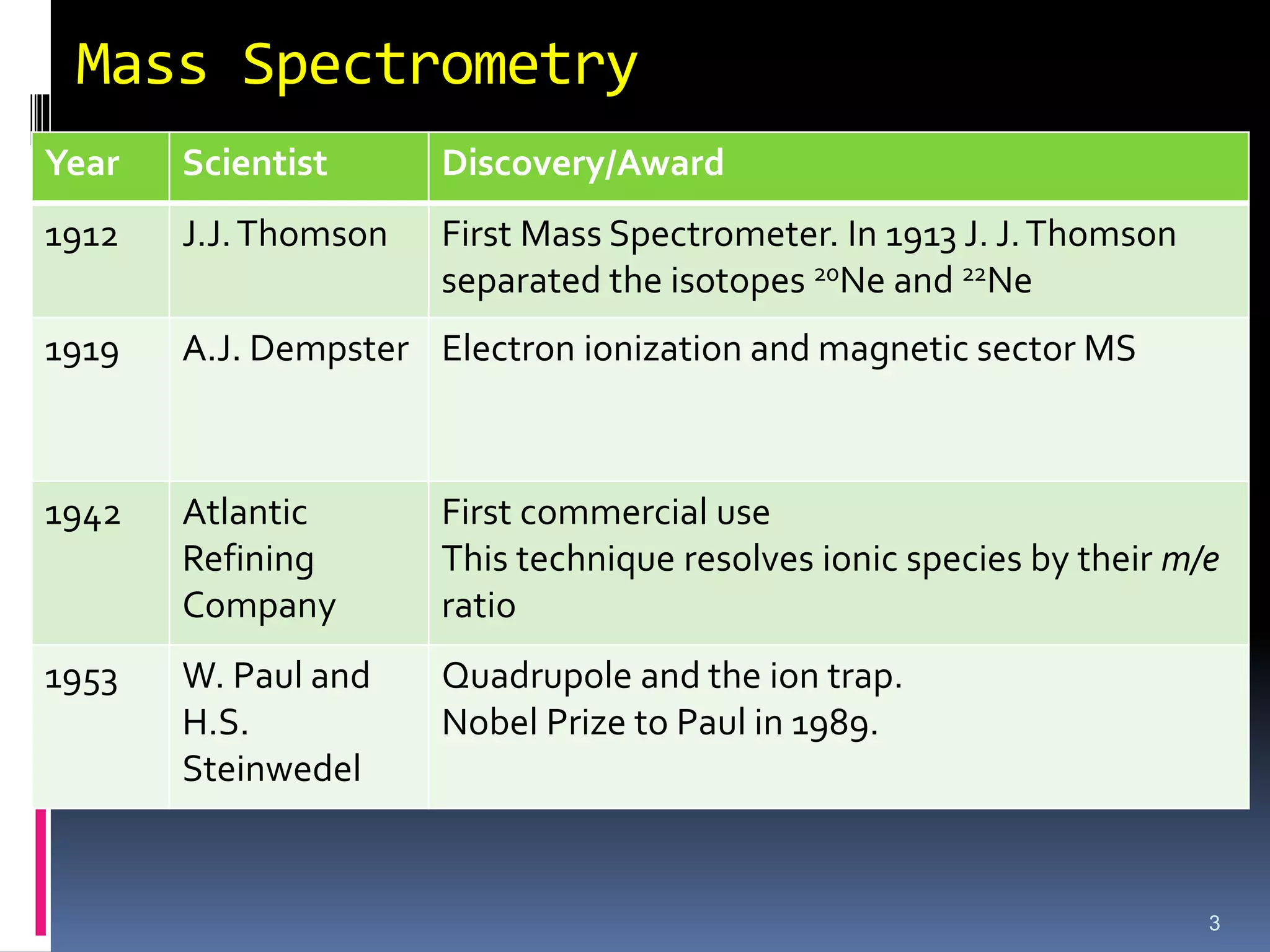
Year ScientistDiscovery/Award
1912 J.J.Thomson First Mass Spectrometer. In 1913 J. J.Thomson
separated the isotopes 20Ne and 22Ne
1919 A.J. Dempster Electron ionization and magnetic sector MS
1942 Atlantic
Refining
Company
First commercial use
This technique resolves ionic species by their m/e
ratio
1953 W. Paul and
H.S.
Steinwedel
Quadrupole and the ion trap.
Nobel Prize to Paul in 1989.
4.
Mass Spectrometry
4
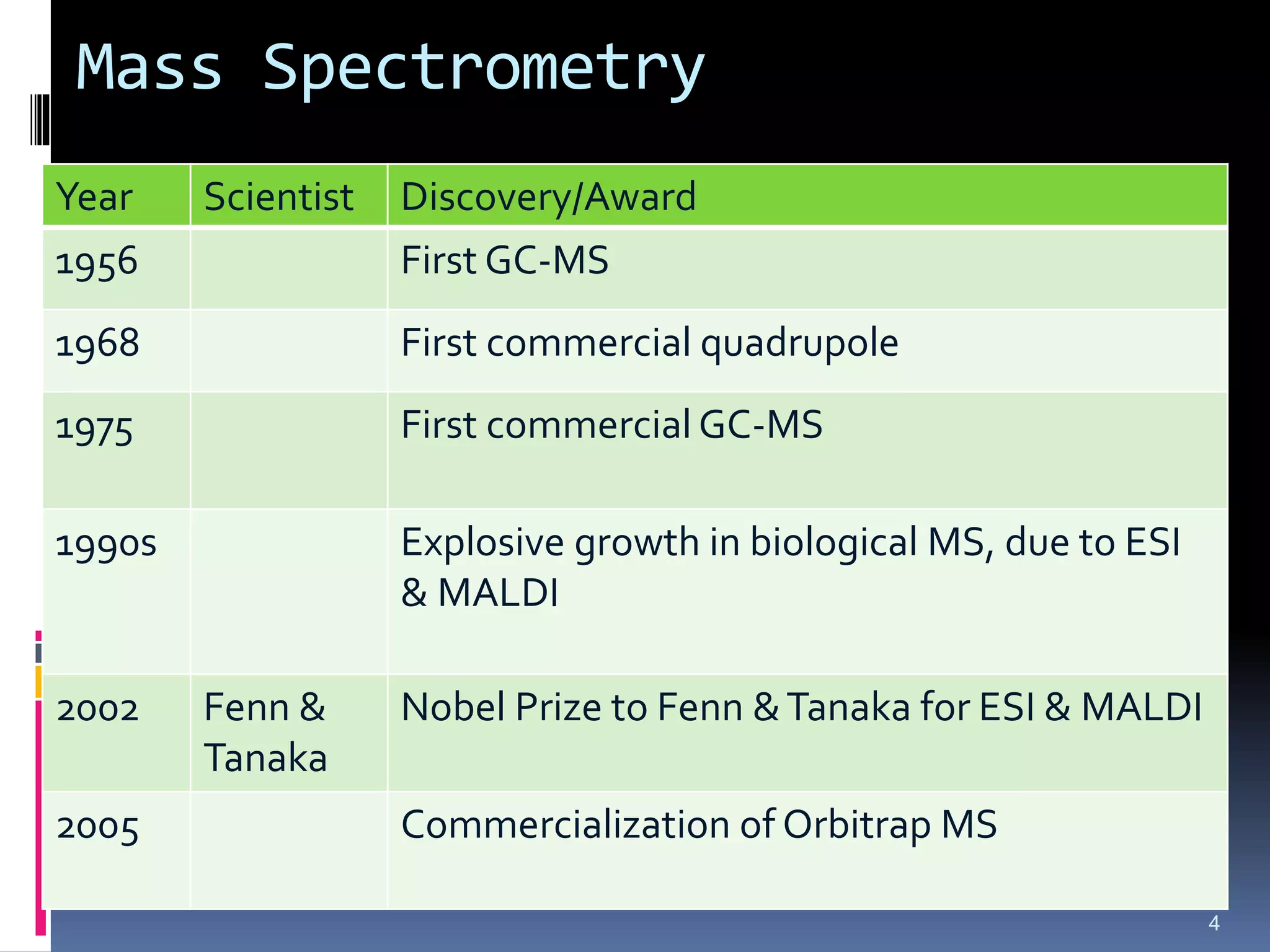
Year ScientistDiscovery/Award
1956 First GC-MS
1968 First commercial quadrupole
1975 First commercial GC-MS
1990s Explosive growth in biological MS, due to ESI
& MALDI
2002 Fenn &
Tanaka
Nobel Prize to Fenn &Tanaka for ESI & MALDI
2005 Commercialization of Orbitrap MS
5.
Mass Spec -Introduction
Very different from IR and NMR
Absorption of electromagnetic energy
Sample can be recovered and reused
Mass spectrometry
Records what happens when an organic molecule is hit
by a beam of high-energy electrons
Sample is completely destroyed
6.
Mass Spec -Introduction
What does a mass spectrum tell us?
1. Molecular weight
2. Molecular formula
Either directly or in conjunction with other kinds
of spectra such as IR or NMR
3. Fragmentation pattern
Key pieces of what the molecule looks like (such
as methyl, ethyl, phenyl, or benzyl groups
7.
Ms spectrometrygives composition of sample.
Structure of inorganic, organic & biological sample
Qualitative & quantitative composition of solid surfaces
Isotopic ratios of atoms in samples
Atomic or Molecular weight expressed in terms of
atomic mass unit (amu) or daltons (Da).
Introduction to Mass Spectrometry
8.
The amuis based upon the relative scale in which the
reference is carbon isotope C-12.
Thus amu is defined as 1/12 the mass of the one neutral
C-12
Molecular weight can be obtained from a very small sample.
It does not involve the absorption or emission of light.
A beam of high-energy electrons breaks the molecule apart.
The masses of the fragments and their relative abundance
reveal information about the structure of the molecule.
8
9.
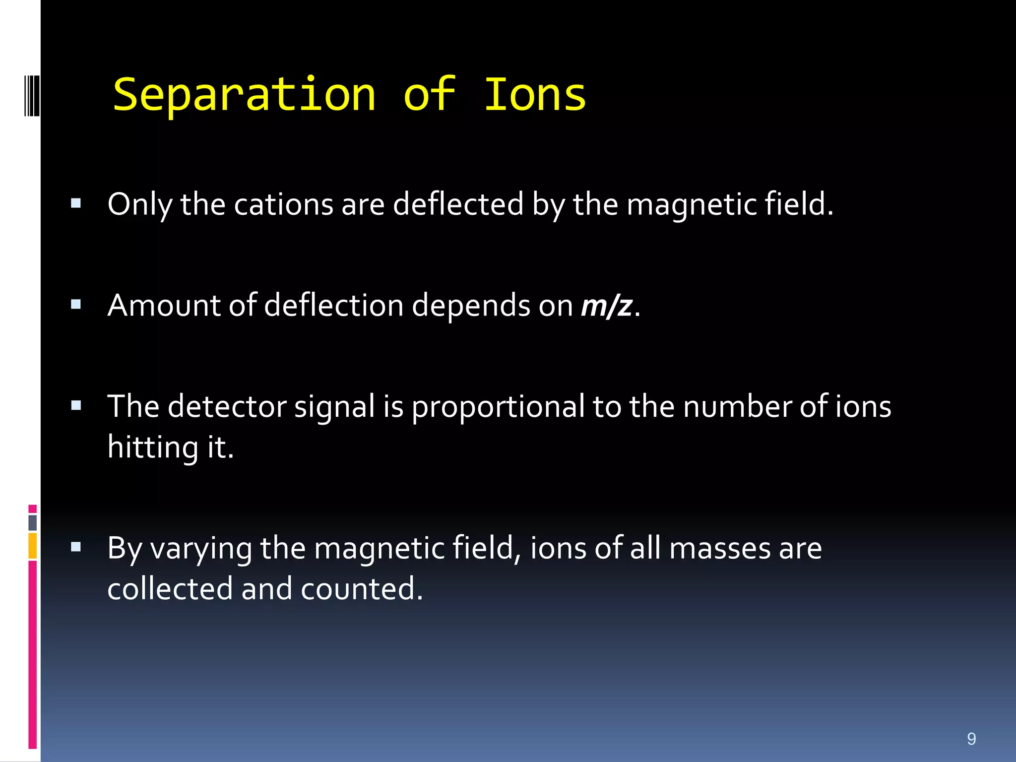
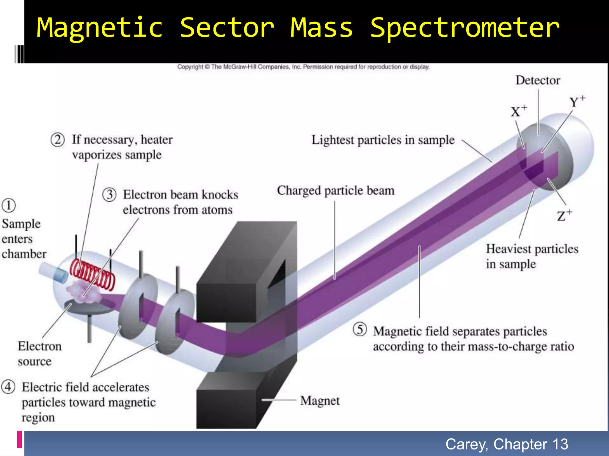
Separation of Ions
Only the cations are deflected by the magnetic field.
Amount of deflection depends on m/z.
The detector signal is proportional to the number of ions
hitting it.
By varying the magnetic field, ions of all masses are
collected and counted.
9
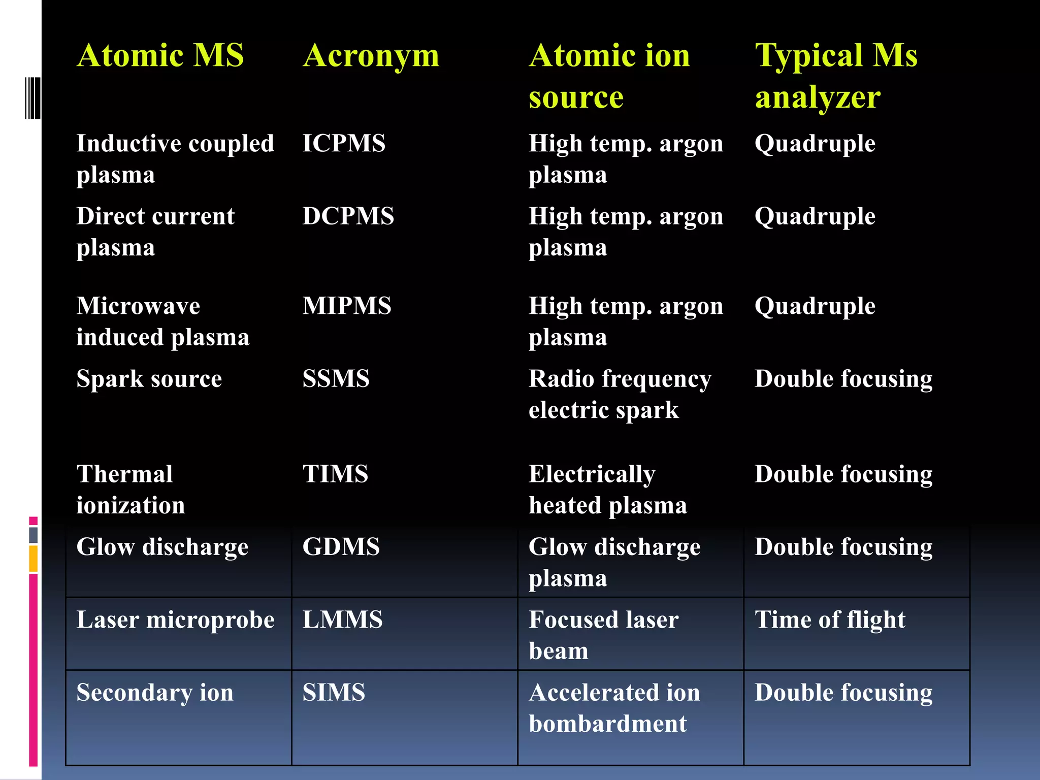
10.
Atomic MS AcronymAtomic ion
source
Typical Ms
analyzer
Inductive coupled
plasma
ICPMS High temp. argon
plasma
Quadruple
Direct current
plasma
DCPMS High temp. argon
plasma
Quadruple
Microwave
induced plasma
MIPMS High temp. argon
plasma
Quadruple
Spark source SSMS Radio frequency
electric spark
Double focusing
Thermal
ionization
TIMS Electrically
heated plasma
Double focusing
Glow discharge GDMS Glow discharge
plasma
Double focusing
Laser microprobe LMMS Focused laser
beam
Time of flight
Secondary ion SIMS Accelerated ion
bombardment
Double focusing
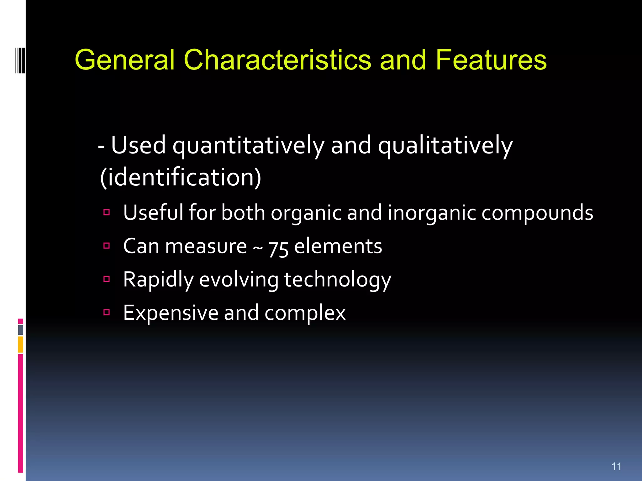
11.
- Used quantitativelyand qualitatively
(identification)
Useful for both organic and inorganic compounds
Can measure ~ 75 elements
Rapidly evolving technology
Expensive and complex
11
General Characteristics and Features
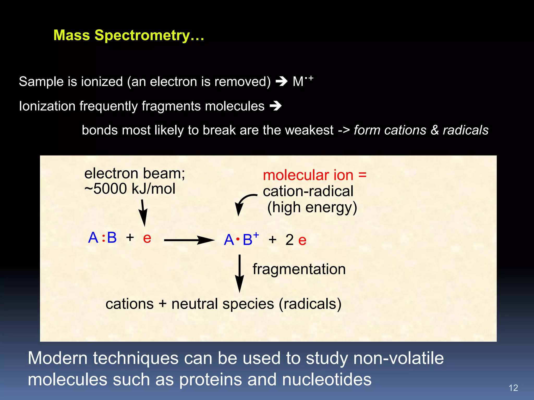
12.
12
A B +e
molecular ion =
cation-radical
(high energy)
electron beam;
~5000 kJ/mol
fragmentation
cations + neutral species (radicals)
: A B+
+ 2 e
Mass Spectrometry…
Sample is ionized (an electron is removed) M
.+
Ionization frequently fragments molecules
bonds most likely to break are the weakest -> form cations & radicals
Modern techniques can be used to study non-volatile
molecules such as proteins and nucleotides
13.
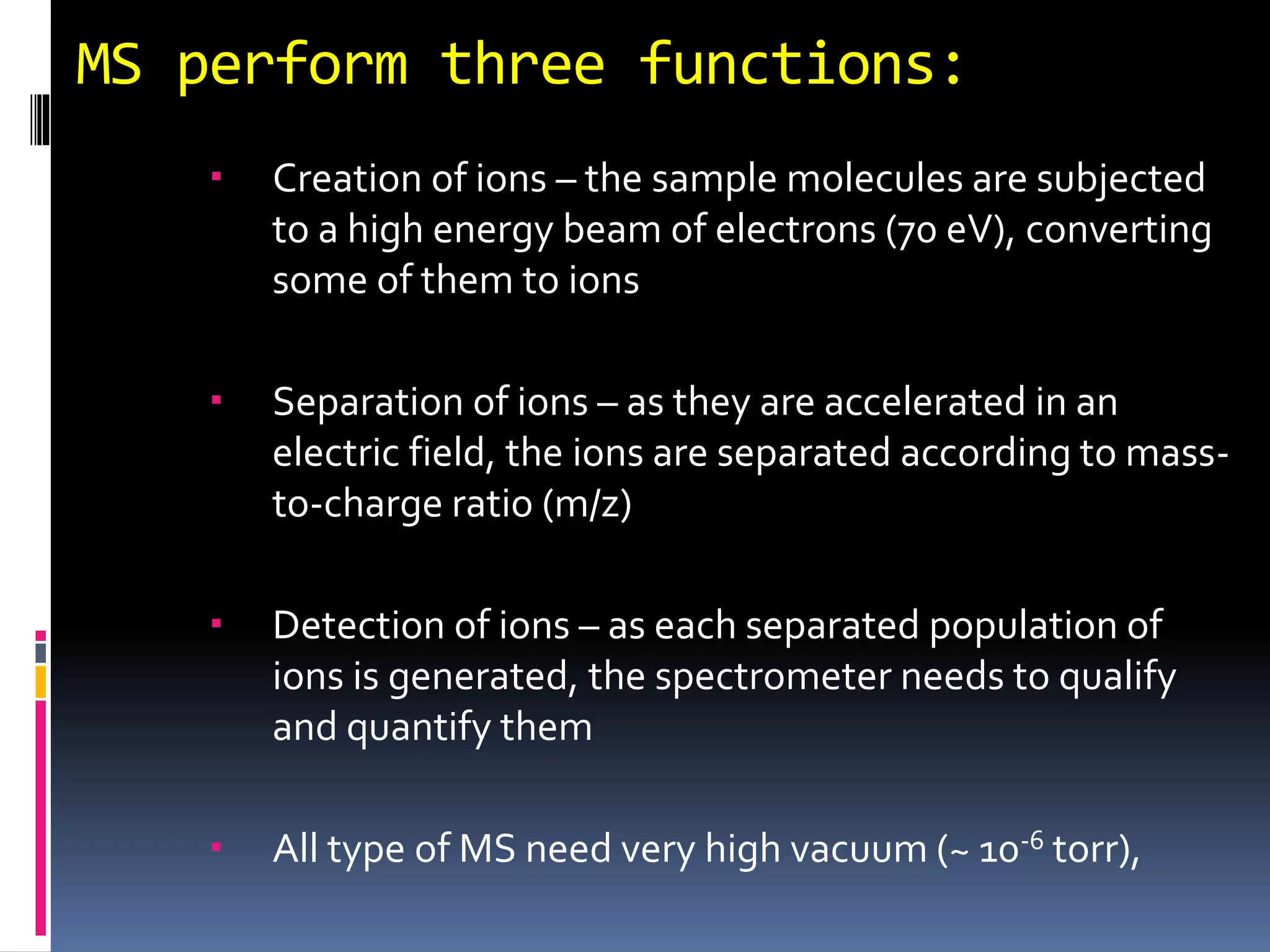
MS perform threefunctions:
Creation of ions – the sample molecules are subjected
to a high energy beam of electrons (70 eV), converting
some of them to ions
Separation of ions – as they are accelerated in an
electric field, the ions are separated according to mass-
to-charge ratio (m/z)
Detection of ions – as each separated population of
ions is generated, the spectrometer needs to qualify
and quantify them
All type of MS need very high vacuum (~ 10-6 torr),
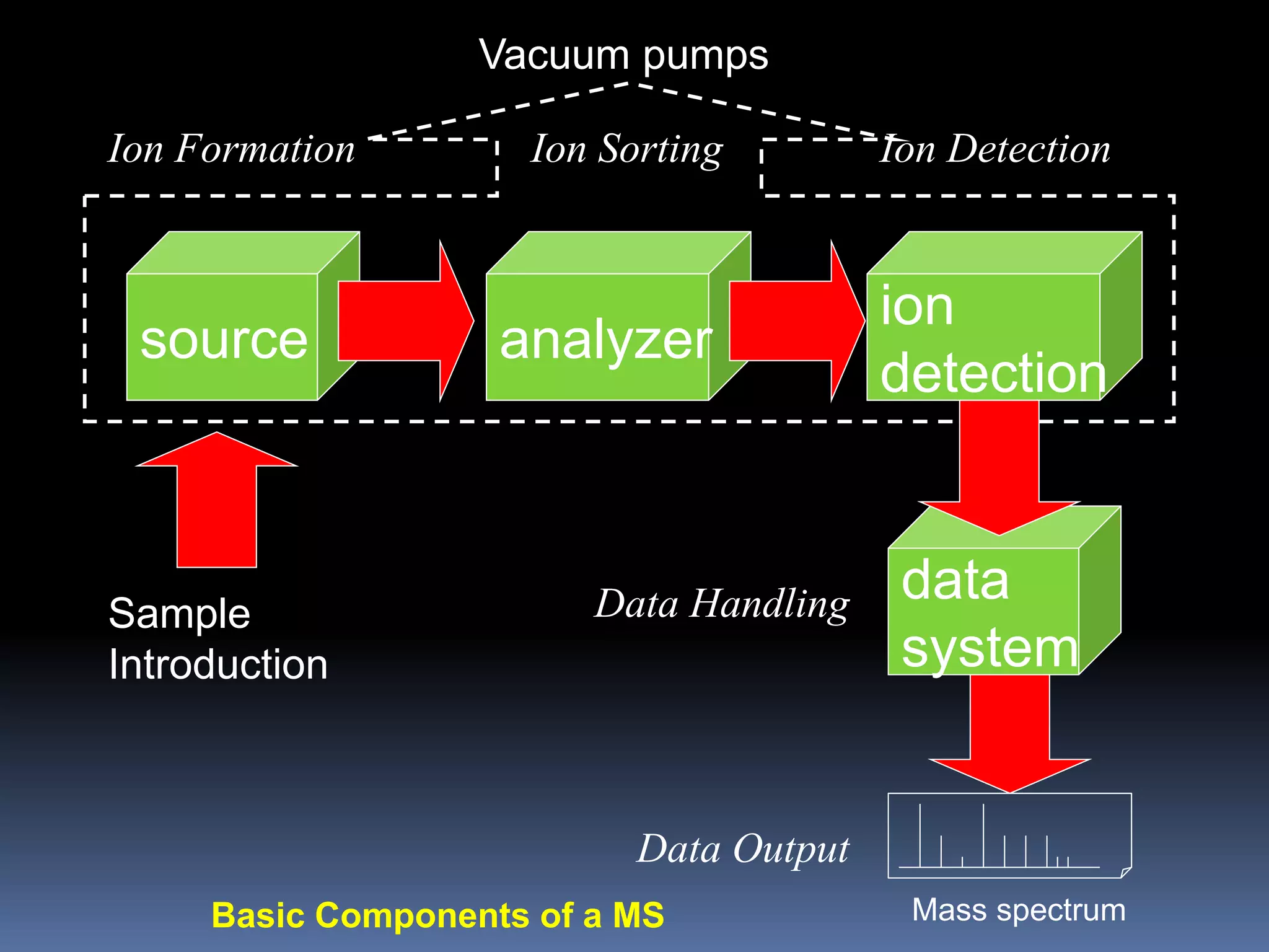
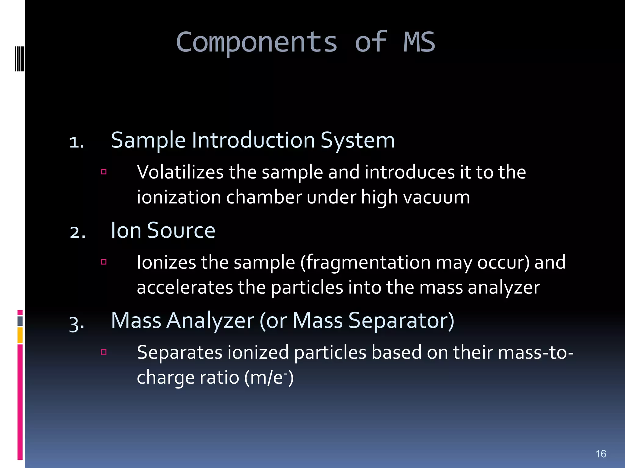
Components of MS
1.Sample Introduction System
Volatilizes the sample and introduces it to the
ionization chamber under high vacuum
2. Ion Source
Ionizes the sample (fragmentation may occur) and
accelerates the particles into the mass analyzer
3. Mass Analyzer (or Mass Separator)
Separates ionized particles based on their mass-to-
charge ratio (m/e-)
16
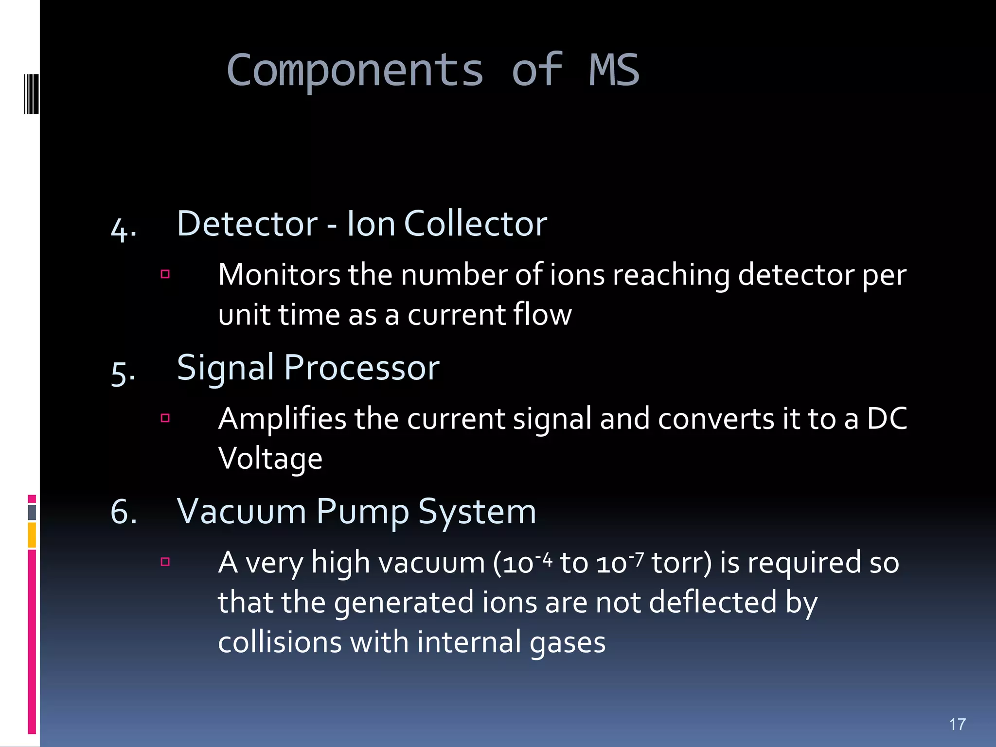
17.
Components of MS
4.Detector - Ion Collector
Monitors the number of ions reaching detector per
unit time as a current flow
5. Signal Processor
Amplifies the current signal and converts it to a DC
Voltage
6. Vacuum Pump System
A very high vacuum (10-4 to 10-7 torr) is required so
that the generated ions are not deflected by
collisions with internal gases
17
18.
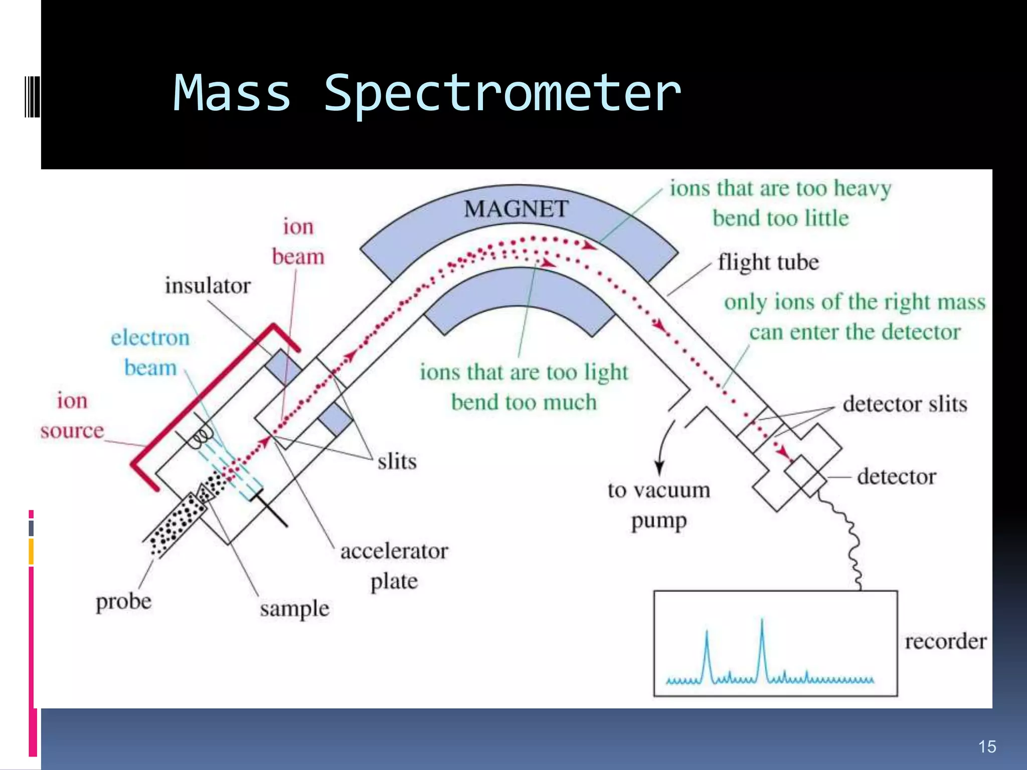
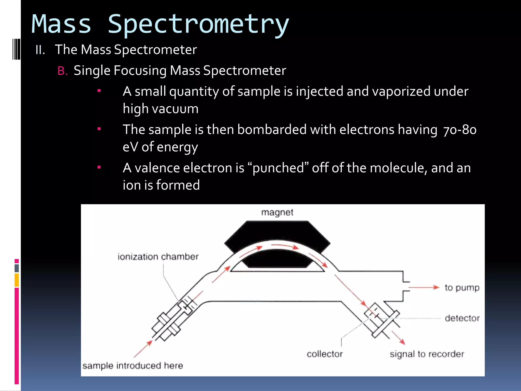
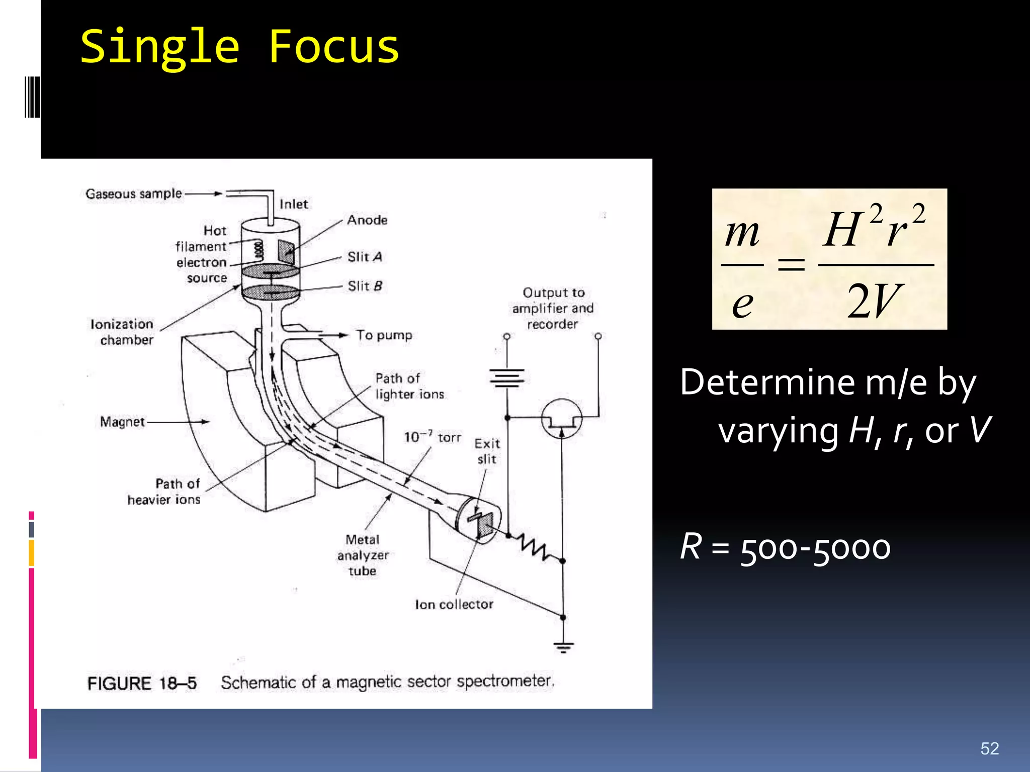
Mass Spectrometry
II. TheMass Spectrometer
B. Single Focusing Mass Spectrometer
A small quantity of sample is injected and vaporized under
high vacuum
The sample is then bombarded with electrons having 70-80
eV of energy
A valence electron is “punched” off of the molecule, and an
ion is formed
19.
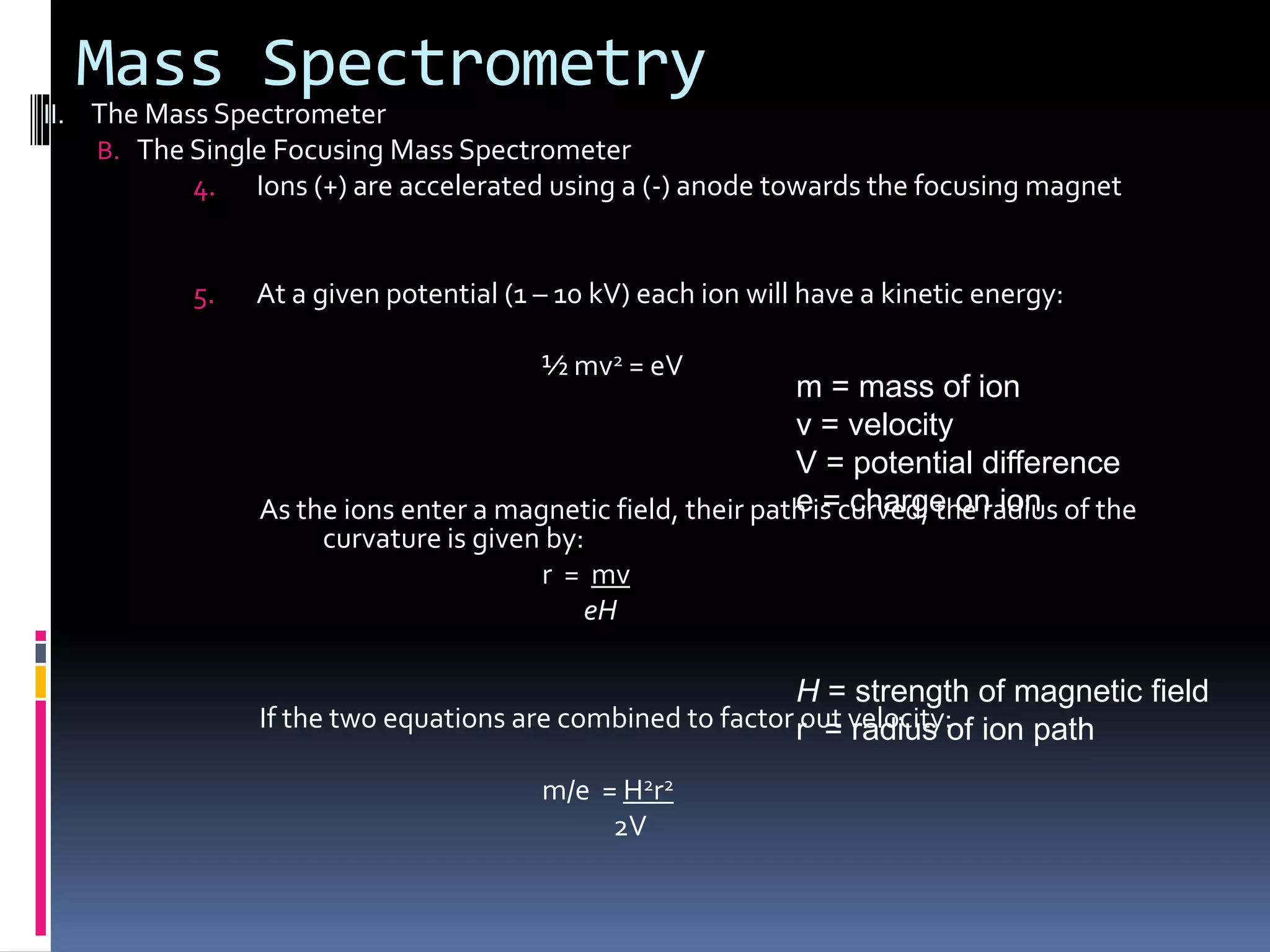
Mass Spectrometry
II. TheMass Spectrometer
B. The Single Focusing Mass Spectrometer
4. Ions (+) are accelerated using a (-) anode towards the focusing magnet
5. At a given potential (1 – 10 kV) each ion will have a kinetic energy:
½ mv2 = eV
As the ions enter a magnetic field, their path is curved; the radius of the
curvature is given by:
r = mv
eH
If the two equations are combined to factor out velocity:
m/e = H2r2
2V
m = mass of ion
v = velocity
V = potential difference
e = charge on ion
H = strength of magnetic field
r = radius of ion path
20.
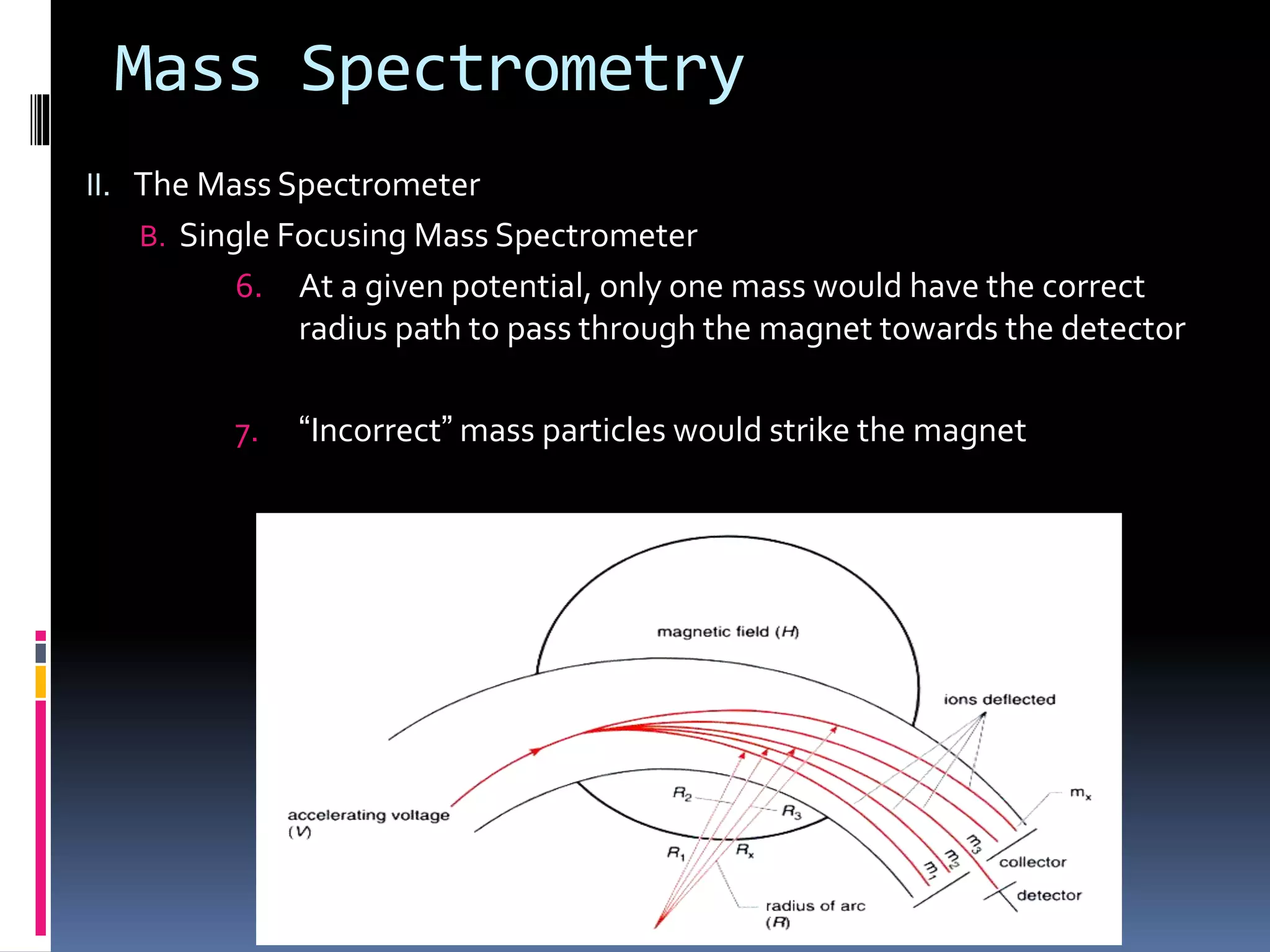
Mass Spectrometry
II. TheMass Spectrometer
B. Single Focusing Mass Spectrometer
6. At a given potential, only one mass would have the correct
radius path to pass through the magnet towards the detector
7. “Incorrect” mass particles would strike the magnet
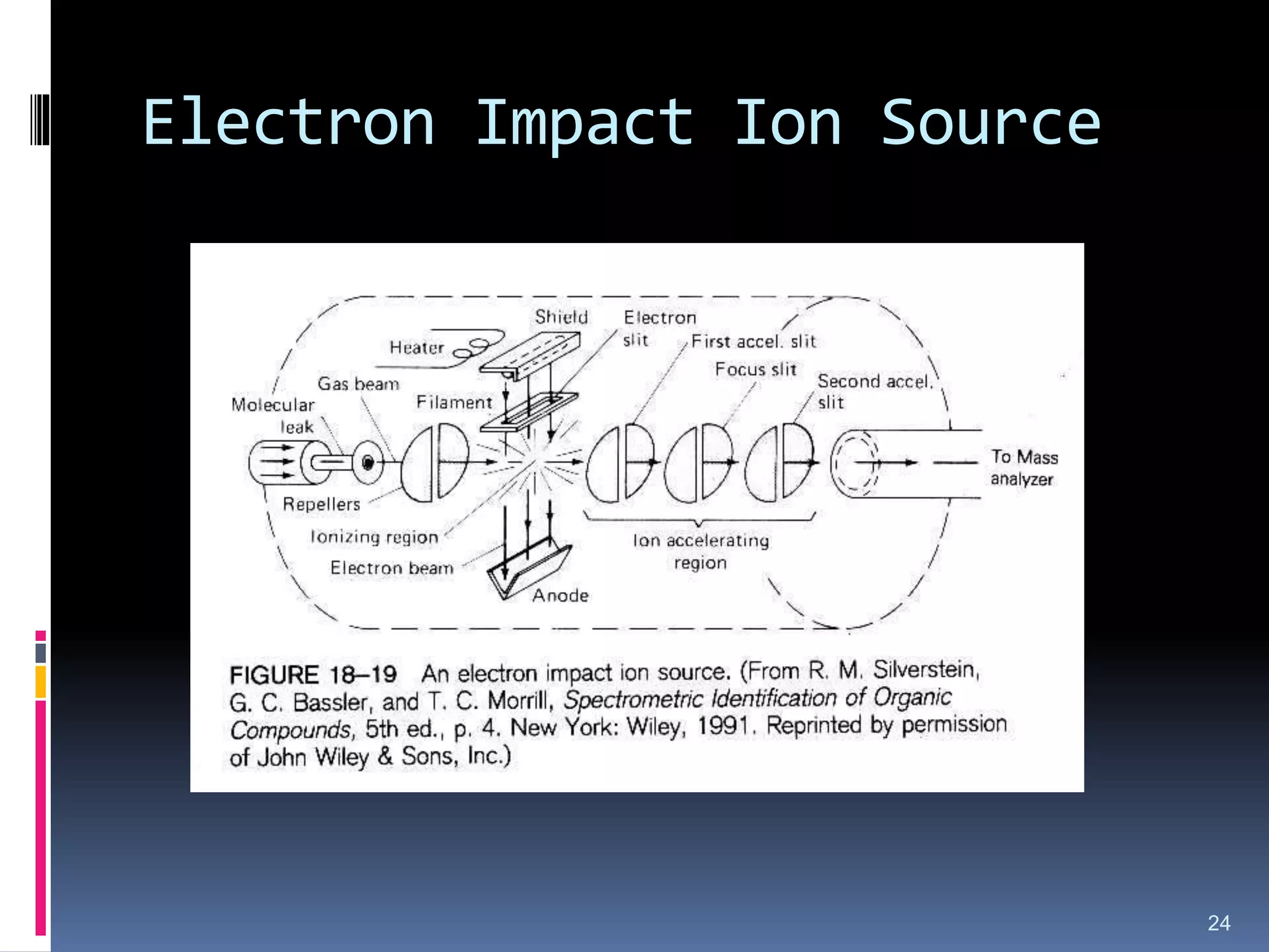
21.
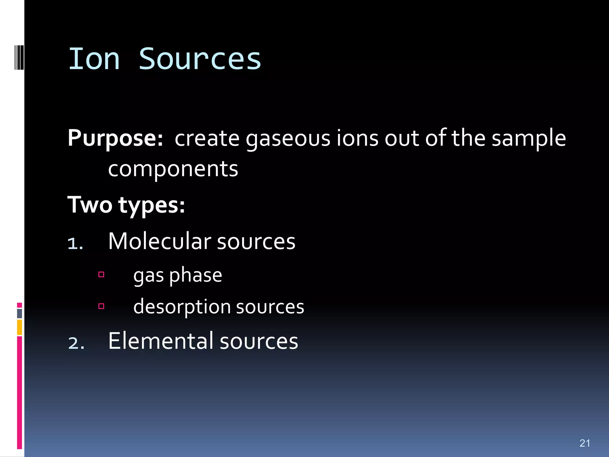
Ion Sources
Purpose: creategaseous ions out of the sample
components
Two types:
1. Molecular sources
gas phase
desorption sources
2. Elemental sources
21
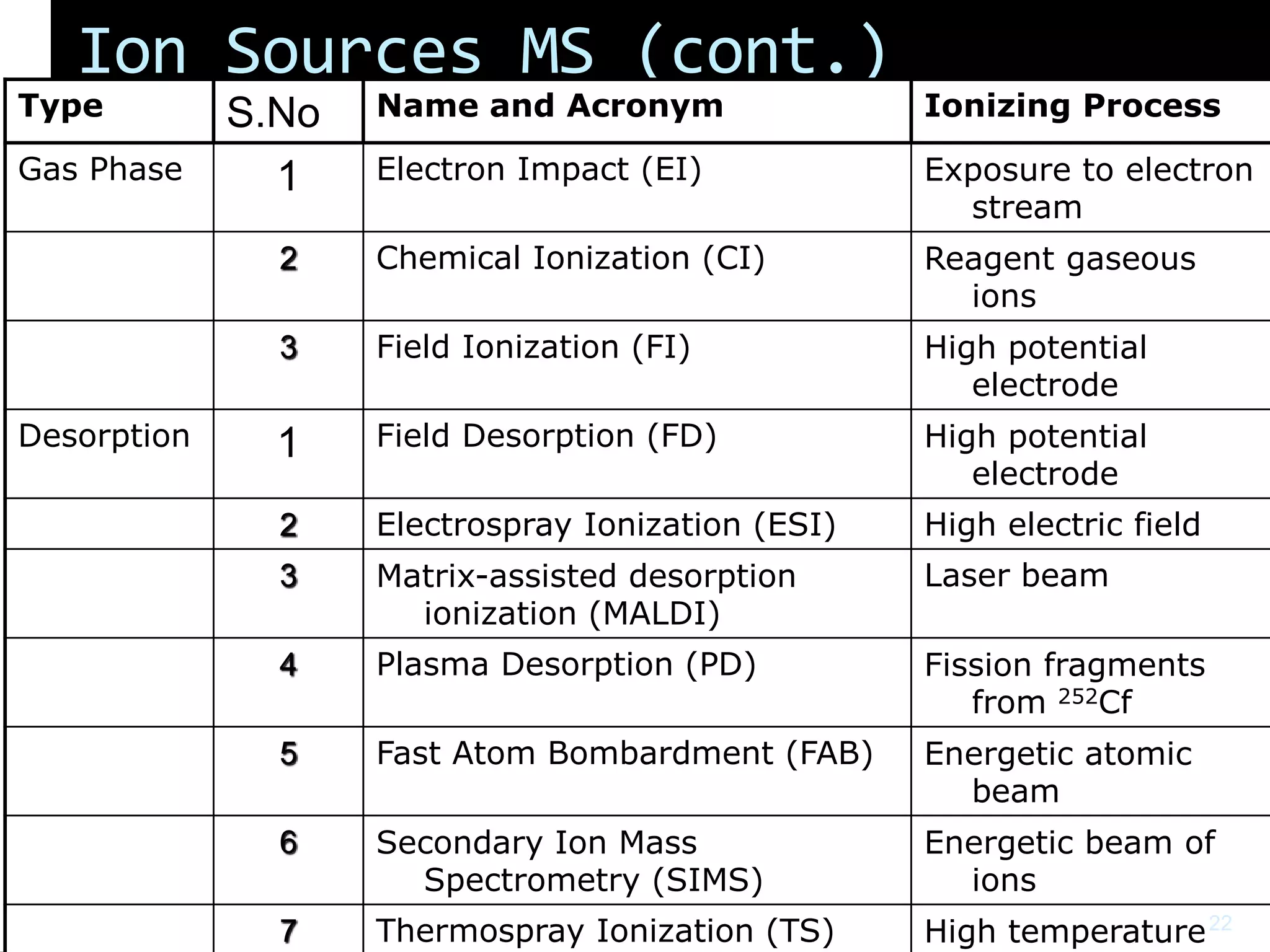
22.
Ion Sources MS(cont.)
Type S.No Name and Acronym Ionizing Process
Gas Phase 1 Electron Impact (EI) Exposure to electron
stream
2 Chemical Ionization (CI) Reagent gaseous
ions
3 Field Ionization (FI) High potential
electrode
Desorption 1 Field Desorption (FD) High potential
electrode
2 Electrospray Ionization (ESI) High electric field
3 Matrix-assisted desorption
ionization (MALDI)
Laser beam
4 Plasma Desorption (PD) Fission fragments
from 252Cf
5 Fast Atom Bombardment (FAB) Energetic atomic
beam
6 Secondary Ion Mass
Spectrometry (SIMS)
Energetic beam of
ions
7 Thermospray Ionization (TS) High temperature22
23.
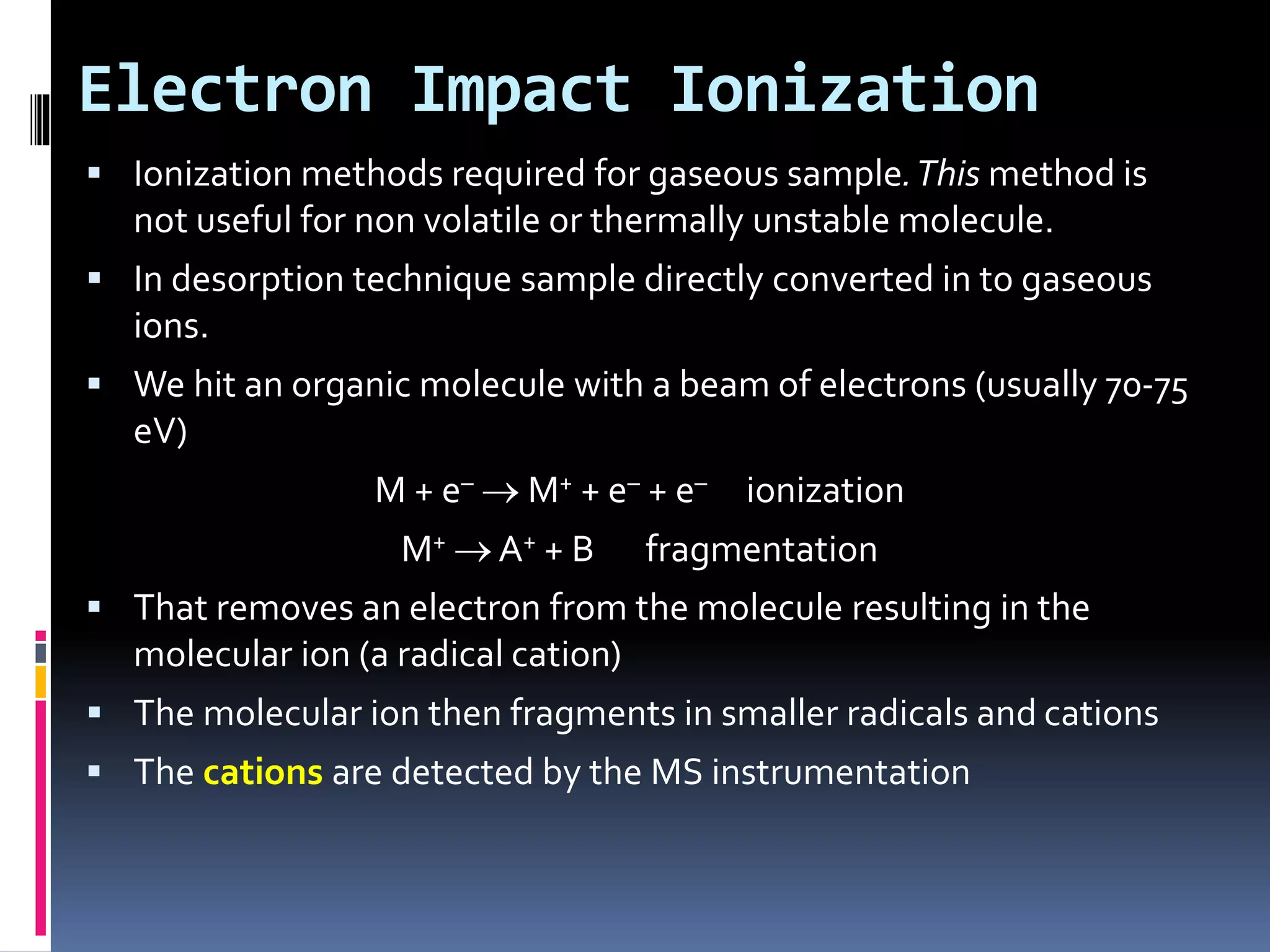
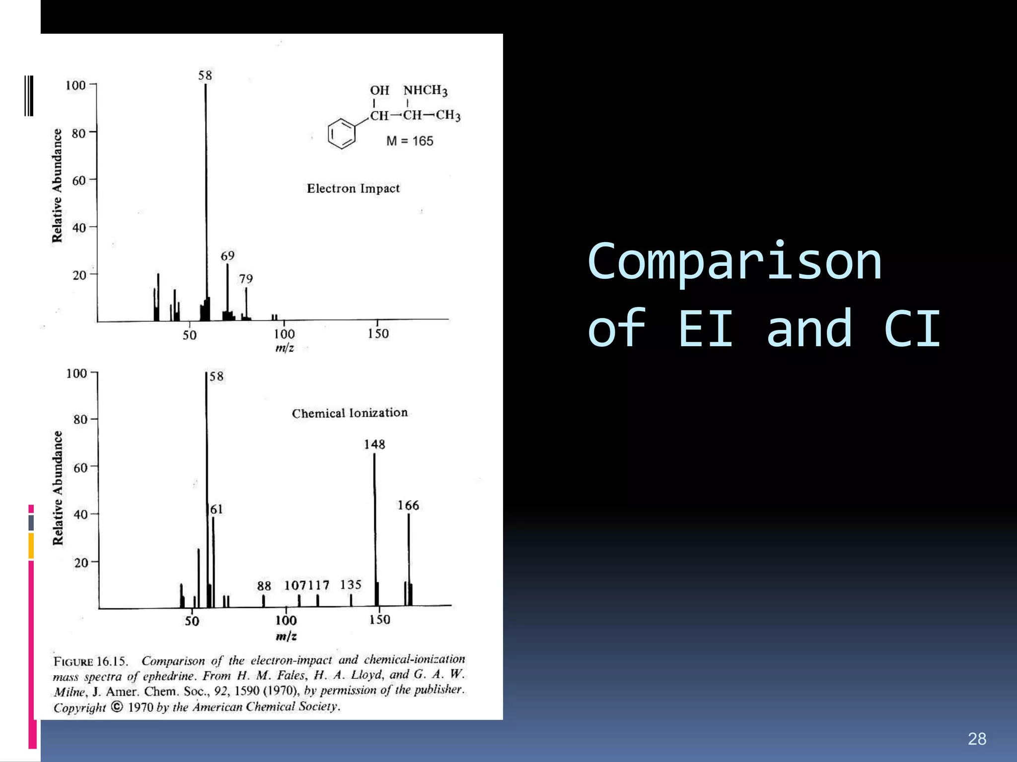
Electron Impact Ionization
Ionization methods required for gaseous sample.This method is
not useful for non volatile or thermally unstable molecule.
In desorption technique sample directly converted in to gaseous
ions.
We hit an organic molecule with a beam of electrons (usually 70-75
eV)
M + e– M+ + e– + e– ionization
M+ A+ + B fragmentation
That removes an electron from the molecule resulting in the
molecular ion (a radical cation)
The molecular ion then fragments in smaller radicals and cations
The cations are detected by the MS instrumentation
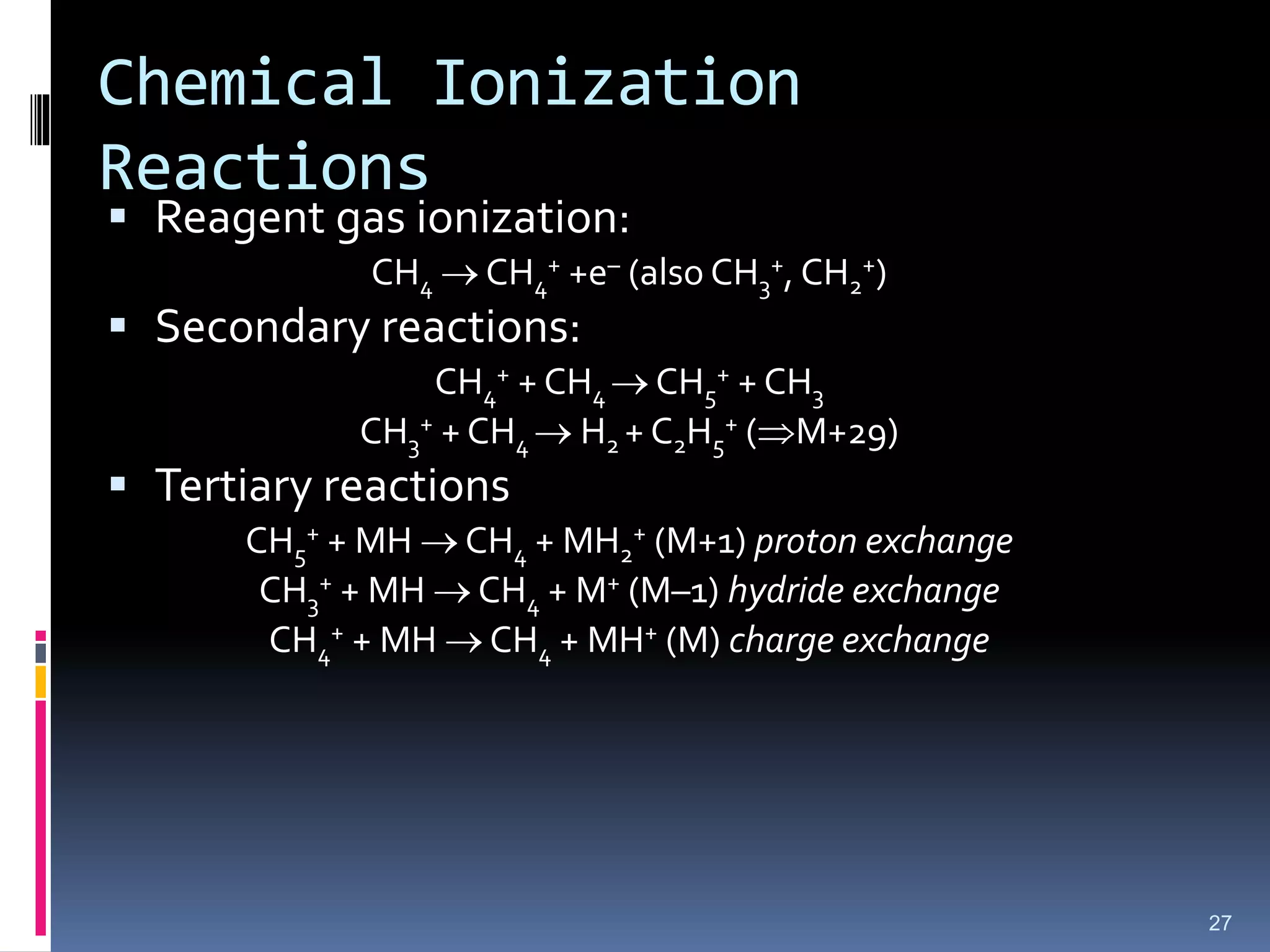
Chemical Ionization
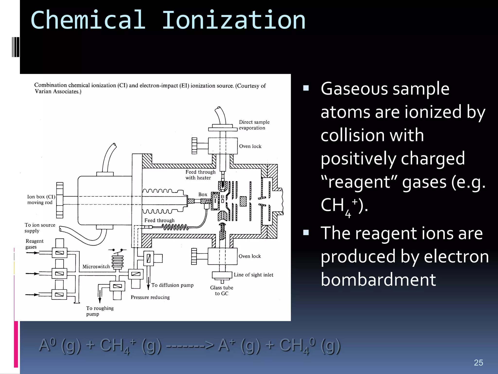
25
Gaseoussample
atoms are ionized by
collision with
positively charged
“reagent” gases (e.g.
CH4
+).
The reagent ions are
produced by electron
bombardment
A0 (g) + CH4
+ (g) -------> A+ (g) + CH4
0 (g)
26.
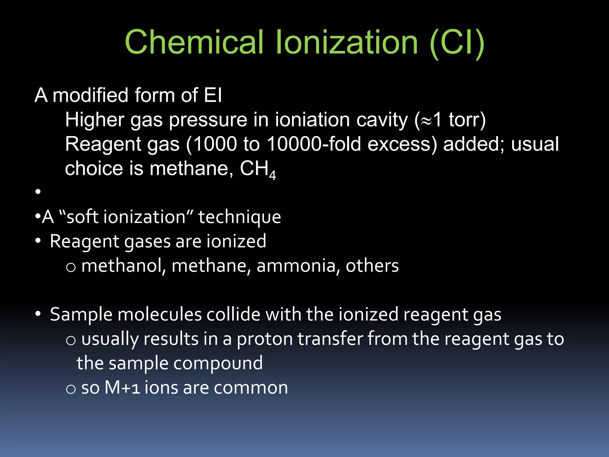
Chemical Ionization (CI)
Amodified form of EI
Higher gas pressure in ioniation cavity (1 torr)
Reagent gas (1000 to 10000-fold excess) added; usual
choice is methane, CH4
•
•A “soft ionization” technique
• Reagent gases are ionized
o methanol, methane, ammonia, others
• Sample molecules collide with the ionized reagent gas
o usually results in a proton transfer from the reagent gas to
the sample compound
o so M+1 ions are common
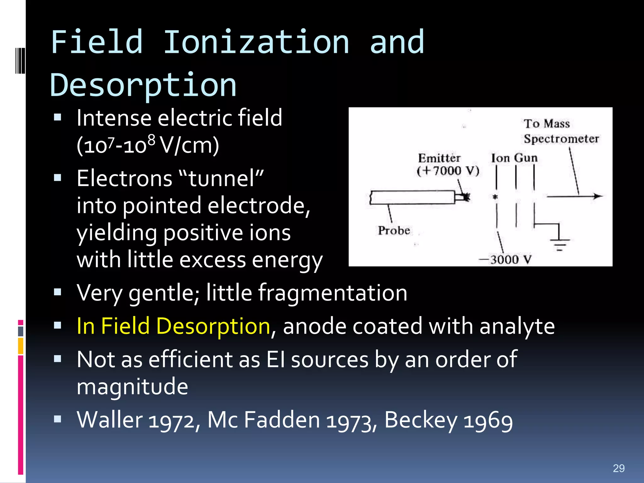
Field Ionization and
Desorption
Intense electric field
(107-108 V/cm)
Electrons “tunnel”
into pointed electrode,
yielding positive ions
with little excess energy
Very gentle; little fragmentation
In Field Desorption, anode coated with analyte
Not as efficient as EI sources by an order of
magnitude
Waller 1972, Mc Fadden 1973, Beckey 1969
29
30.
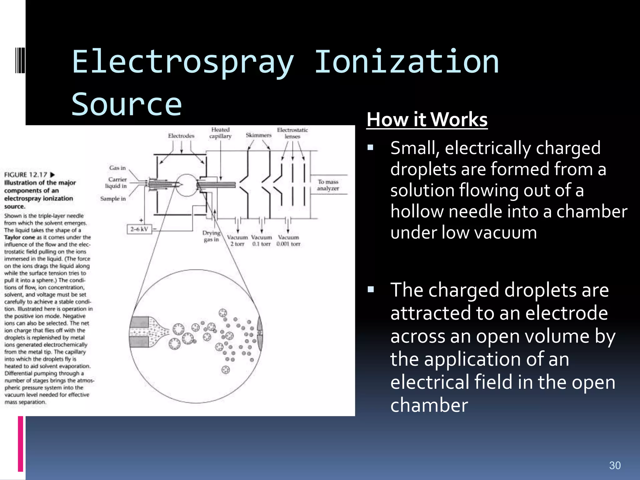
Electrospray Ionization
Source
30
How itWorks
Small, electrically charged
droplets are formed from a
solution flowing out of a
hollow needle into a chamber
under low vacuum
The charged droplets are
attracted to an electrode
across an open volume by
the application of an
electrical field in the open
chamber
31.
Electrospray Ionization Source
Some of the solvent is evaporated (and concentration occurs)
during transit across the chamber
As the droplets shrink, ions are forced closer together. At
some point the repulsive forces between the ions is greater
than surface tension and small droplets break off the larger
drops.
This process continues several times as the droplets transit
across the chamber
Eventually the solvent disappears and ions are generated, a
process called ion evaporation& analysed by quadrupole Mass
analyser
31
32.
Matrix-Assisted Laser
Desorption/Ionization (MALDI)
Analyte mixed with radiation-absorbing material such as
Nicotinic acid, Benzoic acid deriv., Pyrazine –carboxylic acid,
cinnamic acid deriv., Nitrobenzyl alcohol
The resulting solution was evaporated on the metallic probe
surface and dried
Sample mixture was exposed to pulsed laser beam, which
result in the sublimation of analyte ion and were drawn into
time-of-flight (TOF) analyser for analysis
Excellent for larger molecules, e.g. peptides, polymers
32
33.
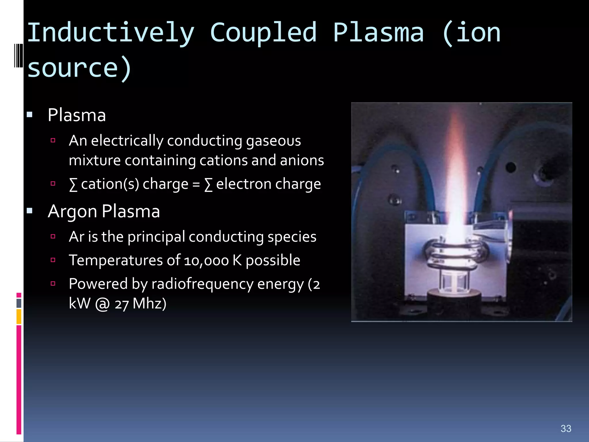
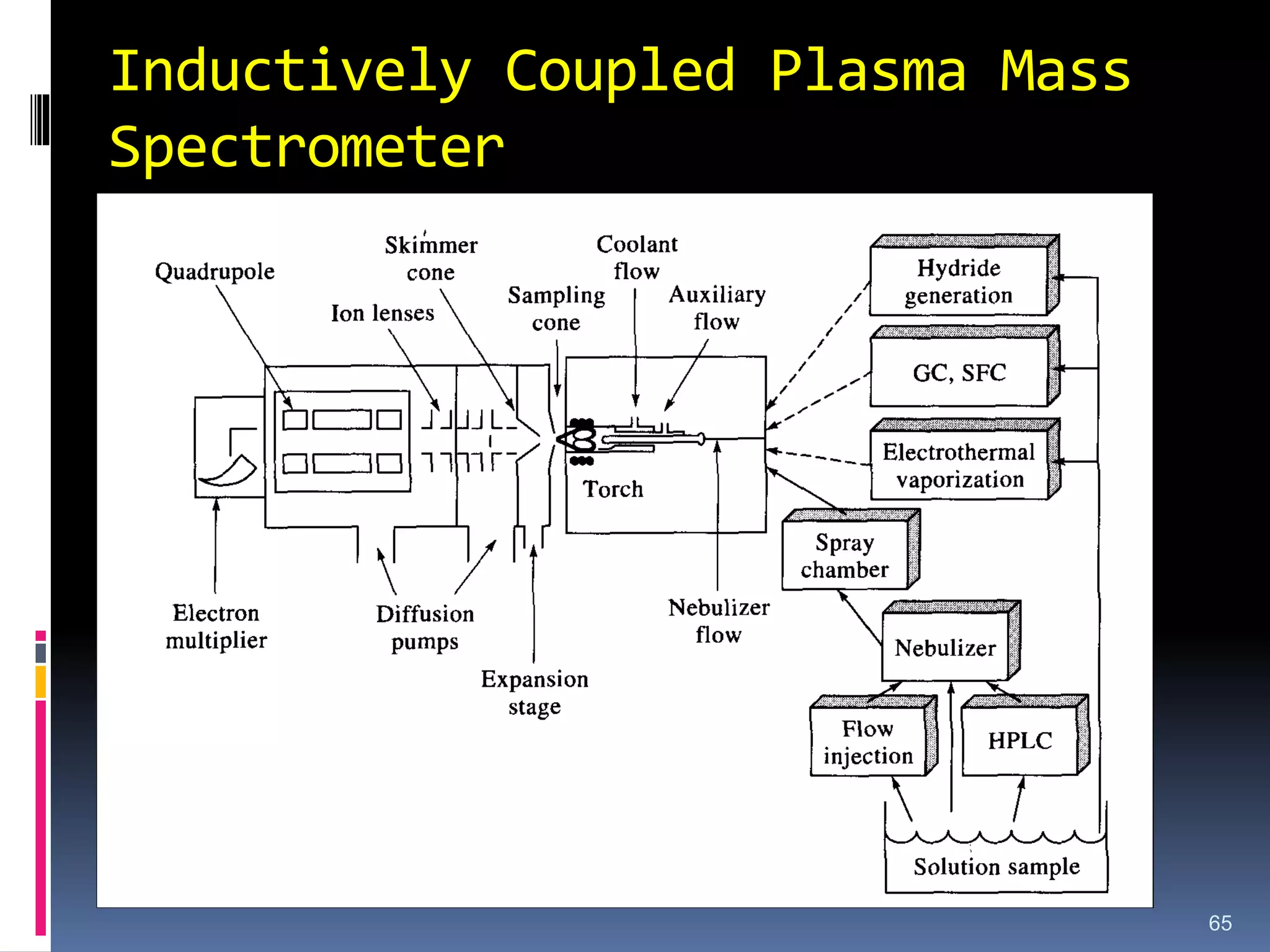
Inductively Coupled Plasma(ion
source)
Plasma
An electrically conducting gaseous
mixture containing cations and anions
∑ cation(s) charge = ∑ electron charge
Argon Plasma
Ar is the principal conducting species
Temperatures of 10,000 K possible
Powered by radiofrequency energy (2
kW @ 27 Mhz)
33
34.
Inductively Coupled Plasma(ion
source)
An ICP “torch” consists of:
Three concentric quartz tubes
through which a stream of argon
flows at a rate of 5-20 L/min
The three concentric rings are
constructed to eliminate
atmospheric gases from contacting
the sample stream in the inner-
most ring
34
35.
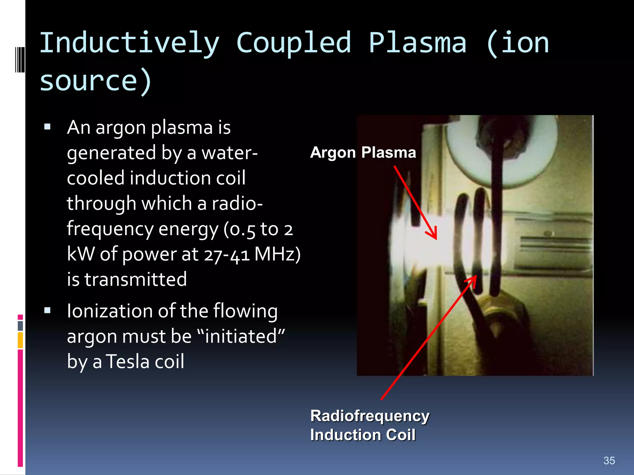
Inductively Coupled Plasma(ion
source)
An argon plasma is
generated by a water-
cooled induction coil
through which a radio-
frequency energy (0.5 to 2
kW of power at 27-41 MHz)
is transmitted
Ionization of the flowing
argon must be “initiated”
by aTesla coil
35
Radiofrequency
Induction Coil
Argon Plasma
36.
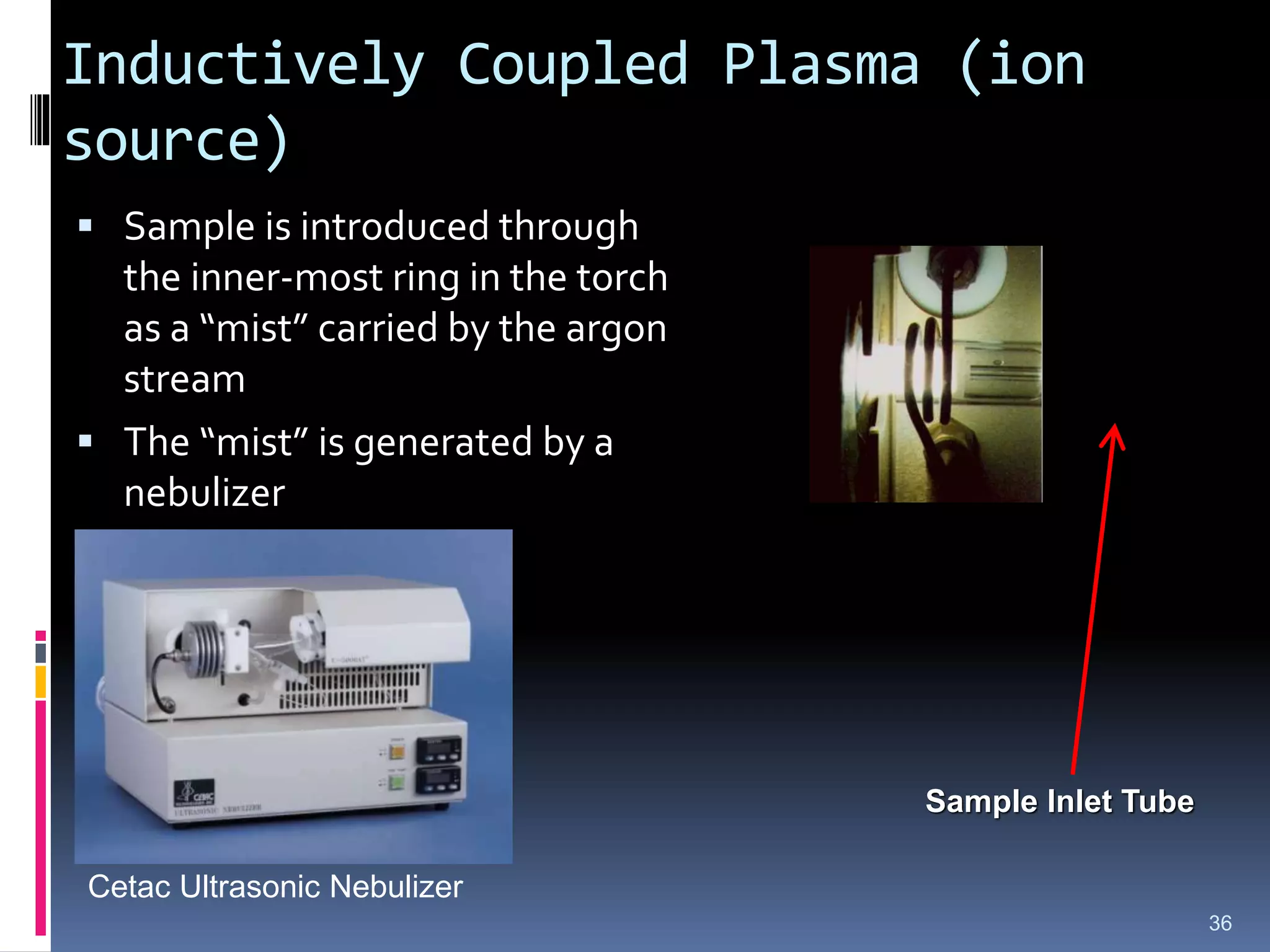
Inductively Coupled Plasma(ion
source)
Sample is introduced through
the inner-most ring in the torch
as a “mist” carried by the argon
stream
The “mist” is generated by a
nebulizer
36
Sample Inlet Tube
Cetac Ultrasonic Nebulizer
37.
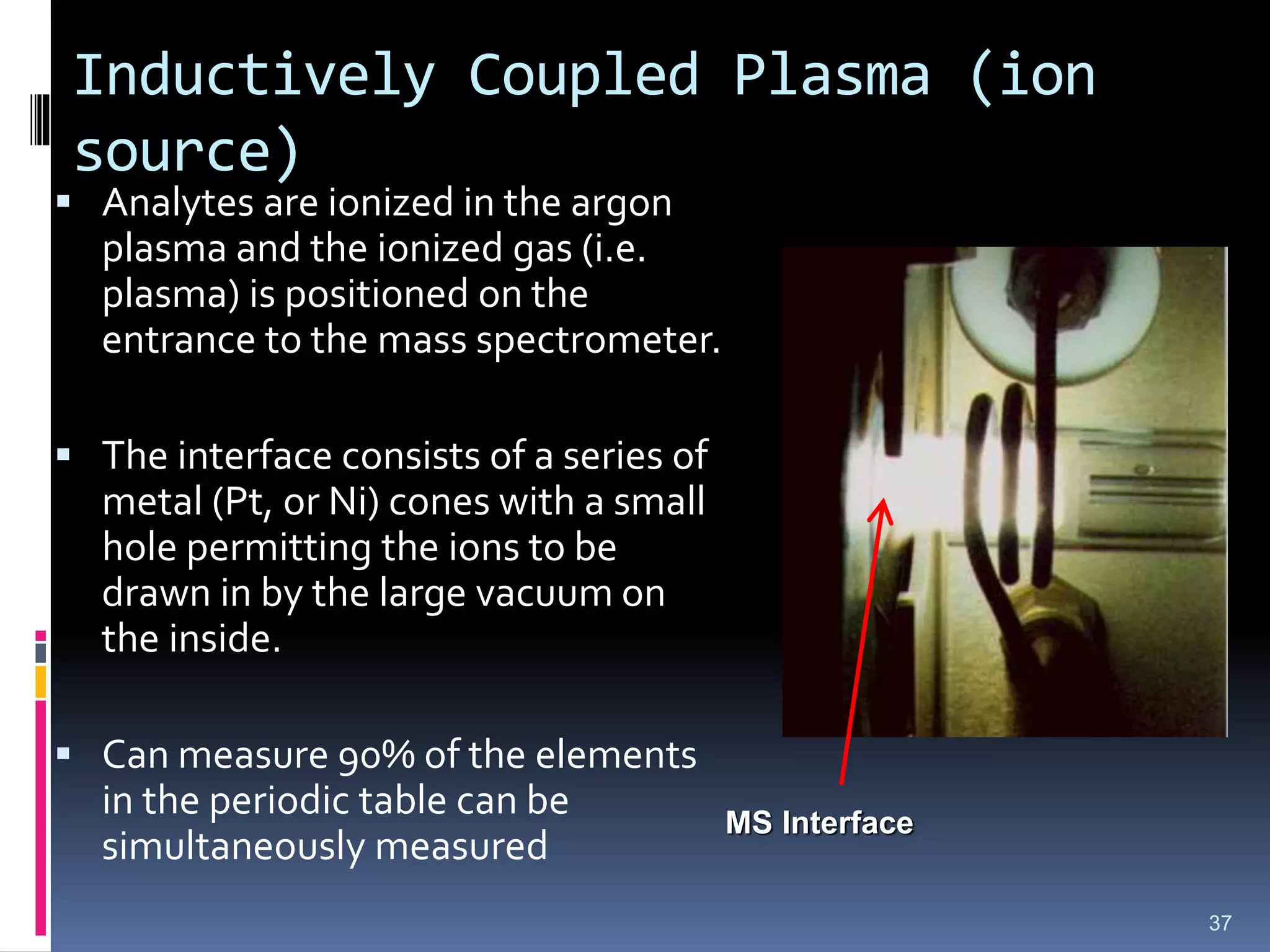
Inductively Coupled Plasma(ion
source)
Analytes are ionized in the argon
plasma and the ionized gas (i.e.
plasma) is positioned on the
entrance to the mass spectrometer.
The interface consists of a series of
metal (Pt, or Ni) cones with a small
hole permitting the ions to be
drawn in by the large vacuum on
the inside.
Can measure 90% of the elements
in the periodic table can be
simultaneously measured
37
MS Interface
38.
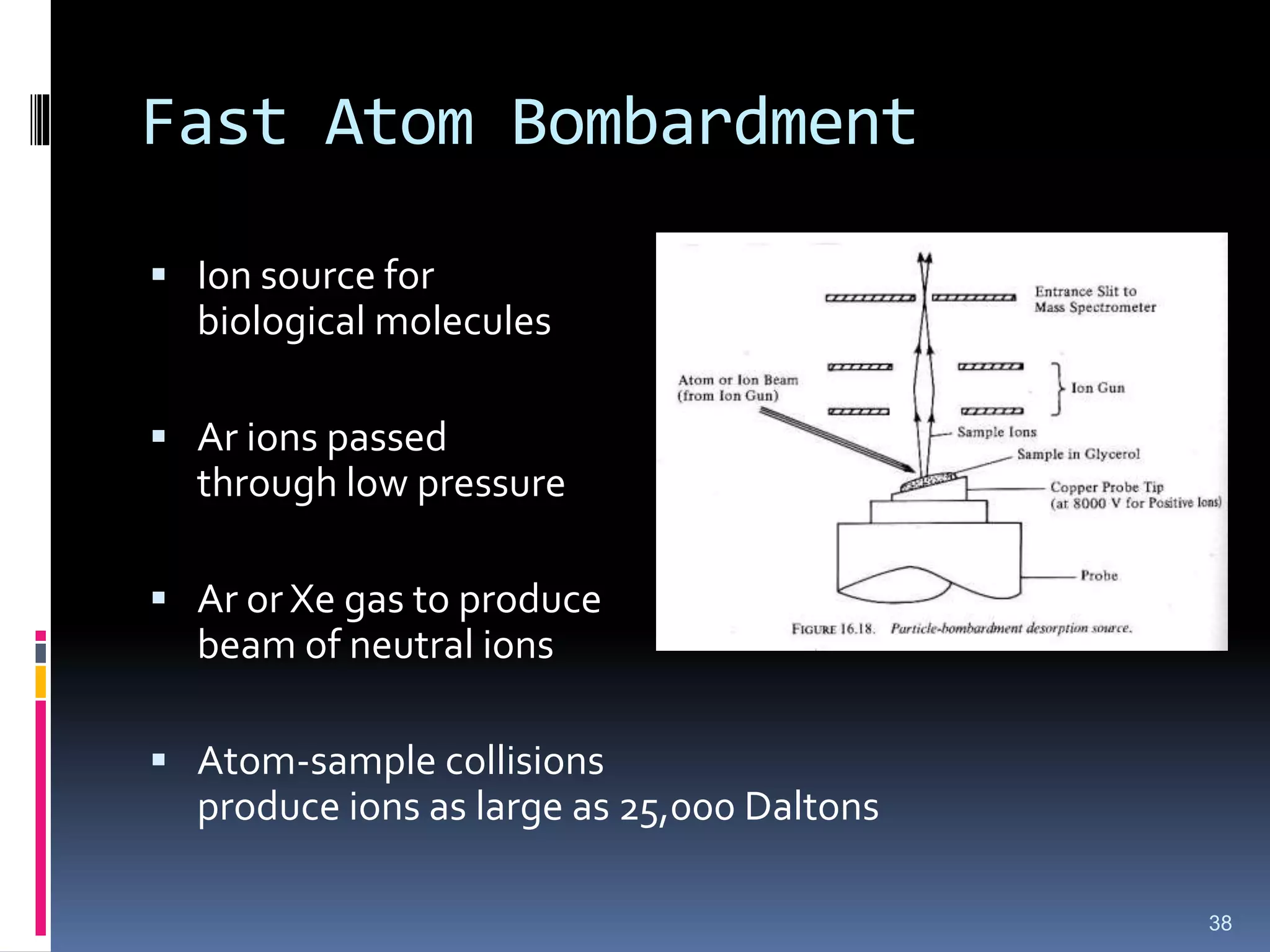
Fast Atom Bombardment
Ion source for
biological molecules
Ar ions passed
through low pressure
Ar or Xe gas to produce
beam of neutral ions
Atom-sample collisions
produce ions as large as 25,000 Daltons
38
39.
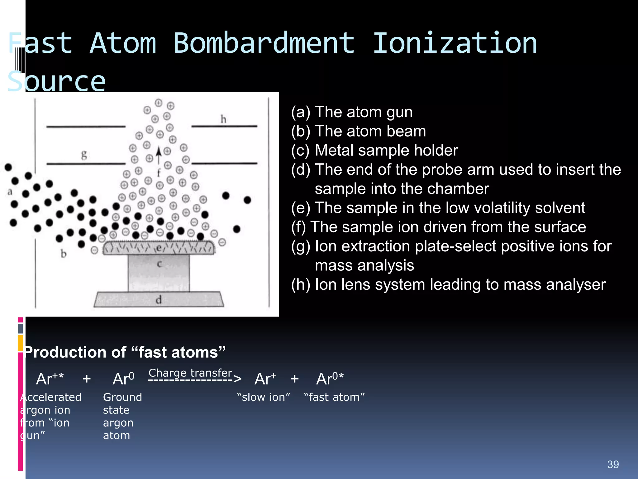
Fast Atom BombardmentIonization
Source
39
Ar+* + Ar0 ----------------> Ar+ + Ar0*
Production of “fast atoms”
Charge transfer
Accelerated
argon ion
from “ion
gun”
Ground
state
argon
atom
“slow ion” “fast atom”
(a) The atom gun
(b) The atom beam
(c) Metal sample holder
(d) The end of the probe arm used to insert the
sample into the chamber
(e) The sample in the low volatility solvent
(f) The sample ion driven from the surface
(g) Ion extraction plate-select positive ions for
mass analysis
(h) Ion lens system leading to mass analyser
40.
FAB Characteristics
Usedwith high molecular weight organic molecules
The fast atom interacts with analyte on a “target” to produce
ions by “sputtering” (i.e. transfer of energy from argon to
analyte)
Analyte ions are accelerated into the MS by application of an
electric field (ion extraction plate and lenses)
40
41.
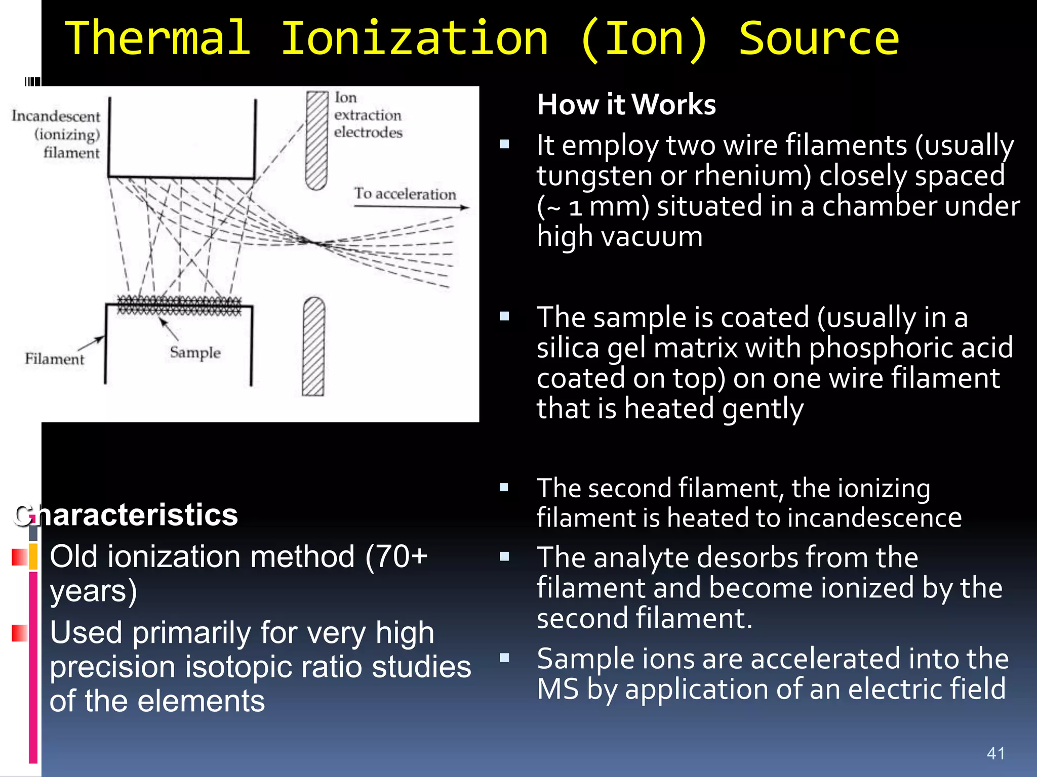
Thermal Ionization (Ion)Source
41
How it Works
It employ two wire filaments (usually
tungsten or rhenium) closely spaced
(~ 1 mm) situated in a chamber under
high vacuum
The sample is coated (usually in a
silica gel matrix with phosphoric acid
coated on top) on one wire filament
that is heated gently
The second filament, the ionizing
filament is heated to incandescence
The analyte desorbs from the
filament and become ionized by the
second filament.
Sample ions are accelerated into the
MS by application of an electric field
Characteristics
Old ionization method (70+
years)
Used primarily for very high
precision isotopic ratio studies
of the elements
42.
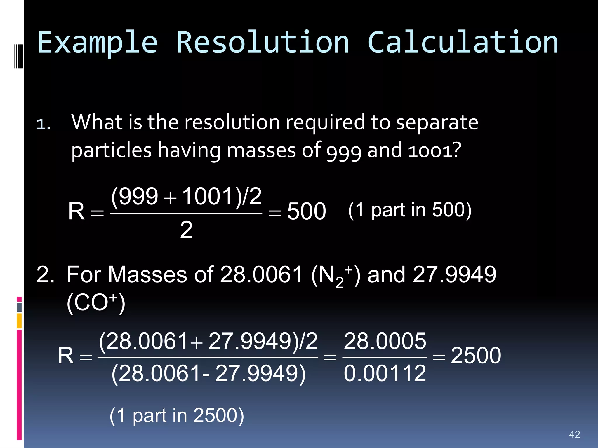
Example Resolution Calculation
1.What is the resolution required to separate
particles having masses of 999 and 1001?
500
2
1001)/2
(999
R
42
(1 part in 500)
2. For Masses of 28.0061 (N2
+) and 27.9949
(CO+)
2500
0.00112
28.0005
27.9949)
-
(28.0061
27.9949)/2
(28.0061
R
(1 part in 2500)
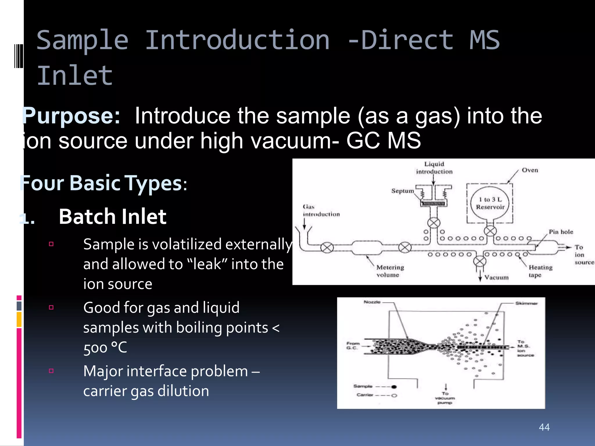
Sample Introduction -DirectMS
Inlet
Four BasicTypes:
1. Batch Inlet
Sample is volatilized externally
and allowed to “leak” into the
ion source
Good for gas and liquid
samples with boiling points <
500 °C
Major interface problem –
carrier gas dilution
44
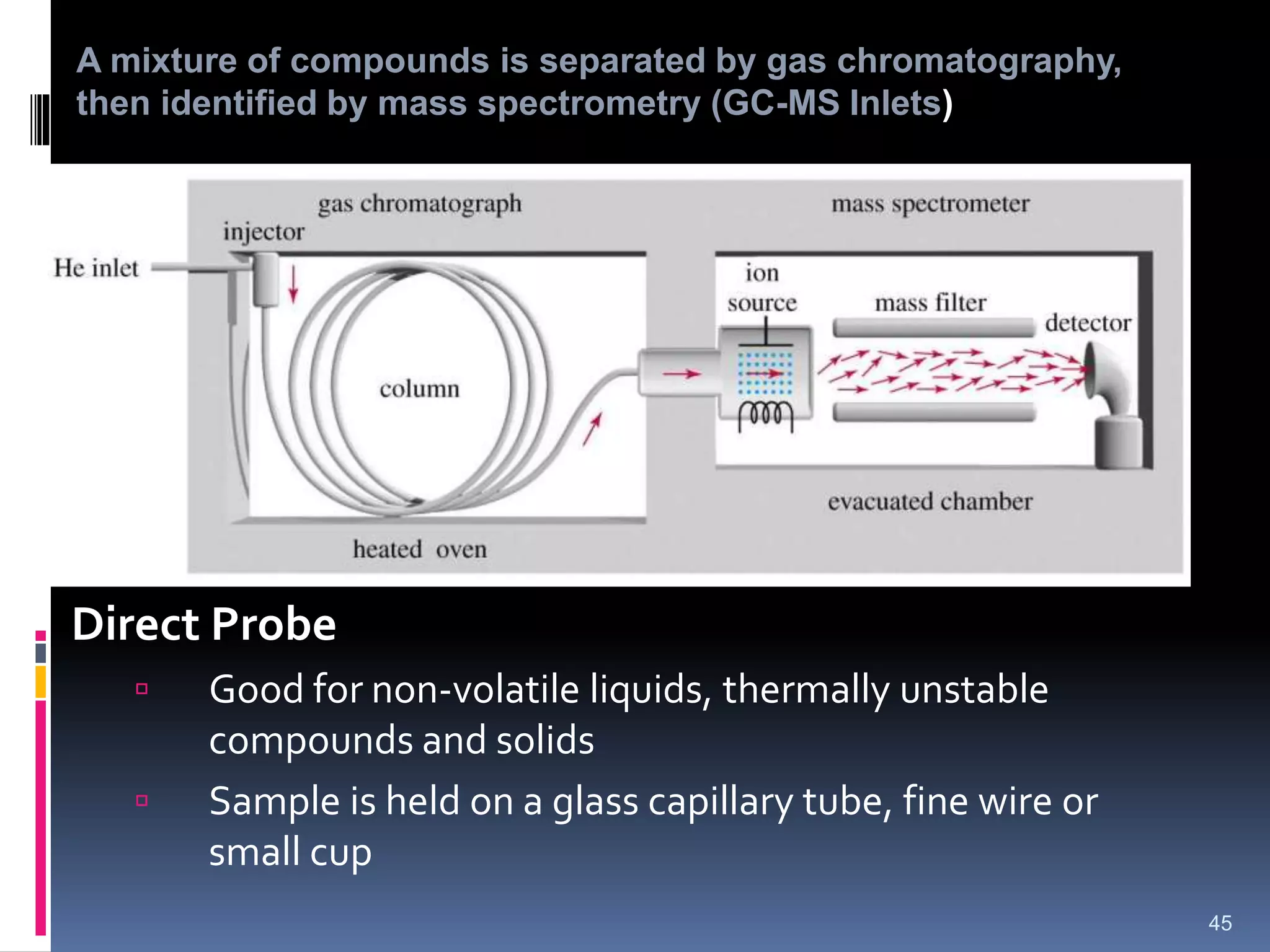
Purpose: Introduce the sample (as a gas) into the
ion source under high vacuum- GC MS
45.
Direct Probe
Goodfor non-volatile liquids, thermally unstable
compounds and solids
Sample is held on a glass capillary tube, fine wire or
small cup
45
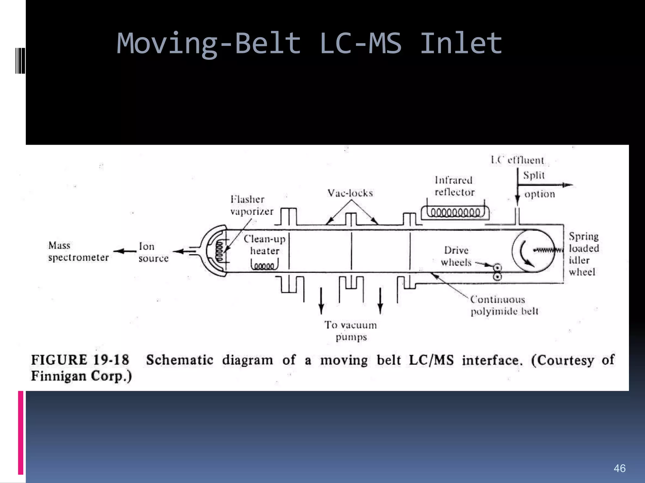
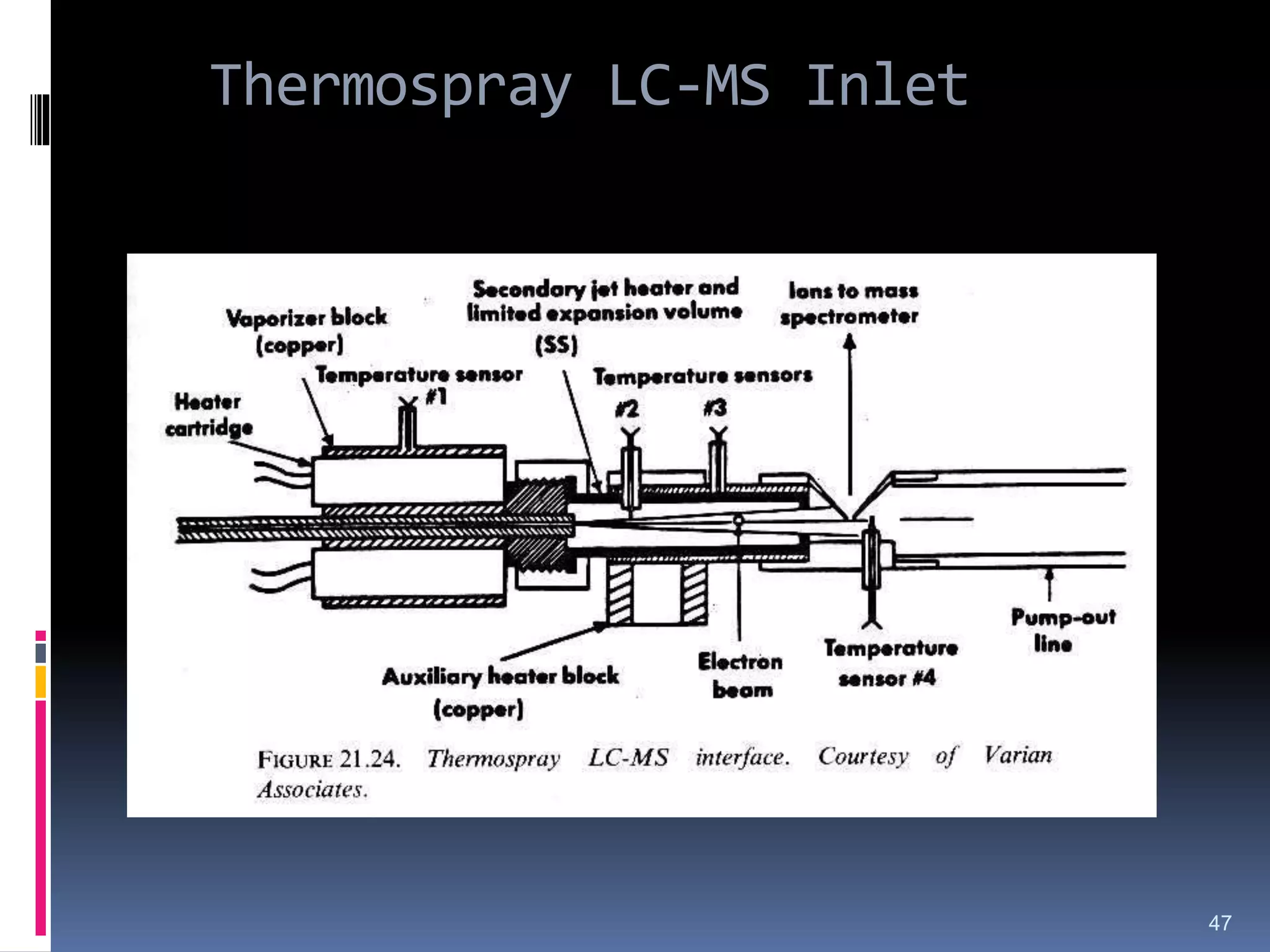
A mixture of compounds is separated by gas chromatography,
then identified by mass spectrometry (GC-MS Inlets)
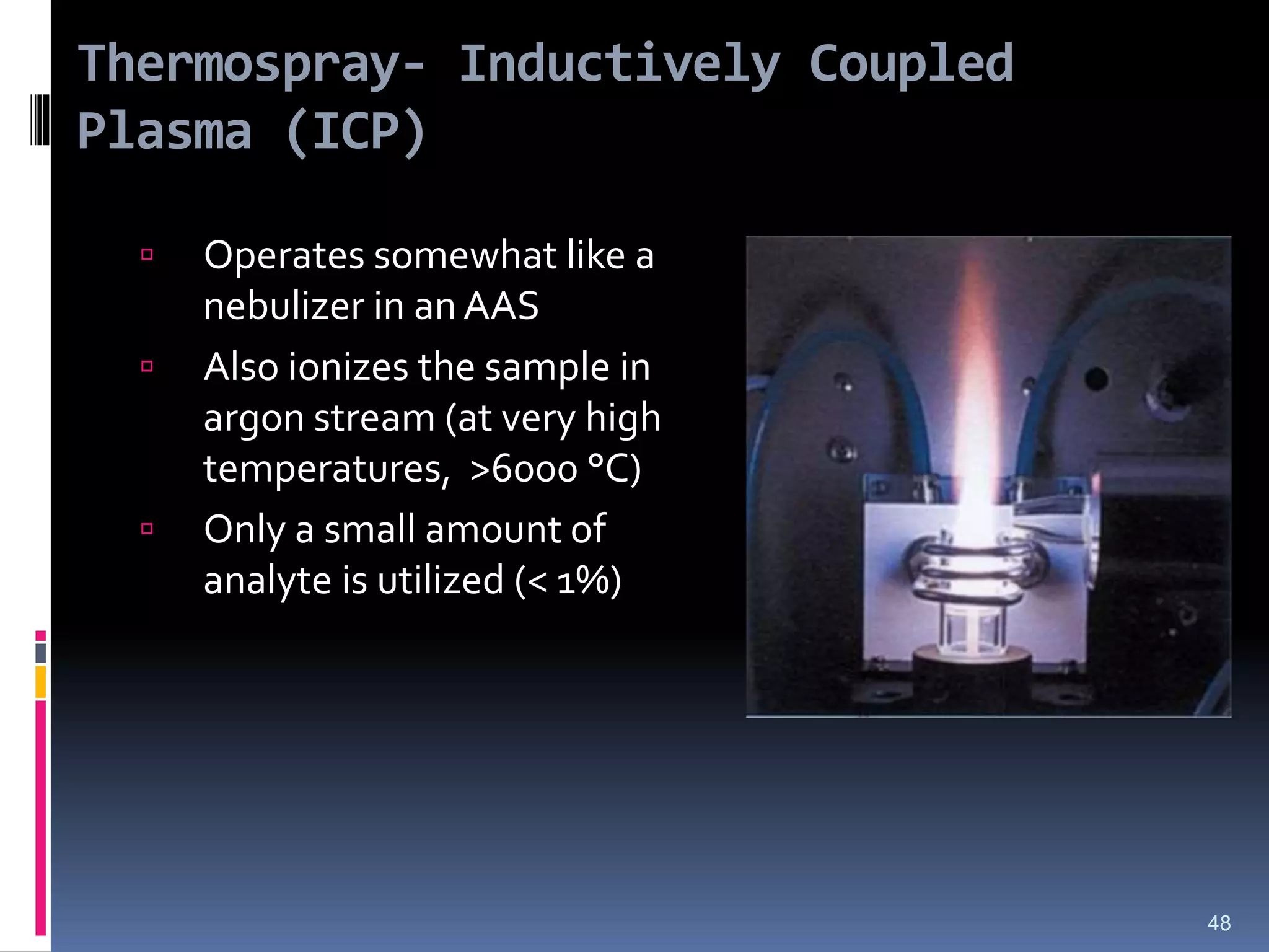
Thermospray- Inductively Coupled
Plasma(ICP)
Operates somewhat like a
nebulizer in an AAS
Also ionizes the sample in
argon stream (at very high
temperatures, >6000 °C)
Only a small amount of
analyte is utilized (< 1%)
48
49.
Mass Analyzer
The functionof the MS analyzer like monochromator in
an optical spectrometer.
Transducer converts the beam of ions to an electrical
signal that can be then Processed, stored in memory.
MS require an elaborate vacuum system to maintain a
low pressure in all of the components except signal
processor
50.
Mass Analyzers
Type MassRange Resolution Sensitivity Advantage Disadvantage
Magnetic
Sector
1-15,000
m/z
0.0001 Low High
resolution
Expensive
Quadrupole 1-5000 m/z Unit High Easy to use;
inexpensive
Low res; low
mass
Ion trap 1-5000 m/z Unit High Easy to use;
inexpensive
Low res; low
mass
Time of
Flight
Unlimited 0.0001 High High mass;
simple design
Fourier
Transform
Up to 70
kDa
0.0001 High Very high res
and mass
Very expensive
Silverstein, et. al., Spectrometric Identification of Organic Compounds, 7th Ed, p 13.
Single Focus
Double Focus
51.
Mass Analyzers
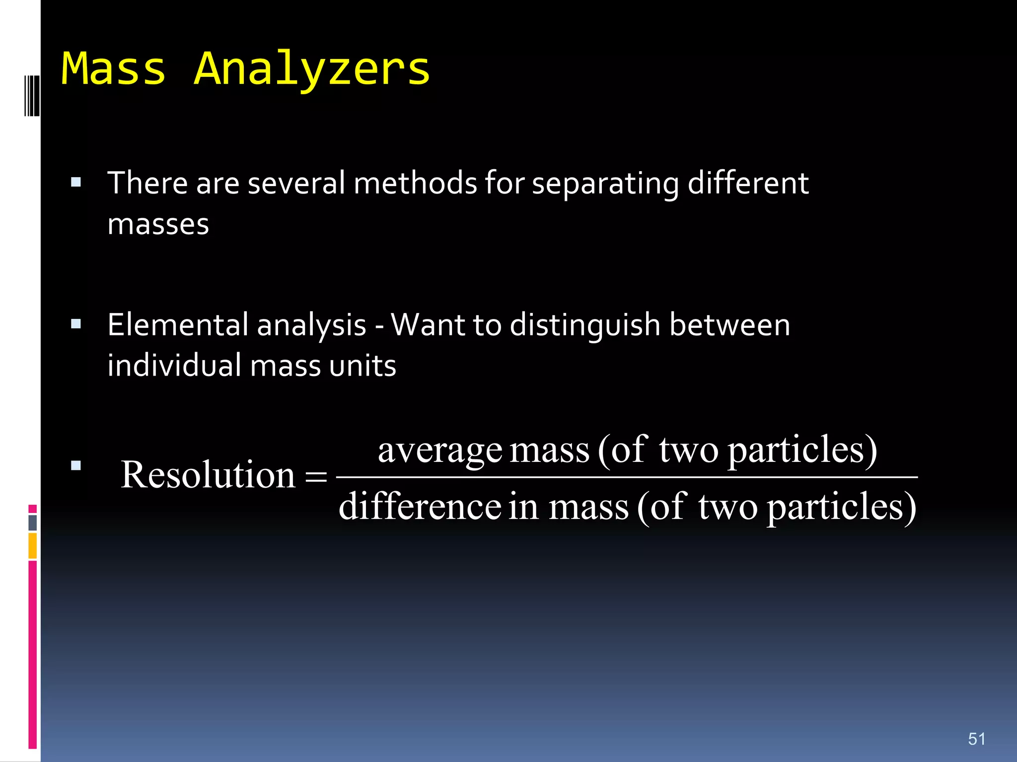
Thereare several methods for separating different
masses
Elemental analysis -Want to distinguish between
individual mass units
particles)
two
(of
mass
in
difference
particles)
two
(of
mass
average
Resolution
51
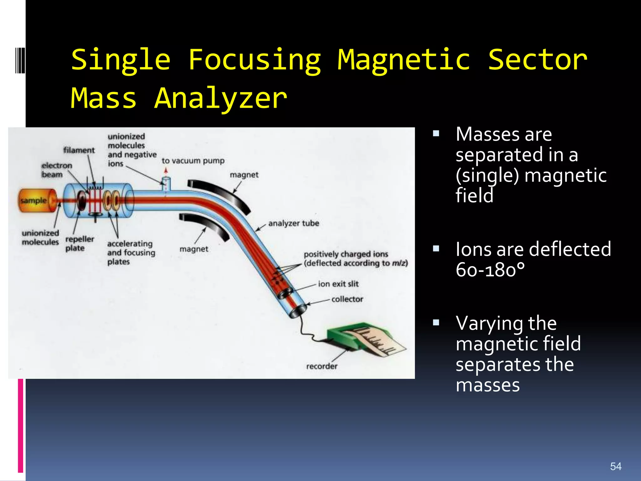
Single Focusing MagneticSector
Mass Analyzer
54
Masses are
separated in a
(single) magnetic
field
Ions are deflected
60-180°
Varying the
magnetic field
separates the
masses
55.
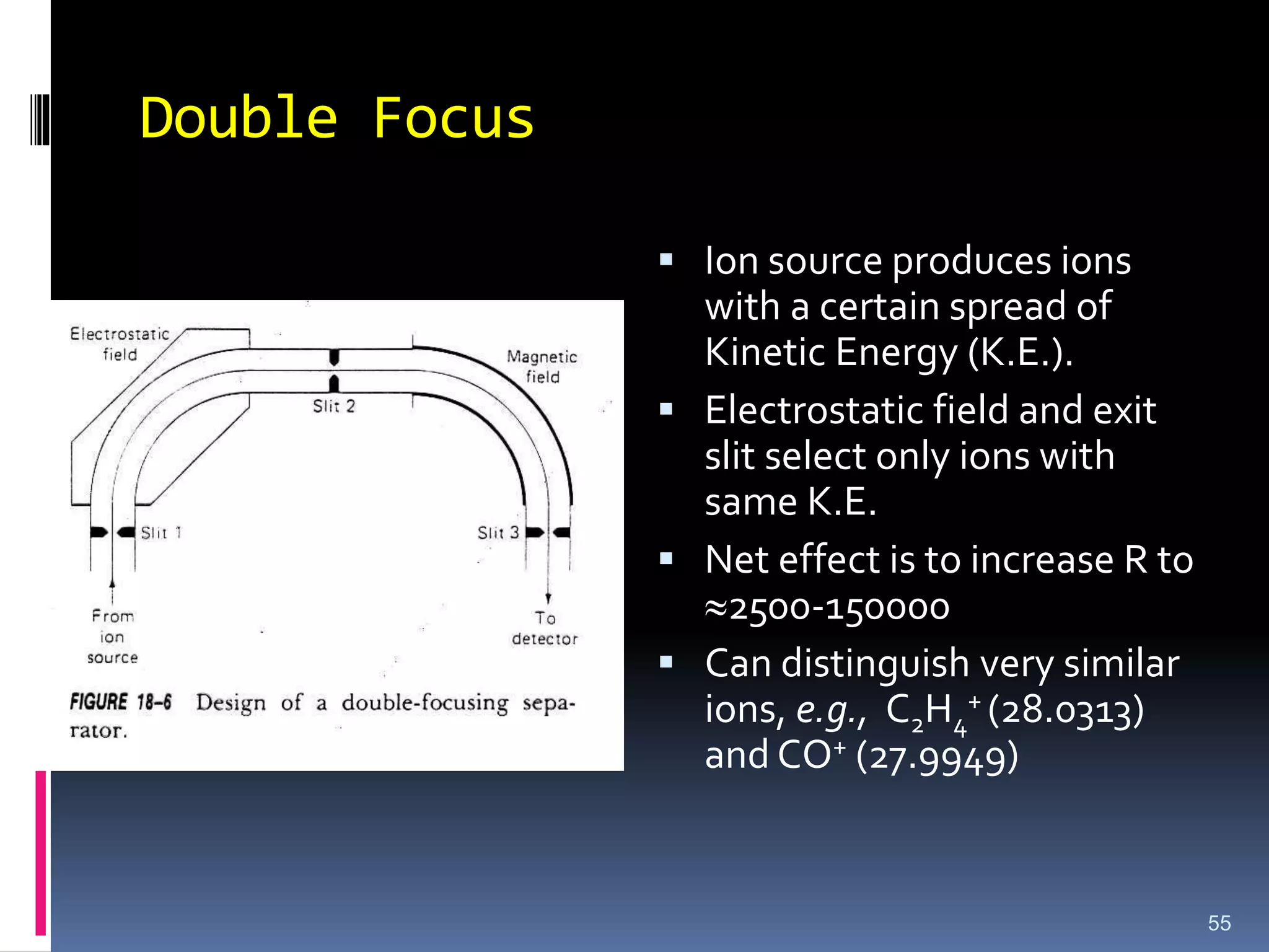
Double Focus
Ionsource produces ions
with a certain spread of
Kinetic Energy (K.E.).
Electrostatic field and exit
slit select only ions with
same K.E.
Net effect is to increase R to
2500-150000
Can distinguish very similar
ions, e.g., C2H4
+ (28.0313)
and CO+ (27.9949)
55
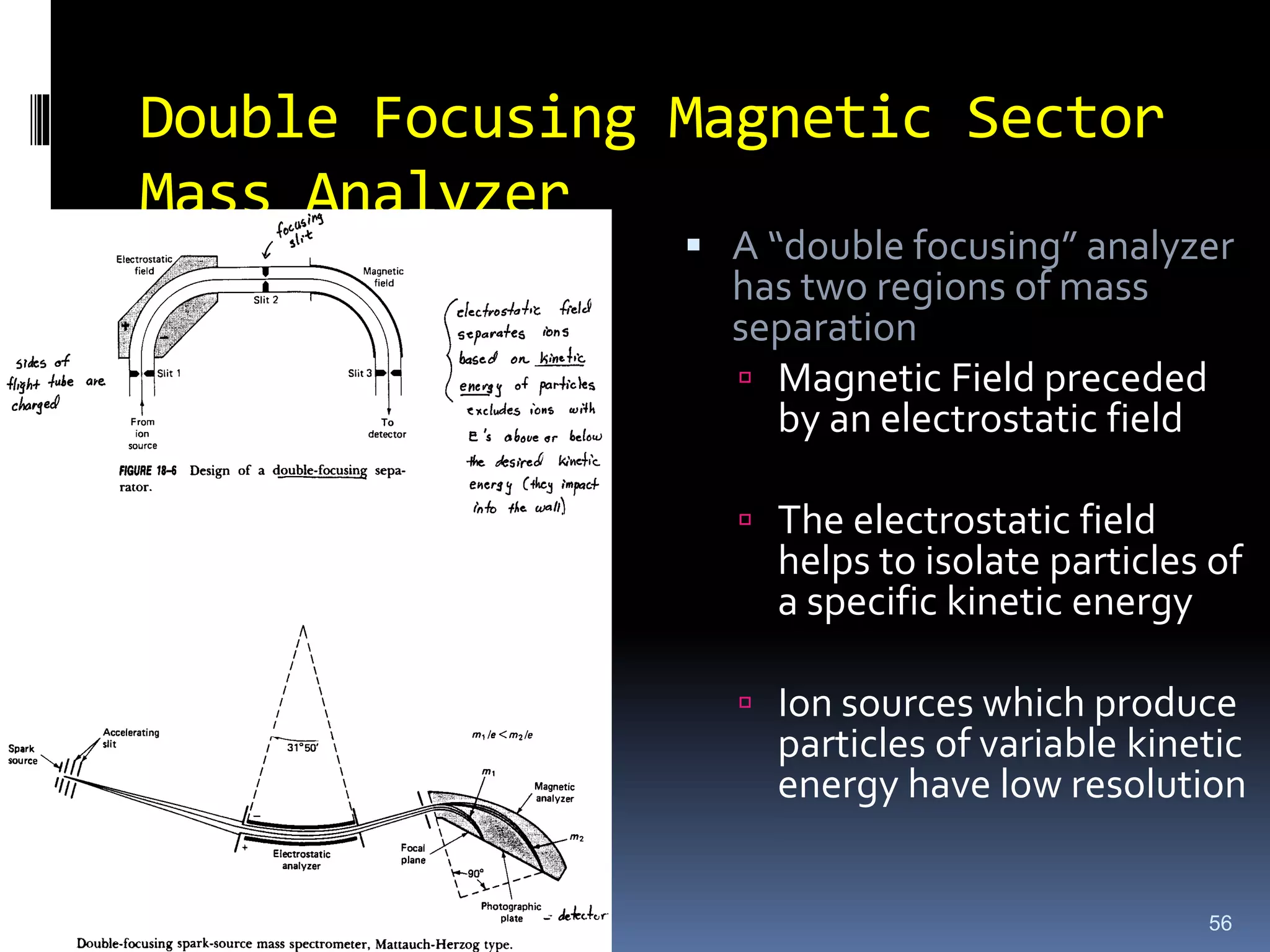
56.
Double Focusing MagneticSector
Mass Analyzer
56
A “double focusing” analyzer
has two regions of mass
separation
Magnetic Field preceded
by an electrostatic field
The electrostatic field
helps to isolate particles of
a specific kinetic energy
Ion sources which produce
particles of variable kinetic
energy have low resolution
57.
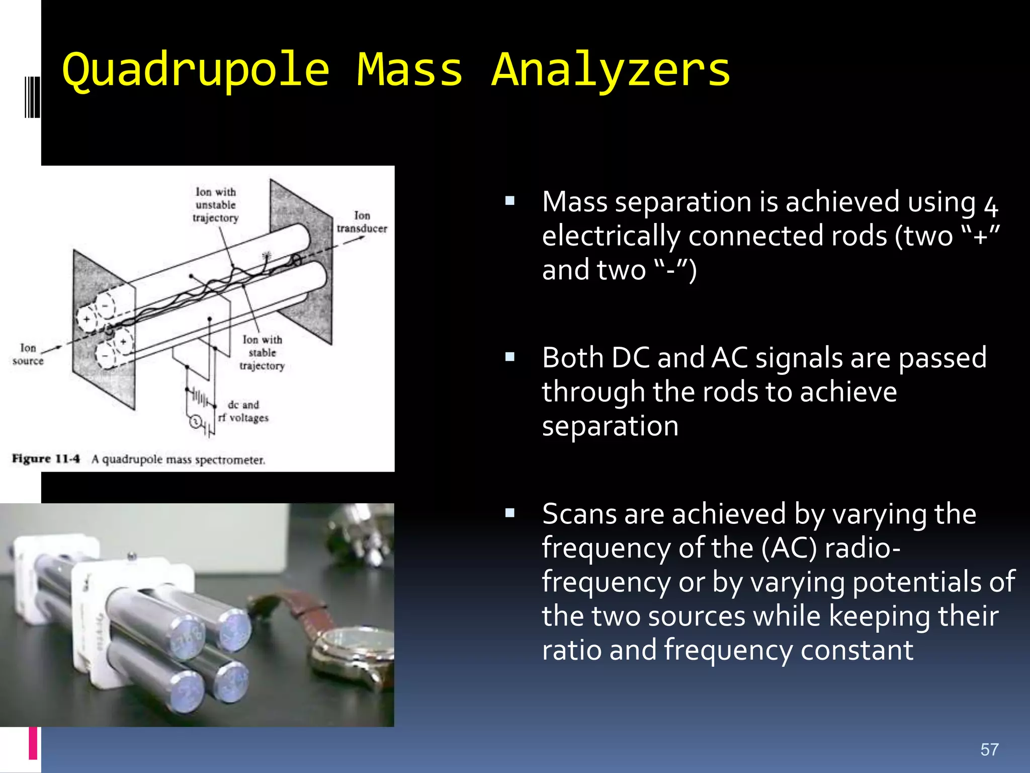
Quadrupole Mass Analyzers
57
Mass separation is achieved using 4
electrically connected rods (two “+”
and two “-”)
Both DC and AC signals are passed
through the rods to achieve
separation
Scans are achieved by varying the
frequency of the (AC) radio-
frequency or by varying potentials of
the two sources while keeping their
ratio and frequency constant
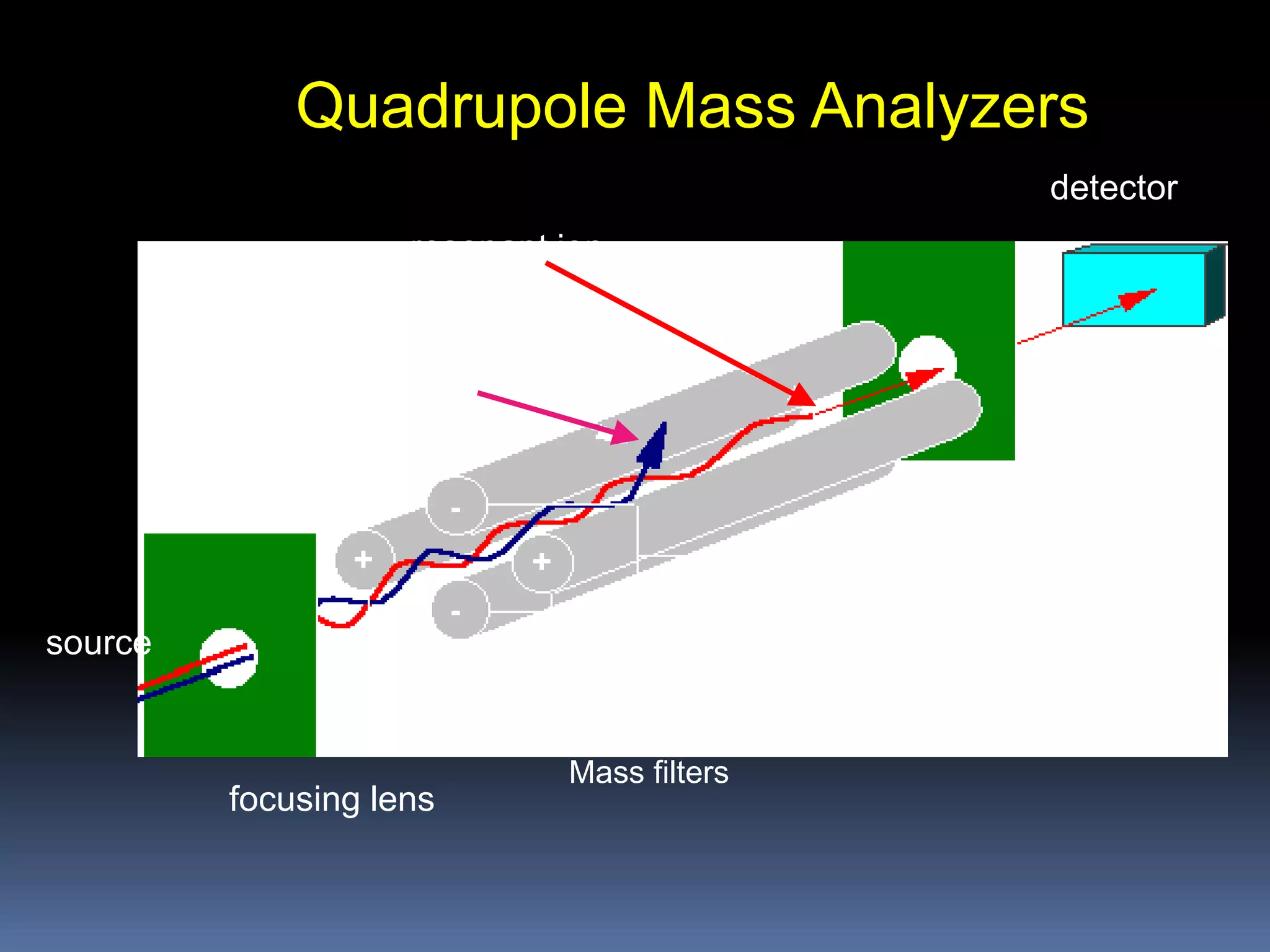
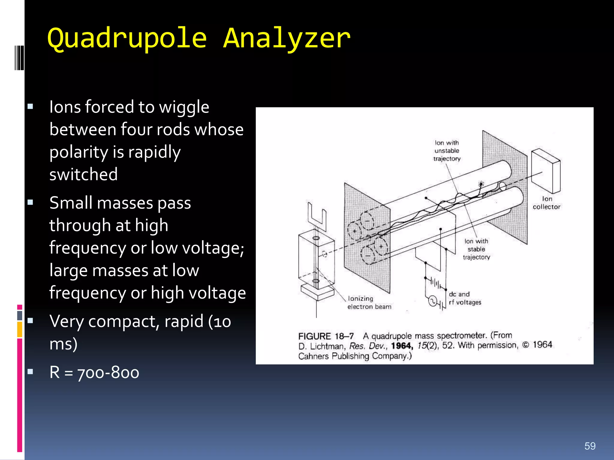
Quadrupole Analyzer
Ionsforced to wiggle
between four rods whose
polarity is rapidly
switched
Small masses pass
through at high
frequency or low voltage;
large masses at low
frequency or high voltage
Very compact, rapid (10
ms)
R = 700-800
59
60.
Merit and Demerit
Classicalmass spectra
Good reproducibility
Relatively small/ compact,
Relatively low-cost systems
Limited resolution
Peak heights variable as a function of mass (mass
discrimination). Peak height vs. mass response must be
'tuned'.
Not well suited for pulsed ionization methods
60
61.
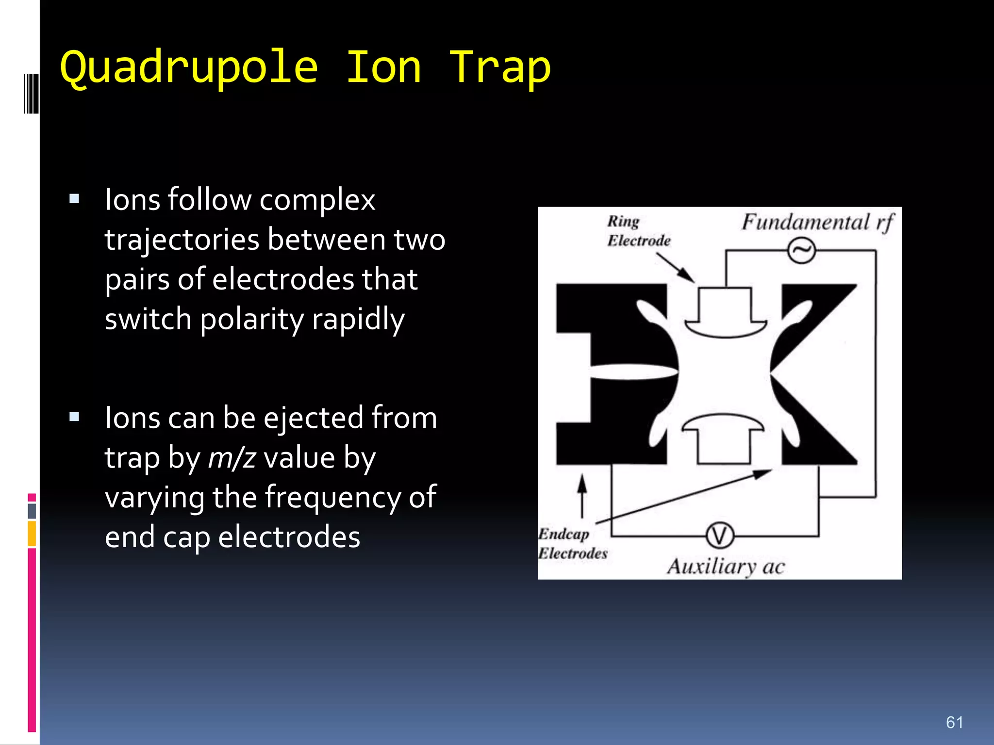
Quadrupole Ion Trap
Ions follow complex
trajectories between two
pairs of electrodes that
switch polarity rapidly
Ions can be ejected from
trap by m/z value by
varying the frequency of
end cap electrodes
61
62.
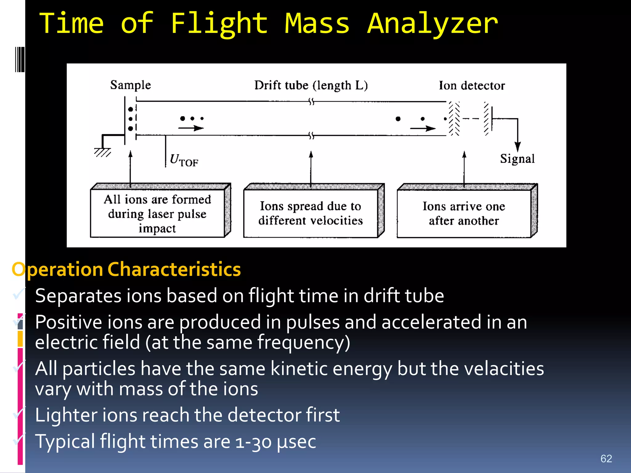
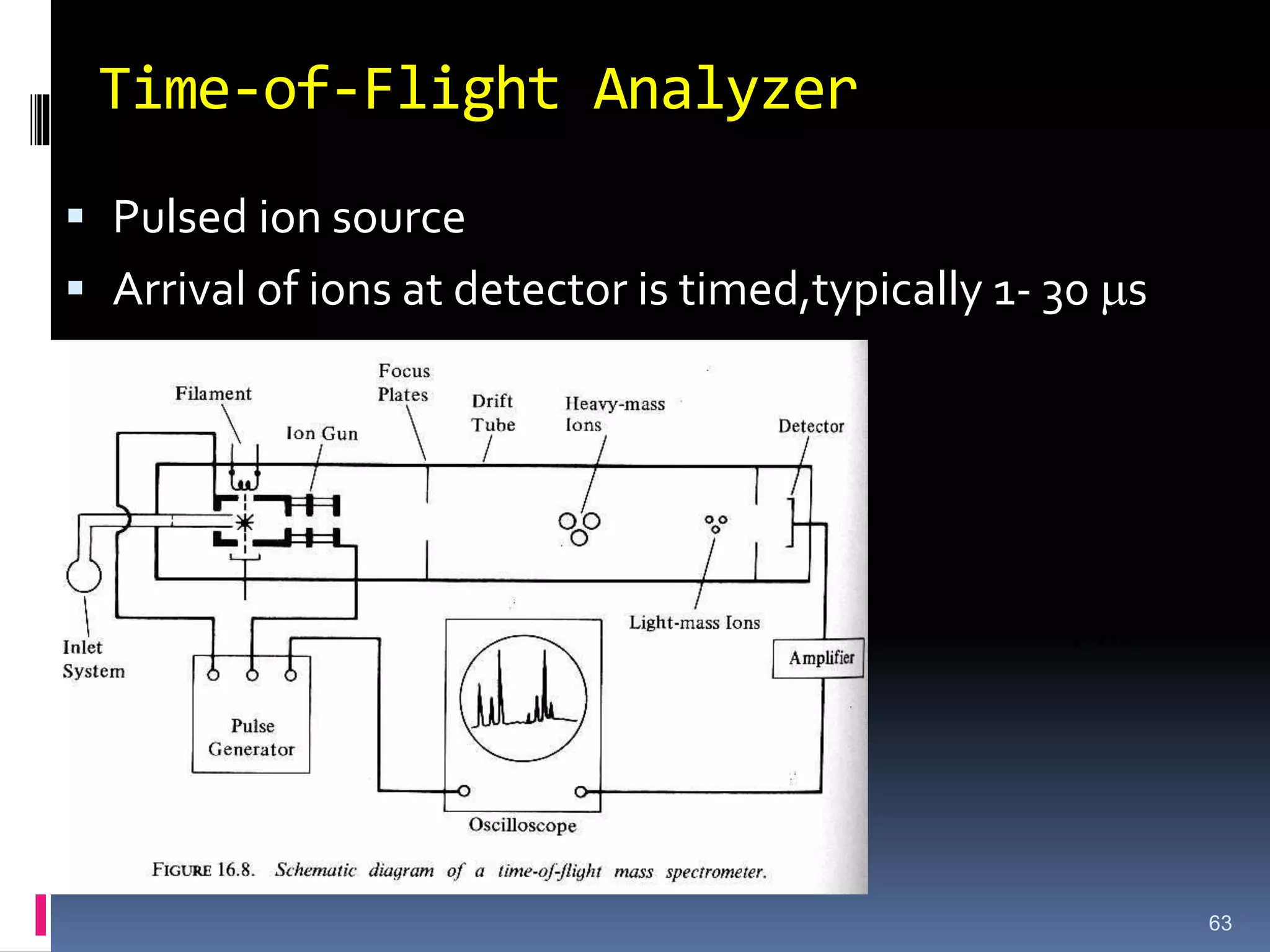
Time of FlightMass Analyzer
62
Operation Characteristics
Separates ions based on flight time in drift tube
Positive ions are produced in pulses and accelerated in an
electric field (at the same frequency)
All particles have the same kinetic energy but the velacities
vary with mass of the ions
Lighter ions reach the detector first
Typical flight times are 1-30 µsec
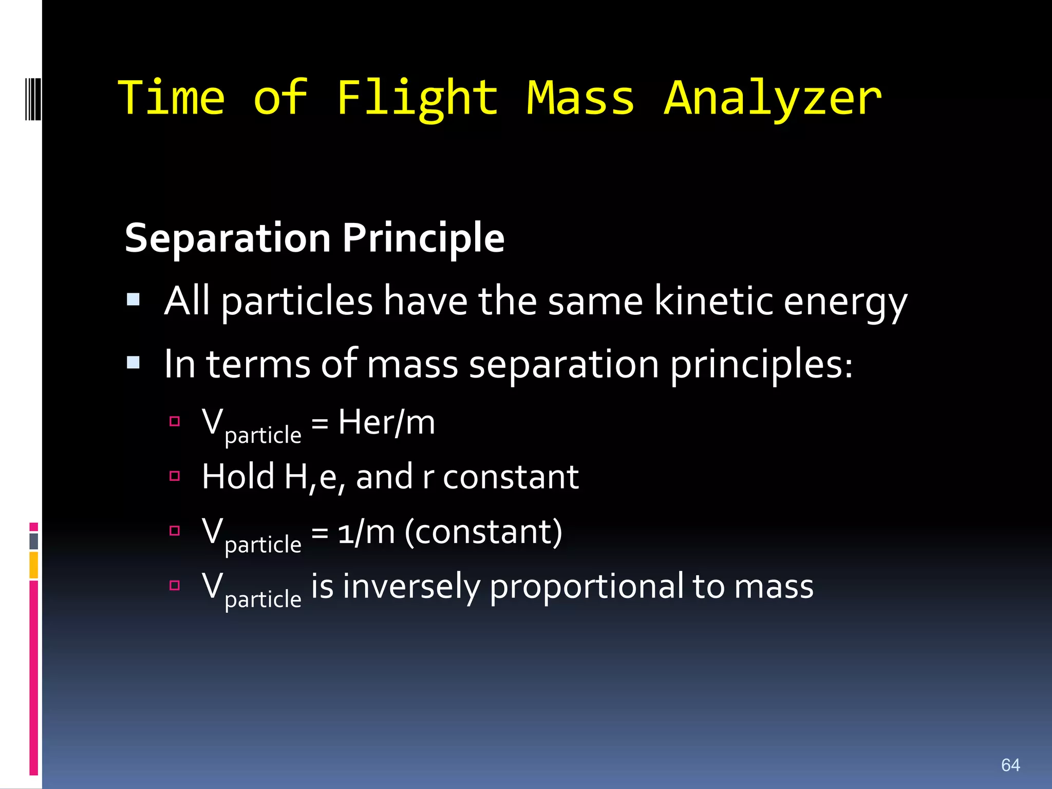
Time of FlightMass Analyzer
Separation Principle
All particles have the same kinetic energy
In terms of mass separation principles:
Vparticle = Her/m
Hold H,e, and r constant
Vparticle = 1/m (constant)
Vparticle is inversely proportional to mass
64
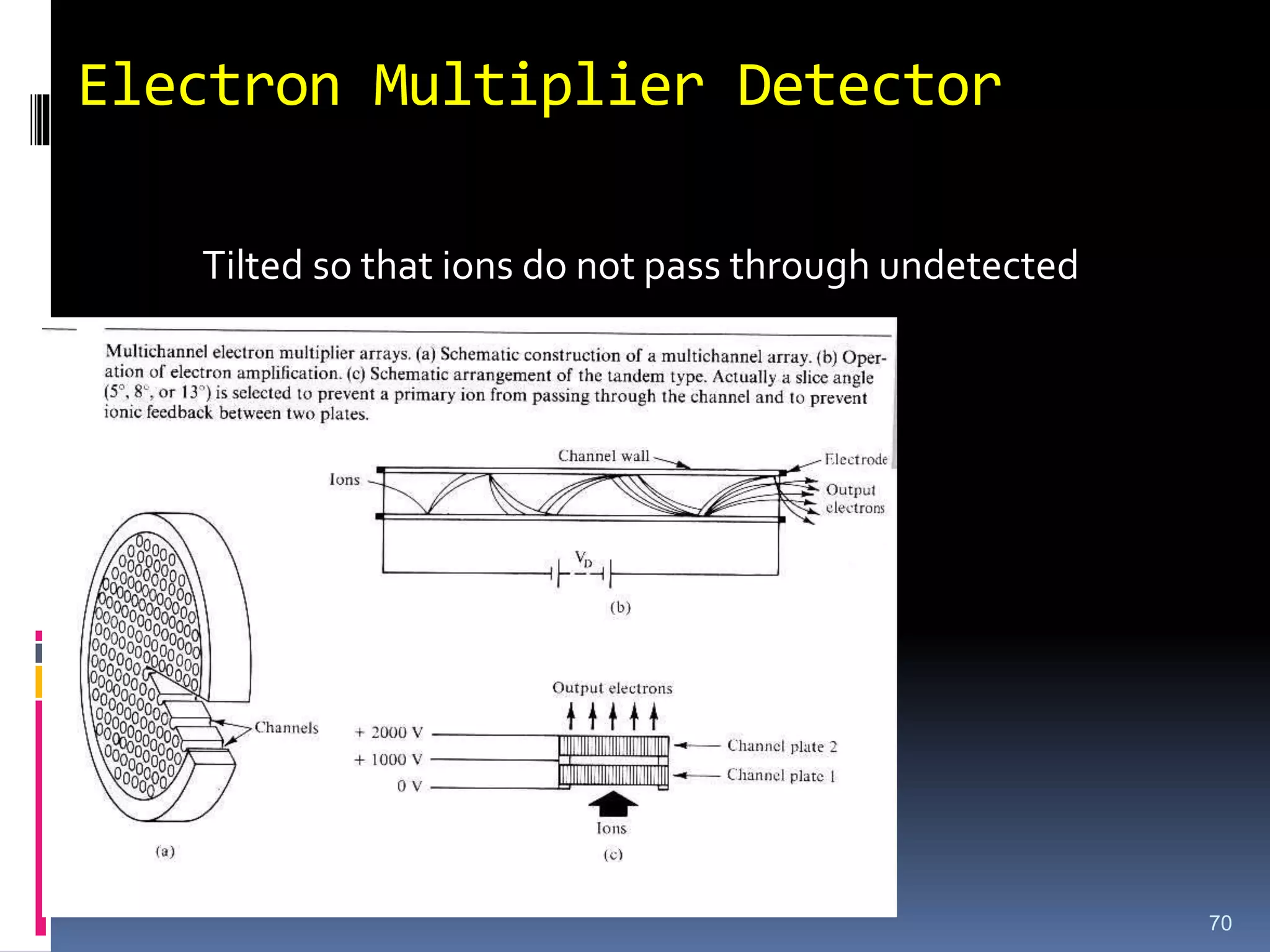
Detectors for MS
Two BasicTypes
1. Electron Multipliers
2. Faraday Cup
Time of Flight (TOF) and FourierTransform Ion-
Cyclotron Resonance (FTICR) instruments can
separate more than one m/e- ratio
simultaneously
Multiple detectors are required in this case
66
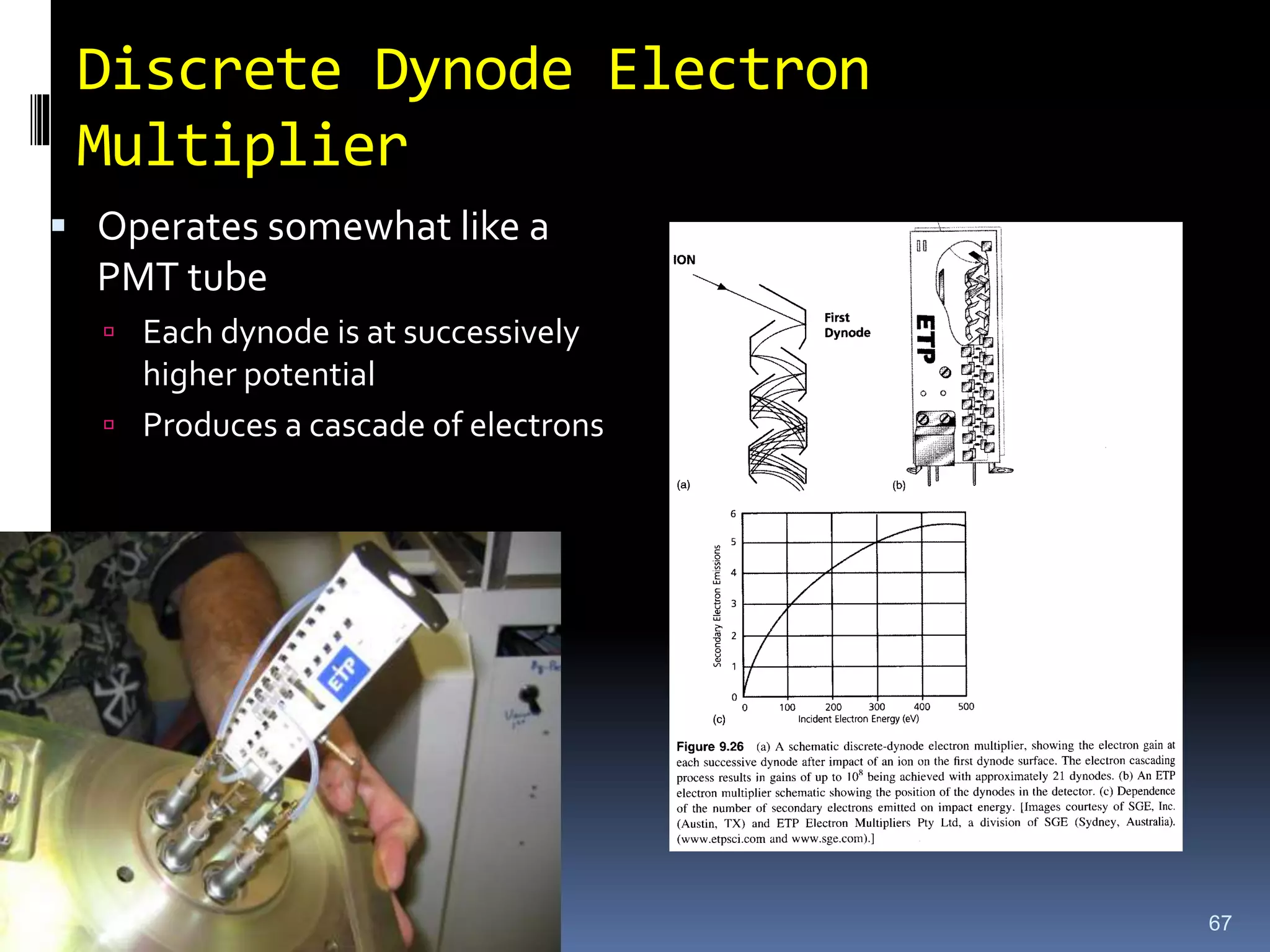
67.
Discrete Dynode Electron
Multiplier
Operates somewhat like a
PMT tube
Each dynode is at successively
higher potential
Produces a cascade of electrons
67
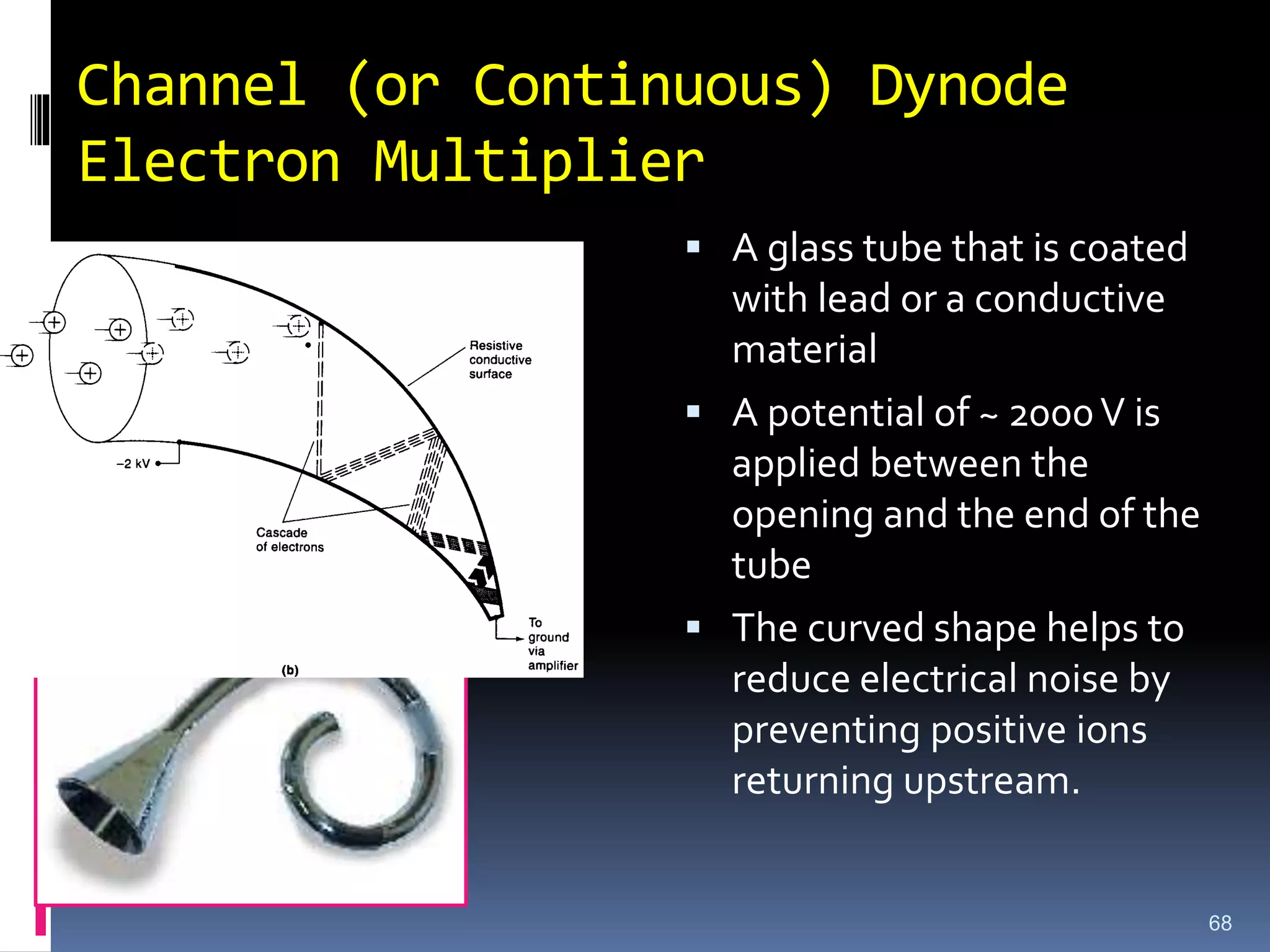
68.
Channel (or Continuous)Dynode
Electron Multiplier
A glass tube that is coated
with lead or a conductive
material
A potential of ~ 2000V is
applied between the
opening and the end of the
tube
The curved shape helps to
reduce electrical noise by
preventing positive ions
returning upstream.
68
69.
Dynode-Based Detectors
Adisadvantage of dynode-based detectors is
that the number of secondary electrons released
in a detector depends on the type of primary
particle, its energy and its incident angle,
Mass discrimination occurs when ions enter the
detector with different velocities.
69
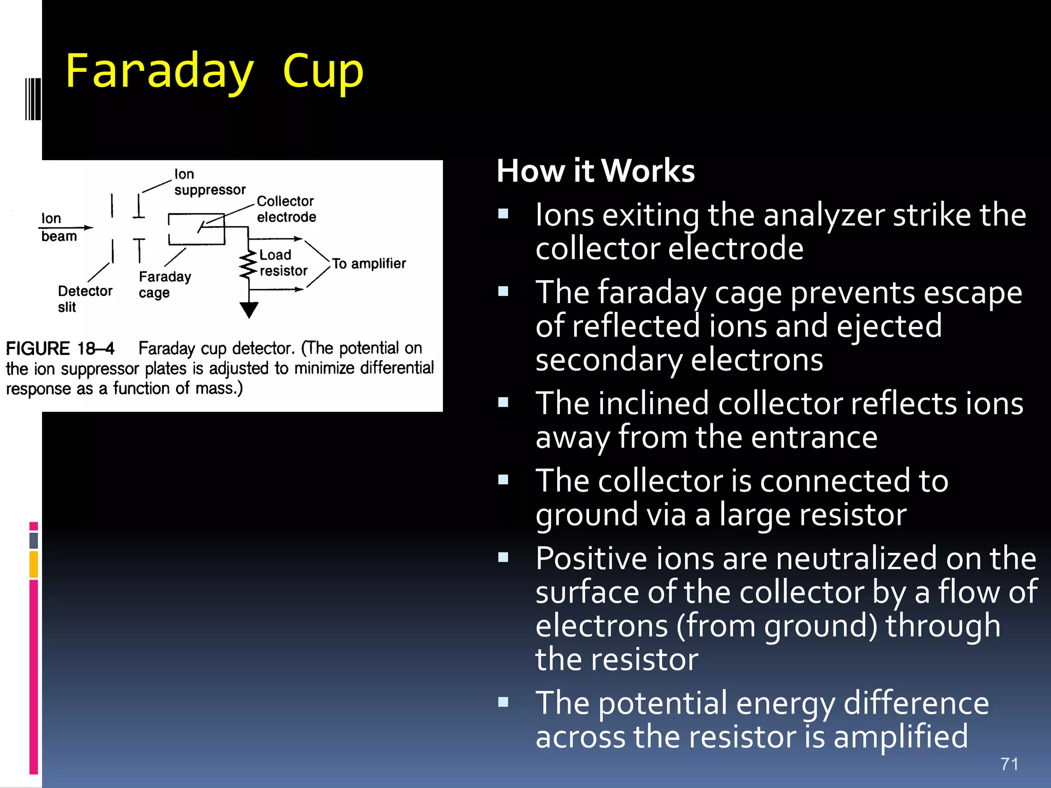
Faraday Cup
71
How itWorks
Ions exiting the analyzer strike the
collector electrode
The faraday cage prevents escape
of reflected ions and ejected
secondary electrons
The inclined collector reflects ions
away from the entrance
The collector is connected to
ground via a large resistor
Positive ions are neutralized on the
surface of the collector by a flow of
electrons (from ground) through
the resistor
The potential energy difference
across the resistor is amplified
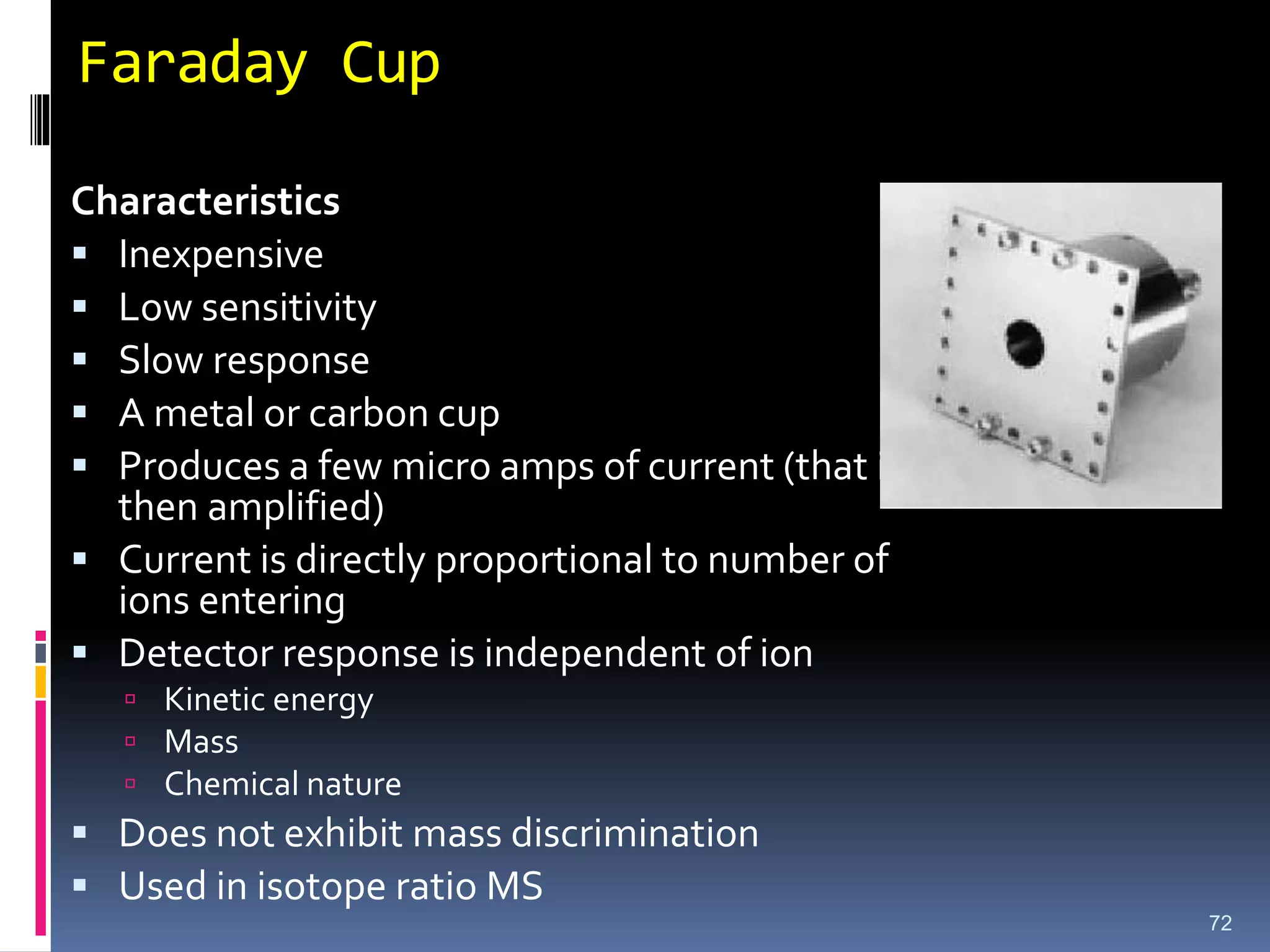
72.
Faraday Cup
Characteristics
Inexpensive
Low sensitivity
Slow response
A metal or carbon cup
Produces a few micro amps of current (that is
then amplified)
Current is directly proportional to number of
ions entering
Detector response is independent of ion
Kinetic energy
Mass
Chemical nature
Does not exhibit mass discrimination
Used in isotope ratio MS
72
73.
Application of MS
1.Drug discovery, combinatorial chemistry, Drug
testing/Pharmacokinetics
2. Antiterror/Security (e.g. bomb molecule ‘sniffers’)
3. Environmental Analysis (e.g. water quality testing)
4. Quality Control (food, pharmaceuticals)
5. Medical Testing (various blood illnesses and… cancer?)
6. Validation of art/History/Anthropology etc.
7. Validation during chemical synthesis
8. Biochemical research (proteomics, interact…omics)
9. Tissue imaging (with MALDI)
10. Analysis of Proteins, peptides, olegonucleotides
11. Clinical testing etc 73
-