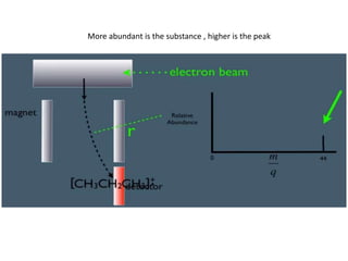
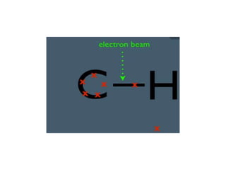
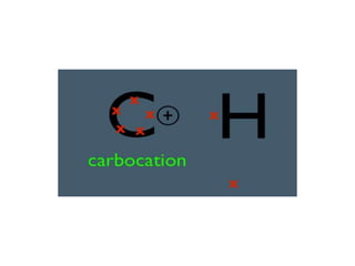
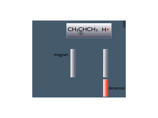
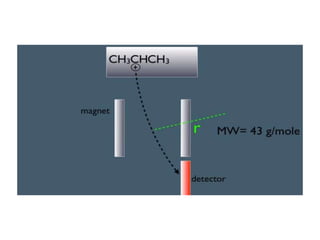
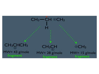
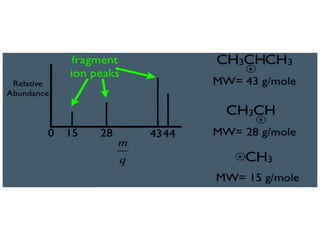
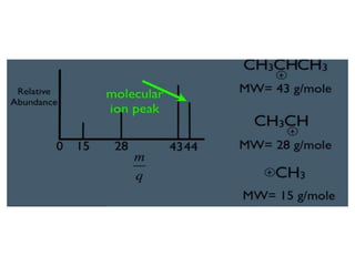
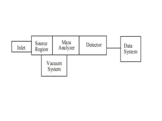
• Mass Spectrometry is a powerful technique
for identifying unknowns, studying molecular
structure.
• mass spectrometry is based upon the motion
of a charged particle, called an ion, in an
electric or magnetic field. The mass to charge
ratio (m/z) of the ion effects this motion.
More abundant is the substance , higher is the peak
Hydrogen carries 1 proton and 1 electron so it is neutral and move straight
Base peak is due to most stable fragment
Methyl gives least abundant peak and is not stable or you amy not see any peak on mass 15
• The mass to charge ratio (m/z) is used to describe ions
observed in mass spectrometry. By convention, m is the
numerical value for the mass of the ion and z is the
numerical value for the charge of the ion.
• The elementary charge unit is defined as z is an integer
equal to the number of electrons lost (or gained for
negative ions).
• For most experiments one electron is lost during ionization
so z is +1 and the m/z value is equivalent to the relative
molecular mass of the ion. Because the unified atomic
mass and the charge number are pure numbers the mass-
to-charge ratio is a number and does not have any units.
• A variety of ionization techniques are used for
mass spectrometry. Most ionization techniques
excite the neutral analyte molecule which then
ejects an electron to form a radical cation (M+ ) .
Other ionization techniques involve ion molecule
reactions that produce adduct MH+ ions (MH ).
• The most important considerations are the
physical state of the analyte and the ionization
energy
• Electron ionization and chemical ionization are only
suitable for gas phase ionization. Fast atom
bombardment, secondary ion mass spectrometry,
electrospray, and matrix assisted laser desorption are
used to ionize condensed phase samples.
• Some ionization techniques are very soft and only
produce molecular ions, other techniques are very
energetic and cause ions to undergo extensive *
fragmentation. Although this fragmentation
complicates the mass spectrum, it provides structural
information for the identification of unknown
compounds
Electron Ionization
• The electrons used for ionization are produced by passing a current
through a wire filament. The amount of current controls the number of
electrons emitted by the filament. An electric field accelerates these
electrons across the source region to produce a beam of high energy
electrons. When an analyte molecule passes through this electron beam, a
valence shell electron can be removed from the molecule to produce an
ion
• EI produces positive ions by knocking a valence electron off
the analyte molecule. As the electron passes close to the
molecule the negative charge of the electron repels and
distorts the electron cloud surrounding the molecule. This
distortion transfers kinetic energy from the fast-moving
electron to the electron cloud of the molecule. If enough
energy is transferred by the process, the molecule will eject
a valence electron and form a radical cation (M ).
• The abundance of the resulting fragments,
often called product ions * , is determined by
the kinetics of the fragmentation pathways
and the ionization energy.
• Changing the ionization energy changes the
observed distribution of fragment ions.
• This distribution provides the structural
information for interpreting mass spectra
Chemical Ionization
• The reagent gas in the CI source is ionized with an electron
beam to produce a cloud of ions.
• The reagent gas ions in this cloud react and produce
adduct ions like CH5+ (Figure 4), which are excellent
proton donors.
• When analyte molecules (M) are introduced to a source
region with this cloud of ions, the reagent gas ions donate
a proton to the analyte molecule and produce MH ion.
• The energetics of the proton transfer is controlled by using
different reagent gases. The most common reagent gases
are methane, isobutane and ammonia.
• Methane is the strongest proton donor
Fast Atom Bombardment and
Secondary Ion Mass Spectrometry
Fast Atom Bombardment (FAB) and Secondary Ion Mass
Spectrometry (SIMS) both use high energy atoms to
sputter and ionize the sample in a single step.
In these techniques, a beam of rare gas neutrals (FAB) or
ions (SIMS) is focused on the liquid or solid sample.
The impact of this high energy beam causes the
analyte molecules to sputter into the gas phase and
ionize in a single step.
Since no heating is required, sputtering techniques
(especially FAB) are useful for studying thermally
labile compounds that decompose in conventional
inlets
• The most significant difference between FAB and SIMS is
the sample preparation.
• In FAB the analyte is dissolved in a liquid matrix. A drop of
the sample/matrix mixture is placed at the end of an
insertion probe and introduced to the source region. The
fast atom beam is focused on this droplet to produce
analyte ions.
• Glycerol or similar low vapor pressure liquids are typically
used for the matrix. Ideally, the analyte is soluble in the
liquid matrix and a monolayer of analyte forms on the
surface of the droplet.
• SIMS experiments are used to study surface species and
solid samples.
• No matrix is used and the ionizing beam is focused
directly on the sample. Although this makes sampling more
difficult, it is useful for studying surface chemistry
• Atmospheric Pressure Ionization (API) sources
ionize the sample at atmospheric pressure
and then transfer the ions into the mass
spectrometer. These techniques are used to
ionize thermally labile samples such as
peptides, proteins and polymers directly from
the condensed phase. The sample is dissolved
in an appropriate solvent and this solution is
introduced into the mass spectrometer.
ElectroSpray Ionization (ESI) is the most
common API application
• As the liquid leaves the nozzle, the electric field
induces a net charge on the small droplets.
• As the solvent evaporates, the droplet shrinks
and the charge density at the surface of the
droplet increases. The droplet finally reaches a
point where the coulombic repulsion from this
electric charge is greater than the surface
tension holding it together.
• This causes the droplet to explode and produce
multiply charged analyte ions.
Matrix Assisted Laser Desorption/Ionization
• Matrix Assisted Laser Desorption/Ionization
(MALDI) is used to analyze extremely large
molecules. This technique directly ionizes and
vaporizes the analyte from the condensed
phase.
• MALDI is often used for the analysis of
synthetic and natural polymers, proteins, and
peptides.
• Analysis of compounds with molecular
weights up to 200,000 dalton is possible and
this high mass limit is continually increasing.
• In MALDI, both desorption and ionization are
induced by a single laser pulse.
• The sample is prepared by mixing the analyte
and a matrix compound chosen to absorb the
laser wavelength.
• This is placed on a probe tip and dried.
• A vacuum lock is used to insert the probe into the
source region of the mass spectrometer.
• A laser beam is then focused on this dried
mixture and the energy from a laser pulse is
absorbed by the matrix.
• This energy ejects analyte ions from the surface
so that a mass spectrum is acquired for each
laser pulse.
MALDI Ionization
• After ions are formed in the source region they are
accelerated into the mass analyzer by an electric field.
• The mass analyzer separates these ions according to
their m/z value.
• The selection of a mass analyzer depends upon the
resolution, mass range, scan rate and detection limits
required for an application.
• Analyzers transmit a single selected m/z to the
detector and the mass spectrum is obtained by
scanning the analyzer so that different mass to charge
ratio ions are detected.
Mass Analyzer
• The quadrupole mass spectrometer is the most common
mass analyzer. Its compact size, fast scan rate, high
transmission efficiency, and modest vacuum requirements
are ideal for small inexpensive instruments.
• In the mass spectrometer, an electric field accelerates ions
out of the source region and into the quadrupole analyzer.
The analyzer consists of four rods or electrodes arranged
across from each other (Figure 9). As the ions travel
through the quadrupole they are filtered according to their
m/z value so that only a single m/z value ion can strike the
detector. The m/z value transmitted by the quadrupole is
determined by the Radio Frequency (RF) and Direct Current
(DC) voltages applied to the electrodes
• Time-of-Flight. The time-of-flight (TOF) mass
analyzer separates ions in time as they travel
down a flight tube.
• This is a very simple mass spectrometer that uses
fixed voltages and does not require a magnetic
field. The greatest drawback is that TOF
instruments have poor mass resolution, usually
less than 500. These instruments have high
transmission efficiency, no upper m/z limit, very
low detection limits, and fast scan rates.

Mass spectrometry.pptx

  • 1.
    • Mass Spectrometryis a powerful technique for identifying unknowns, studying molecular structure. • mass spectrometry is based upon the motion of a charged particle, called an ion, in an electric or magnetic field. The mass to charge ratio (m/z) of the ion effects this motion.
  • 8.
    More abundant isthe substance , higher is the peak
  • 12.
    Hydrogen carries 1proton and 1 electron so it is neutral and move straight
  • 18.
    Base peak isdue to most stable fragment
  • 19.
    Methyl gives leastabundant peak and is not stable or you amy not see any peak on mass 15
  • 21.
    • The massto charge ratio (m/z) is used to describe ions observed in mass spectrometry. By convention, m is the numerical value for the mass of the ion and z is the numerical value for the charge of the ion. • The elementary charge unit is defined as z is an integer equal to the number of electrons lost (or gained for negative ions). • For most experiments one electron is lost during ionization so z is +1 and the m/z value is equivalent to the relative molecular mass of the ion. Because the unified atomic mass and the charge number are pure numbers the mass- to-charge ratio is a number and does not have any units.
  • 22.
    • A varietyof ionization techniques are used for mass spectrometry. Most ionization techniques excite the neutral analyte molecule which then ejects an electron to form a radical cation (M+ ) . Other ionization techniques involve ion molecule reactions that produce adduct MH+ ions (MH ). • The most important considerations are the physical state of the analyte and the ionization energy
  • 23.
    • Electron ionizationand chemical ionization are only suitable for gas phase ionization. Fast atom bombardment, secondary ion mass spectrometry, electrospray, and matrix assisted laser desorption are used to ionize condensed phase samples. • Some ionization techniques are very soft and only produce molecular ions, other techniques are very energetic and cause ions to undergo extensive * fragmentation. Although this fragmentation complicates the mass spectrum, it provides structural information for the identification of unknown compounds
  • 24.
    Electron Ionization • Theelectrons used for ionization are produced by passing a current through a wire filament. The amount of current controls the number of electrons emitted by the filament. An electric field accelerates these electrons across the source region to produce a beam of high energy electrons. When an analyte molecule passes through this electron beam, a valence shell electron can be removed from the molecule to produce an ion
  • 25.
    • EI producespositive ions by knocking a valence electron off the analyte molecule. As the electron passes close to the molecule the negative charge of the electron repels and distorts the electron cloud surrounding the molecule. This distortion transfers kinetic energy from the fast-moving electron to the electron cloud of the molecule. If enough energy is transferred by the process, the molecule will eject a valence electron and form a radical cation (M ).
  • 26.
    • The abundanceof the resulting fragments, often called product ions * , is determined by the kinetics of the fragmentation pathways and the ionization energy. • Changing the ionization energy changes the observed distribution of fragment ions. • This distribution provides the structural information for interpreting mass spectra
  • 27.
    Chemical Ionization • Thereagent gas in the CI source is ionized with an electron beam to produce a cloud of ions. • The reagent gas ions in this cloud react and produce adduct ions like CH5+ (Figure 4), which are excellent proton donors. • When analyte molecules (M) are introduced to a source region with this cloud of ions, the reagent gas ions donate a proton to the analyte molecule and produce MH ion. • The energetics of the proton transfer is controlled by using different reagent gases. The most common reagent gases are methane, isobutane and ammonia. • Methane is the strongest proton donor
  • 28.
    Fast Atom Bombardmentand Secondary Ion Mass Spectrometry Fast Atom Bombardment (FAB) and Secondary Ion Mass Spectrometry (SIMS) both use high energy atoms to sputter and ionize the sample in a single step. In these techniques, a beam of rare gas neutrals (FAB) or ions (SIMS) is focused on the liquid or solid sample. The impact of this high energy beam causes the analyte molecules to sputter into the gas phase and ionize in a single step. Since no heating is required, sputtering techniques (especially FAB) are useful for studying thermally labile compounds that decompose in conventional inlets
  • 29.
    • The mostsignificant difference between FAB and SIMS is the sample preparation. • In FAB the analyte is dissolved in a liquid matrix. A drop of the sample/matrix mixture is placed at the end of an insertion probe and introduced to the source region. The fast atom beam is focused on this droplet to produce analyte ions. • Glycerol or similar low vapor pressure liquids are typically used for the matrix. Ideally, the analyte is soluble in the liquid matrix and a monolayer of analyte forms on the surface of the droplet. • SIMS experiments are used to study surface species and solid samples. • No matrix is used and the ionizing beam is focused directly on the sample. Although this makes sampling more difficult, it is useful for studying surface chemistry
  • 30.
    • Atmospheric PressureIonization (API) sources ionize the sample at atmospheric pressure and then transfer the ions into the mass spectrometer. These techniques are used to ionize thermally labile samples such as peptides, proteins and polymers directly from the condensed phase. The sample is dissolved in an appropriate solvent and this solution is introduced into the mass spectrometer.
  • 31.
    ElectroSpray Ionization (ESI)is the most common API application • As the liquid leaves the nozzle, the electric field induces a net charge on the small droplets. • As the solvent evaporates, the droplet shrinks and the charge density at the surface of the droplet increases. The droplet finally reaches a point where the coulombic repulsion from this electric charge is greater than the surface tension holding it together. • This causes the droplet to explode and produce multiply charged analyte ions.
  • 32.
    Matrix Assisted LaserDesorption/Ionization • Matrix Assisted Laser Desorption/Ionization (MALDI) is used to analyze extremely large molecules. This technique directly ionizes and vaporizes the analyte from the condensed phase. • MALDI is often used for the analysis of synthetic and natural polymers, proteins, and peptides. • Analysis of compounds with molecular weights up to 200,000 dalton is possible and this high mass limit is continually increasing.
  • 33.
    • In MALDI,both desorption and ionization are induced by a single laser pulse. • The sample is prepared by mixing the analyte and a matrix compound chosen to absorb the laser wavelength. • This is placed on a probe tip and dried. • A vacuum lock is used to insert the probe into the source region of the mass spectrometer. • A laser beam is then focused on this dried mixture and the energy from a laser pulse is absorbed by the matrix. • This energy ejects analyte ions from the surface so that a mass spectrum is acquired for each laser pulse.
  • 34.
  • 35.
    • After ionsare formed in the source region they are accelerated into the mass analyzer by an electric field. • The mass analyzer separates these ions according to their m/z value. • The selection of a mass analyzer depends upon the resolution, mass range, scan rate and detection limits required for an application. • Analyzers transmit a single selected m/z to the detector and the mass spectrum is obtained by scanning the analyzer so that different mass to charge ratio ions are detected.
  • 36.
    Mass Analyzer • Thequadrupole mass spectrometer is the most common mass analyzer. Its compact size, fast scan rate, high transmission efficiency, and modest vacuum requirements are ideal for small inexpensive instruments. • In the mass spectrometer, an electric field accelerates ions out of the source region and into the quadrupole analyzer. The analyzer consists of four rods or electrodes arranged across from each other (Figure 9). As the ions travel through the quadrupole they are filtered according to their m/z value so that only a single m/z value ion can strike the detector. The m/z value transmitted by the quadrupole is determined by the Radio Frequency (RF) and Direct Current (DC) voltages applied to the electrodes
  • 37.
    • Time-of-Flight. Thetime-of-flight (TOF) mass analyzer separates ions in time as they travel down a flight tube. • This is a very simple mass spectrometer that uses fixed voltages and does not require a magnetic field. The greatest drawback is that TOF instruments have poor mass resolution, usually less than 500. These instruments have high transmission efficiency, no upper m/z limit, very low detection limits, and fast scan rates.