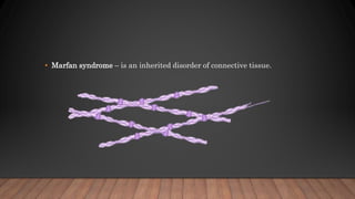
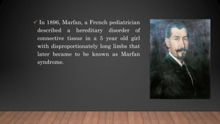
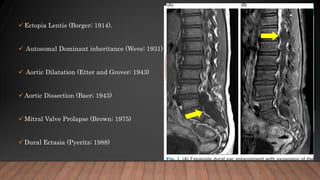
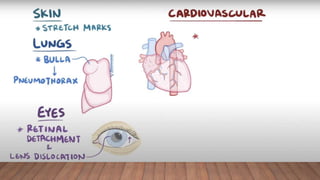
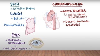
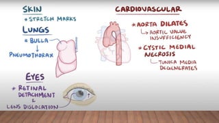
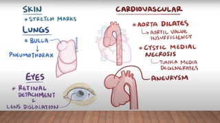
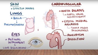
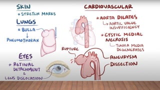
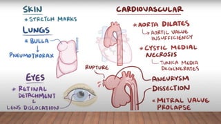
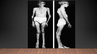
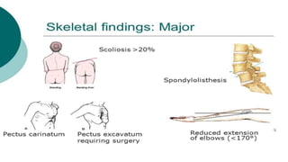
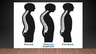
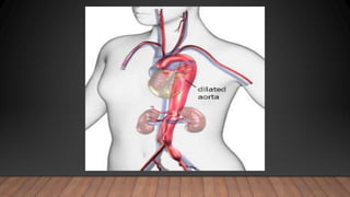
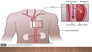
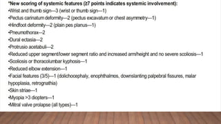
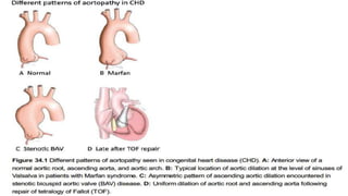
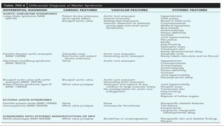
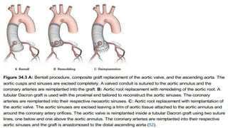
7. In 1896, Marfan, a French pediatrician
described a hereditary disorder of
connective tissue in a 5 year old girl
with disproportionately long limbs that
later became to be known as Marfan
syndrome.
43. The incidence is reported to be 1 in 10,000 live births.
Approximately one-fourth of cases are sporadic.
The disorder shows autosomal dominant inheritance.
There is no racial or gender preference.
45. MFS is associated with abnormal production,
matrix deposition and/ or stability of fibrillin-
1(FBN1), a 350-kDa ECM protein.
FBN1 mutations have been identified in over
90 percent patients.
More than 1,000 disease-causing mutations
identified to date.
46. The fibrillin-1 (FBN1) locus resides on the long arm of
chromosome 15 (15q21), and the gene is composed of 65
exons.
47.
48.
49.
50. MFS was traditionally considered to result from a
structural deficiency of connective tissues.
Mutations in the fibrillin-1 gene result in the
production of an abnormal fibrillin protein, leading to
abnormalities in the mechanical stability and elastic
properties of connective tissue.
51. Reduced fibrillin-1 was thought to lead to a primary
derangement of elastic fiber deposition, because both skin and
aorta from affected patients show decreased elastin, along with
elastic fiber fragmentation.
52.
53.
54. CONTINUED…….
More recently, research suggests that transforming growth
factor-beta is implicated in the failure of normal elastic
tissue formation.
TGFBR 1 and 2 mutations – may have similar
manifestations.
In response to stress (such as hemodynamic forces in the
proximal aorta), affected organs were thought to manifest
this structural insufficiency with accelerated degeneration.
55. CONTINUED…….
Cystic medial necrosis :
The term cystic medial necrosis was coined by Erdheim to describe the lacunar
appearance of medial degeneration in MFS; however no actual cysts or overt
necrosis is present.
Cystic medial necrosis and the other histologic findings are not specific for MFS,
although greater elastin fragmentation has been described in patients with aortic root
aneurysms with MFS.
The histologic changes are thought to reflect injury and repair
61. SKELETAL SYSTEM
• Overgrowth of the long bones (dolichostenomelia) is often the most
obvious manifestation of MFS.
• Individuals taller than predicted by their genetic background
• Joint laxity
• Results in reduced upper segment: lower segment ratio (US : LS)
• US : LS <1 for ages 0-5 yr
• US : LS <0.95 for ages 6-7 yr
• US : LS <0.9 for ages 8-9 yr
• US : LS <0.85 above age 10 yr
• Arm span to height ratio >1.05 times.
62.
63.
64.
65. CONTINUED
• Paradoxically, some individuals with MFS have reduced joint mobility, particularly of
the elbow and digits - reduced elbow extension (≤180 degrees with full extension).
67. CONTINUED
• Thumb sign - entire distal phalanx protrudes beyond the ulnar
border of a clenched fist with or without the assistance of the patient or
examiner to achieve maximum adduction.
68. CONTINUED
• Wrist sign - the top of the thumb covers the entire fingernail of the
fifth finger when wrapped around the contralateral wrist.
69. CONTINUED
• Pectus deformity — Pectus carinatum - more specific for MFS than pectus excavatum or
chest asymmetry.
• Hindfoot valgus — occurs with forefoot abduction and lowering of the midfoot and should
be evaluated from anterior and posterior views.
• Pes planus (flat foot) without hindfoot valgus is less specific
• Generalized joint hypermobility also may occur, producing findings that overlap with the
much more common benign joint hypermobility syndrome.
72. CONTINUED
Scoliosis and kyphosis
• A Cobb’s angle of at least 20 degrees (on an anteriorposterior
radiographic view of the spine, the angle between a line drawn along
the superior end plate of the superior end vertebra and a second line
drawn along the inferior end plate of the inferior end vertebra of the
scoliosis) Exaggerated kyphotic thoracolumbar spinal curvature.
73.
74.
75.
76. CONTINUED
• Protrusio acetabuli Can be diagnosed by plain radiograph, computed
tomography (CT), or magnetic resonance imaging (MRI). On an anterior-posterior pelvic
film, medial protrusion of the acetabulum ≥3mm beyond the ilio-ischial (Kohler) line is
diagnostic.
77. CONTINUED
• Facial features — dolichocephaly (reduced cephalic index or head width/length ratio),
enophthalmos, downslanting palpebral fissures, malar hypoplasia, and retrognathia.
78. Dural ectasia
• Enlargement of the spinal canal owing to progressive ectasia of dura and neural
foramina and to erosion of vertebral bone.
• Usually involves the lumbosacral spine 60-90% pts on MRI/CT.
• It is sensitive but not specific sign of MFS, and is commonly seen in Loeys-Dietz
syndrome and Shprintzen-Goldberg syndrome, has been reported in the vascular form
of Ehlers-Danlos syndrome.
79. • MRI most sensitive technique.
• No correlation appears to exist between the severity of dural ectasia and the degree of aortic
dilatation.
80.
81. OCULAR ABNORMALITIES
• Ectopia lentis:
• 50 to 80 percent.
• Detected on slit-lamp examination after
maximal dilatation of the pupil and the
lens is usually displaced upward and
temporally.
• It is caused by failure of the supporting
ciliary zonules.
82. • Myopia >3 diopters - secondary myopia due to increased axis globe length.
83. CONTINUED
• Flat cornea.
• Hypoplastic iris.
• Hypoplastic ciliary muscle causing miosis.
• Retinal detachment.
• Glaucoma and early cataract formation.
• Retinal tears and detachment are commonly bilateral.
84. • Pulmonary disease — Some patients develop emphysematous changes with lung bullae
predominantly in the upper lobes, can predispose to spontaneous pneumothorax.
85. • Skin striae — The presence of striae atrophicae contributes one point to the systemic score if
not associated with pronounced weight changes or pregnancy and if in uncommon location
such as the mid back, lumbar region, upper arm, axillary region, or thigh.
86.
87. • Within the heart, thickening of the atrioventricular valves is common and
often associated with valvular prolapse.
• Variable degrees of regurgitation may be present.
• In children with early onset and severe MFS, insufficiency of the mitral
valve can lead to congestive heart failure, pulmonary hypertension and
death in infancy; this manifestation is the leading cause of morbidity and
mortality in young children with the disorder.
88.
89. • Supraventricular arrhythmias and ventricular dysrhythmias may be seen in
association with mitral valve dysfunction, and there is an increased
prevalence of prolonged QT interval.
• Dilated cardiomyopathy occurs with increased prevalence in patients with
MFS, most often attributed to volume overload imposed by valve
regurgitation.
• Aortic valve dysfunction is generally a late occurrence and attributed to
stretching of the aortic annulus by an expanding aortic root aneurysm.
90. AORTIC DISEASE
• Aortic root disease, leading to aneurysmal dilatation, aortic
regurgitation, and dissection - main cause of morbidity and mortality
• Aortic enlargement is typically located at the level of sinuses of
Valsalva and may eventually extend to the sinotubular junction and
proximal ascending aorta
• Although dilated, the aorta in MFS tends to be stiffer and less
distensible
91. • Dilatation of the aorta, often (about 25%) accompanied by aortic
regurgitation, progresses with time.
• 50 percent of young children with MFS
• 60 to 80 percent of adult patients with MFS
92.
93.
94.
95.
96.
97.
98. CONTINUED
• The incidence of serious cardiovascular complications, including aortic
dissection or rupture, has been estimated to be around 4.3% in childhood
with a rise to approximately 20% in adolescence .
• Aortic regurgitation may develop in 15% to 44% of MFS patients during
childhood and adolescence, and has been strongly associated with acute
cardiovascular events
99. CONTINUED
• Dilatation may also involve other segments of the thoracic aorta,
the abdominal aorta, the root of the pulmonary artery or even the
carotid and intracranial arteries.
• The normal range for aortic diameter varies with body size and
age - nomograms and Z-scores used to identify aortic dilatation.
• Undiagnosed and untreated MFS - frequently associated with
aortic dissection. May have a family history of dissection.
100. CONTINUED
• The frequency with which MFS is responsible for aortic dissection varies
with age.
• 50% of those under age 40
• 2 % of those with age 40 - 70
• No patient over age 70
101. CONTINUED
• Additional predictors of aortic complications include younger age
at presentation and a family history of severe aortic disease .
• Patients diagnosed with MFS during childhood have significantly
fewer adverse cardiac events compared to those diagnosed during
adulthood, highlighting the importance of early identification of
the disease
102. CONTINUED
• Following diagnosis of MFS, an echocardiogram is recommended at 6
months to assess the progression of aortic disease
• If measurements remain stable, echocardiographic follow-up can be
performed every 6 months in children and on an annual basis in Adults.
103. CONTINUED
• Children who do not meet the full diagnostic criteria of the disease should
be screened at least every 5 years, until they reach adulthood
• Baseline echocardiographic imaging requires diameter measurement at the
level of the sinuses of Valsalva and should be indexed to age and body
surface area
• In the instance of poor echocardiographic windows, magnetic resonance
imaging (MRI) or computed tomography (CT) angiograms should be
performed.
104. CONTINUED
• Routine CT or MRI imaging of the entire thoracic aorta is recommended in
patients who present with descending aortic dilation, type B aortic
dissection, or following ascending aortic aneurysm repair
105.
106. CONTINUED
• Mitral valve prolapse (MVP) Common but nonspecific – only 1 point in
systemic scoring 40-54% MFS adults; upto 90% in some series frequency of
MVP increases with age; greater in women. Tricuspid valve prolapse may
also occur.
• On echo mitral leaflets elongated and redundant either or both leaflets
may prolapse most have mild or less regurgitation
107.
108. CONTINUED
• Approximately 25 percent of patients with MVP have progressive disease -
defined by the appearance or worsening of clinical symptoms of mitral
regurgitation or worsening on echocardiography.
• Heart failure attributable to mitral valve prolapse and regurgitation
represents a major source of morbidity and mortality in young children with
the most extreme and rapidly progressive presentation of MFS.
• Some report suggest - some patients may have a cardiomyopathy with
biventricular enlargement and generally asymptomatic mild systolic
dysfunction unrelated to valvular disease
109.
110. STEP BY STEP APPROACH
• History and physical examination (including slit-lamp ophthalmic
examination with pupil dilation) in conjunction with imaging of
the aortic root and the ascending, descending, and abdominal
aorta (echo, CT, MRI) are usually sufficient for diagnosis.
• Family history of Marfan's syndrome, or of aortic dissection or
aneurysm.
• There is also a weak association with high parental age
111. Other historical considerations
• Family history of myopia, astigmatism, strabismus,
amblyopia, premature cataract or other lens abnormalities,
glaucoma, retinal detachment, dental extraction or braces
for dental crowding, hernias, or spontaneous
pneumothorax.
112. PHYSICAL EXAMINATION
• Tall stature, wide arm span, high level of pubic bone, high arched palate,
arachnodactyly, positive wrist and thumb sign, pectus excavatum, pectus
carinatum, scoliosis, striae, flat feet, thick spectacles for myopia, hernias,
aortic or mitral valve murmur may be present.
• Spontaneous pneumothorax or emphysematous bullae may present as
dyspnoea.
113. • There may be signs of heart failure due to valve disease or cardiomyopathy.
• Complete ophthalmic examination, including fundus examination with
pupil dilation - signs of lens subluxation or dislocation, cataract, glaucoma,
or retinal detachment.
• May present with acute aortic dissection or rupture
114. INITIAL INVESTIGATIONS
• Echocardiography, thorax CT, and thorax MRI are used initially for aortic
root imaging.
• Abdominal ultrasound, CT, and MRI are used for visualization of the
descending aorta.
• CXR is performed to exclude the presence of a pneumothorax, and may
reveal emphysematous bullae.
115. SUBSEQUENT INVESTIGATIONS
• Blood screening for mutations in the fibrillin-1 (FBN1) gene confirms the
diagnosis if in doubt.
• Once detected, the mutation can be used to screen other relatives, and
used for antenatal diagnosis and pre-implantation genetic diagnosis.
116. • Lower spine CT scan or MRI can be performed to exclude dural ectasia.
• Plasma homocysteine levels help in unclear cases to differentiate
homocystinuria.
• Skin biopsy is indicated only if Ehlers-Danlos syndrome is suspected.
118. THE GHENT CRITERIA
• A definite diagnosis can be made applying the Ghent criteria, which are based on
demonstration of symptoms from different organ systems and family history of MFS.
• However, Ghent nosology cannot exclude MFS in children, due to the variability in onset
and severity of symptoms in this age group.
• Recently, an international expert board has published a revised Ghent nosology, in
which aortic root aneurysm and ectopia lentis are the cardinal features of MFS with a
new scoring system for other systemic features of the disease
125. • The prognosis of patients with MFS has improved with the use of medical therapy
(beta blockers and angiotensin receptor blockers).
• Routine and noninvasive monitoring of aortic size.
• Elective surgical repair of the aorta.
• Restriction of vigorous physical exercise
127. • Beta-blockers
• Exercise Limitation
• If beta -blockers not well tolerated then calcium channel blockers
• In small number of patients use ARBs significantly slowed the rate of
progression of aortic root dilatation.
128. AORTIC MONITORING
• Monitoring of the thoracic aortic diameter is recommended to identify patients
at high risk for aortic dissection.
132. • Due to the absence of general consensus in the pediatric population, most
centers advise prophylactic surgery using the diameter criterion for adults (∼50
mm) but also take into consideration the presence of accelerated aortic growth
(>10 mm/yr), development of aortic regurgitation, or need for mitral valve
surgery
• If the aortic valve is affected-a composite replacement of the valve and ascending
aorta is performed
133. • In cases with no substantial aortic valve disease, valve-sparing techniques
can be applied, including remodeling of the aortic root or reimplantation of
the aortic valve.
• Following aortic root surgery, close follow-up and continuation of medical
therapy is necessary, since distal segments of the aorta may also be affected
134.
135. RECENT ADVANCES
• Personalized external aortic support (PEARS) is an experimental surgical
alternative being investigated in Europe, which has so far been applied in young
adults only, mostly in the setting of MFS .
• The patient's own aortic dimensions are used to manufacture a replica of the aorta
in plastic on which a bespoke external support is made of a fabric mesh. This can
be positioned around the aortic root and ascending aorta without cardiopulmonary
bypass and involves a shorter operation, than that for the conventional approach
141. • The diagnosis of Marfan syndrome (MFS) in familial and sporadic cases are
based upon the presence of characteristic manifestations, particularly aortic root
dilatation/dissection and ectopia lentis, as well as other systemic features.
• MFS is caused by a variety of mutations in the FBN1 gene. FBN1 mutations
have been identified in over 90 percent of patients with MFS.
• Revised Ghent criteria is used for diagnosing Marfan syndrome.
142. CONTINUED
• The differential diagnosis for MFS includes a variety of conditions with
phenotypic features that partially overlap the Marfan phenotype, including
disorders associated with FBN1/2 or TGFBR1/2 mutations, as well as a variety of
other genetic disorders.
• First degree relatives of patients with a gene mutation associated with aortic
aneurysms and/or dissection (eg, FBN1, TGFBR1, TGFBR2, COL3A1, ACTA2,
MYH11) should undergo counseling and genetic testing. Those found to have the
genetic mutation should then undergo aortic imaging.
143. • For patients with aortic aneurysm and/or dissection without a known mutation, aortic
imaging is recommended for first degree relatives to identify those with asymptomatic
disease. If one or more first degree relatives are found to have thoracic aortic
dilatation, aneurysm, or dissection, then imaging of second degree relatives is
reasonable.
144. CONTINUED
• Echocardiography is recommended at initial diagnosis and at six months to assess
the aortic root and ascending aorta in patients with MFS.
• Monitoring should be performed at least annually in patients with Ao root
diameter more than 4.0 cm, and biannually in patients at higher risk (Ao root
diameter more than 4.5 cm; Ao root enlargement more than 0.5 cm per year ;
family history of Ao dissection).