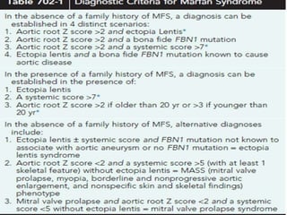
MARFAN SYNDROME
PRESENTER: Dr.SHASIDHAR REDDY
INTRODUCTION
• Marfan syndrome (MFS) is an inherited, systemic, connective
tissue disorder caused by mutations in the gene encoding the
extracellular matrix (ECM) protein fibrillin-1.
• It is primarily associated with skeletal,cardiovascular, and
ocular pathology.
• The diagnosis is based on clinical findings, some of which are
age dependent.
EPIDEMIOLOGY
• The incidence is reported to be 1 in 10,000 live births.
• This disorder shows autosomal dominant inheritance.
PATHOGENESIS
• MFS is associated with abnormal production, matrix
deposition and/or stability of fibrillin-1, which is the major
constituent of microfibrils, with prominent disruption of
microfibrils and elastic fibers in diseased tissues.
CLINICAL MANIFESTATIONS
• MFS is a multisystem disorder, with cardinal manifestations in
the skeletal, cardiovascular, and ocular systems.
SKELETAL MANIFESTATIONS:
• Dolichostenomelia - Overgrowth of the long bones is often
the most obvious manifestation of MFS and may produce a
reduced upper segment: lower segment ratio (US : LS) or an
arm span to height ratio >1.05 times.
• Anterior chest deformity due to excessive rib growthand
pushing sternum either outward-pectus carinatum or inward-
pectus excavatum.
• Thoracolumbar scoliosis.
• Protrusio acetabuli (Inward bulging of acetabulum into pelvic
cavity)
• Pes planus(flat feet)
• Joint hypermobility/contractures
• Arachnodactyly(long and slender fingers)
• Contractures of fingers and elbows
• Craniofacial manifestations- Dolicocephaly(narrow skull)
Enophthalmos(deep set eyes)
• Retrognathia/micrognathia
• Malar hypoplasia (flattening of midface)
• CARDIOVASCULAR MANIFESTATIONS:
• Thickening of atrioventricular valves associated with valvular
prolapse.
• In children with early onset and severe MFS insufficiency of
the mitral valve can lead to congestive heart
failure,pulmonary hypertension and death in infancy.
• Aortic aneurysm, dissection and rupture, principally at the
level of the sinuses of Valsalva (aortic root), remains the most
life-threatening manifestations of MFS.
• OTHER SYSTEMS:
SKIN:
• Stretch marks- pinkish,scar like lesions that later become
white(striae atrophicae)
LUNGS:
• Progressive anterior chest deformity or thoracic scoliosis
• Spontaneous pneumothorax – due to widening of distal air
spaces.
• OCCULAR MANIFESTATIONS:
• Ectopia lentis(Dislocation of the ocular lens)- occurs in
approximately 60-70% of patients.
• Early and severe myopia, flat cornea, increased axial length of
the globe, hypoplastic iris.
• Patients are also predisposed to retinal detachment and early
cataracts or glaucoma.
DIAGNOSIS
• The diagnosis is based on a defined set of clinical criteria
drawn up by an international panel of experts: revised Ghent
nosology for the MFS
MANAGEMENT
• Management focuses on preventing complications and
genetic counseling.
• Regular evaluations for cardiovascular disease, scoliosis, or
ophthalmologic problems to be done.
Mitral valve prolapse:
• Most patients with mitral valve prolapse do not require
treatment unless they have symptoms. If the patient becomes
symptomatic:
o Medication: beta blockers (these reduce blood pressure and
the heart beats with less force) and aspirin (reduces risk of
blood clots)
o Surgery: Surgery is not very common except with severe
prolapse
 Valve repair: Valve repair preserves the patient tissue
 Valve replacement: The valve is replaced by a
mechanical valve or animal tissue.
Aortic dissection:
• Aortic dissection requires surgery.
• Part of the dissected aorta is removed to prevent blood from
entering the aortic wall, and reconstruct the aorta with a
synthetic tube.
Aortic dilation:
• Aortic dilation requires surgical management. It should be
performed when the aorta is 4.5-5 cm, the rate of growth is 0.5
cm or more, and there is the presence of aortic regurgitation.
PROGNOSIS
• The major cause of mortality is aortic root dilation, dissection,
and rupture, with the majority of fatal events occurring in the
3rd and 4th decade of life.
• A reevaluation of life expectancy in MFS suggests that early
diagnosis and refined medical and surgical management has
greatly improved the prognosis for patients with the
condition.
• In the more classic form of MFS, it is estimated that more than
90% of individuals will have a cardiovascular event during
their lifetime, placing both physical and mental stress on
patients and their families.
REFERENCE
• NELSON TEXTBOOK OF PEDIATRICS-FIRST SOUTH ASIA
EDITION.
• IAP TEXTBOOK OF PEDIATRICS.

Marfan syndrome

  • 1.
  • 2.
    INTRODUCTION • Marfan syndrome(MFS) is an inherited, systemic, connective tissue disorder caused by mutations in the gene encoding the extracellular matrix (ECM) protein fibrillin-1. • It is primarily associated with skeletal,cardiovascular, and ocular pathology. • The diagnosis is based on clinical findings, some of which are age dependent.
  • 3.
    EPIDEMIOLOGY • The incidenceis reported to be 1 in 10,000 live births. • This disorder shows autosomal dominant inheritance.
  • 4.
    PATHOGENESIS • MFS isassociated with abnormal production, matrix deposition and/or stability of fibrillin-1, which is the major constituent of microfibrils, with prominent disruption of microfibrils and elastic fibers in diseased tissues.
  • 5.
    CLINICAL MANIFESTATIONS • MFSis a multisystem disorder, with cardinal manifestations in the skeletal, cardiovascular, and ocular systems. SKELETAL MANIFESTATIONS: • Dolichostenomelia - Overgrowth of the long bones is often the most obvious manifestation of MFS and may produce a reduced upper segment: lower segment ratio (US : LS) or an arm span to height ratio >1.05 times. • Anterior chest deformity due to excessive rib growthand pushing sternum either outward-pectus carinatum or inward- pectus excavatum.
  • 6.
    • Thoracolumbar scoliosis. •Protrusio acetabuli (Inward bulging of acetabulum into pelvic cavity) • Pes planus(flat feet) • Joint hypermobility/contractures • Arachnodactyly(long and slender fingers) • Contractures of fingers and elbows • Craniofacial manifestations- Dolicocephaly(narrow skull) Enophthalmos(deep set eyes) • Retrognathia/micrognathia • Malar hypoplasia (flattening of midface)
  • 7.
    • CARDIOVASCULAR MANIFESTATIONS: •Thickening of atrioventricular valves associated with valvular prolapse. • In children with early onset and severe MFS insufficiency of the mitral valve can lead to congestive heart failure,pulmonary hypertension and death in infancy. • Aortic aneurysm, dissection and rupture, principally at the level of the sinuses of Valsalva (aortic root), remains the most life-threatening manifestations of MFS.
  • 8.
    • OTHER SYSTEMS: SKIN: •Stretch marks- pinkish,scar like lesions that later become white(striae atrophicae) LUNGS: • Progressive anterior chest deformity or thoracic scoliosis • Spontaneous pneumothorax – due to widening of distal air spaces.
  • 9.
    • OCCULAR MANIFESTATIONS: •Ectopia lentis(Dislocation of the ocular lens)- occurs in approximately 60-70% of patients. • Early and severe myopia, flat cornea, increased axial length of the globe, hypoplastic iris. • Patients are also predisposed to retinal detachment and early cataracts or glaucoma.
  • 10.
    DIAGNOSIS • The diagnosisis based on a defined set of clinical criteria drawn up by an international panel of experts: revised Ghent nosology for the MFS
  • 13.
    MANAGEMENT • Management focuseson preventing complications and genetic counseling. • Regular evaluations for cardiovascular disease, scoliosis, or ophthalmologic problems to be done.
  • 14.
    Mitral valve prolapse: •Most patients with mitral valve prolapse do not require treatment unless they have symptoms. If the patient becomes symptomatic: o Medication: beta blockers (these reduce blood pressure and the heart beats with less force) and aspirin (reduces risk of blood clots) o Surgery: Surgery is not very common except with severe prolapse  Valve repair: Valve repair preserves the patient tissue  Valve replacement: The valve is replaced by a mechanical valve or animal tissue.
  • 15.
    Aortic dissection: • Aorticdissection requires surgery. • Part of the dissected aorta is removed to prevent blood from entering the aortic wall, and reconstruct the aorta with a synthetic tube.
  • 16.
    Aortic dilation: • Aorticdilation requires surgical management. It should be performed when the aorta is 4.5-5 cm, the rate of growth is 0.5 cm or more, and there is the presence of aortic regurgitation.
  • 17.
    PROGNOSIS • The majorcause of mortality is aortic root dilation, dissection, and rupture, with the majority of fatal events occurring in the 3rd and 4th decade of life. • A reevaluation of life expectancy in MFS suggests that early diagnosis and refined medical and surgical management has greatly improved the prognosis for patients with the condition. • In the more classic form of MFS, it is estimated that more than 90% of individuals will have a cardiovascular event during their lifetime, placing both physical and mental stress on patients and their families.
  • 18.
    REFERENCE • NELSON TEXTBOOKOF PEDIATRICS-FIRST SOUTH ASIA EDITION. • IAP TEXTBOOK OF PEDIATRICS.