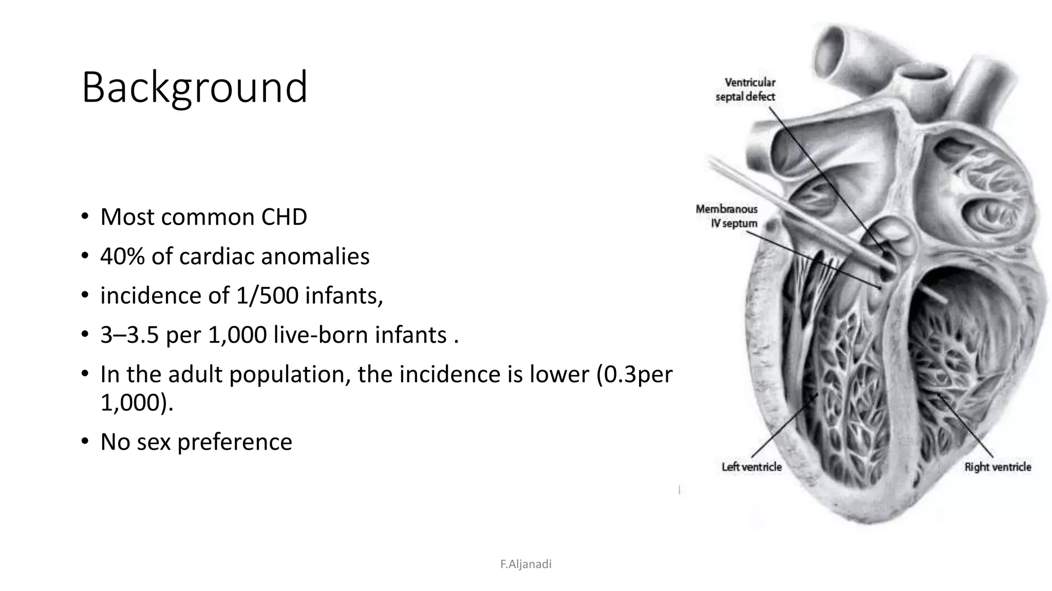
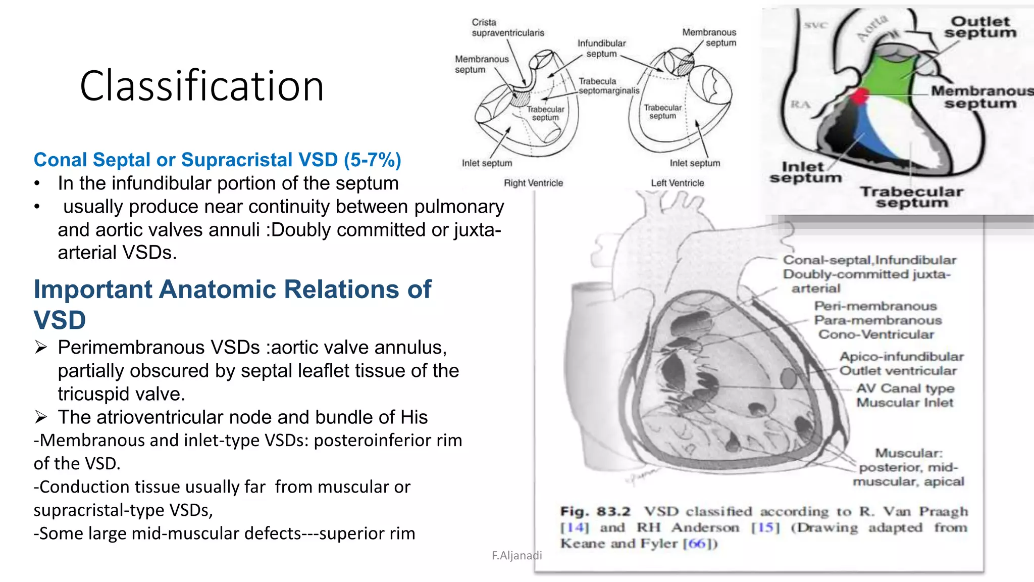
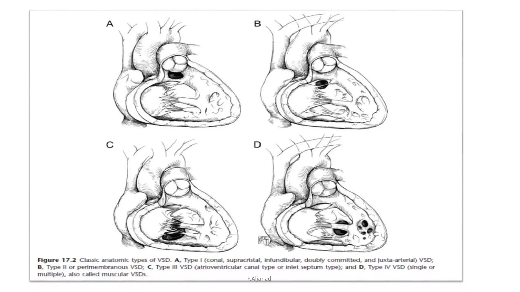
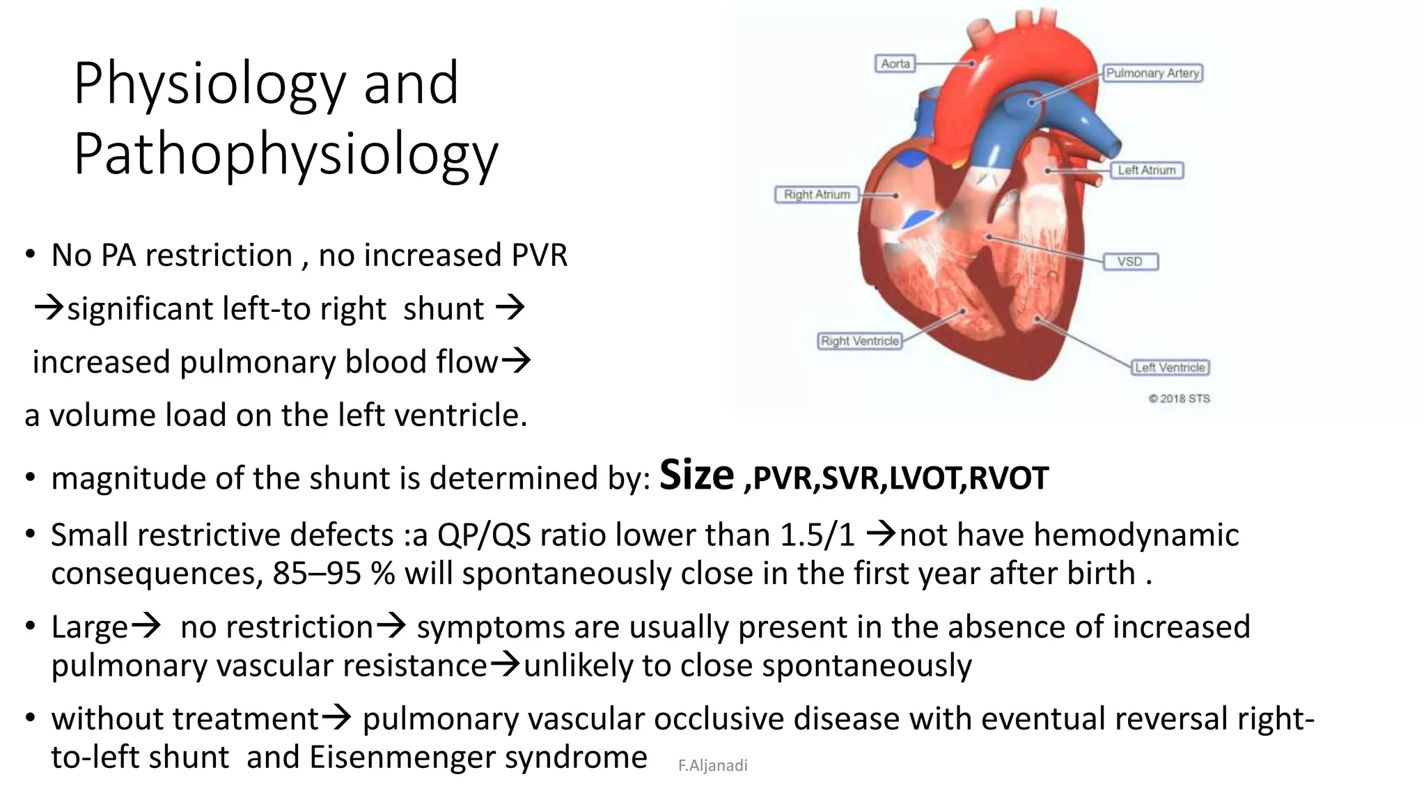
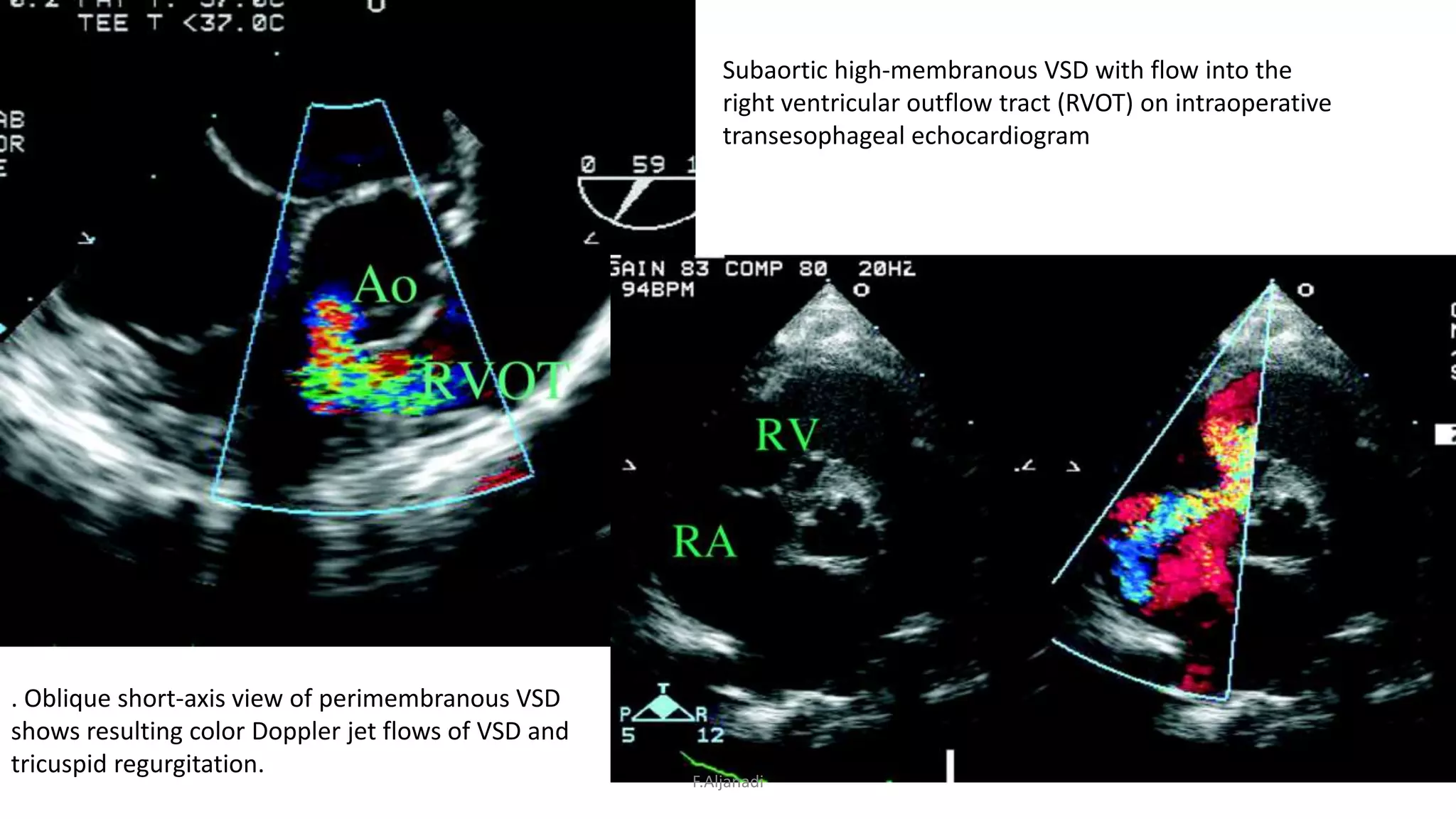
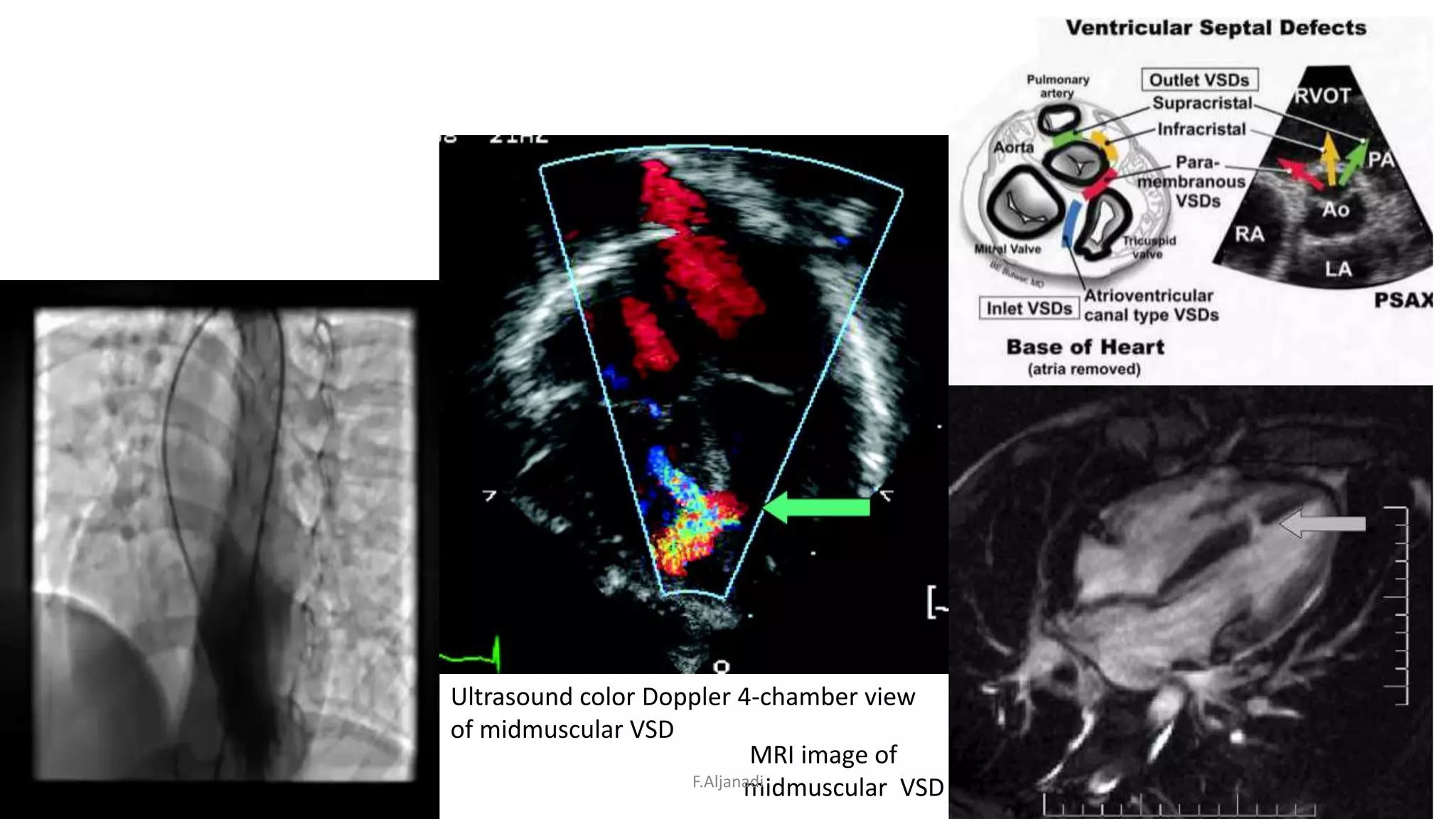
Ventricular Septal Defect
VSD
Firas Aljanadi
RVH
Oct 2019
F.Aljanadi
Background
• Most common CHD
• 40% of cardiac anomalies
• incidence of 1/500 infants,
• 3–3.5 per 1,000 live-born infants .
• In the adult population, the incidence is lower (0.3per
1,000).
• No sex preference
F.Aljanadi
Genetics
• Underlying etiology of VSD is unclear. Chromosomal , Familial ,
Geographic, Environmental
• Trisomy 21 (Down syndrome), 22q11 deletion (Di George syndrome),
and 45X deletion (Turner syndrome).
• Familial forms linked to TBX5, GATA4, and NKX2.5 mutations .
• Children from an adult: a risk of VSD as high as 3 % if father
• a 6 % risk if the mother is affected
F.Aljanadi
Historical Background
• Vesale (sixteenth century), Morgagni,Senac (eighteenth century).
• Roger (nineteenth century) 1897 the correlation with cardiac murmur
• In France, the term “maladie de Roger” (Roger’s disease) : a very small
VSD without any clinical consequence but with a loud murmur
• PA banding : Muller and Damman in 1952 .
• 1954, Lillehei, first to surgically close a VSD with the aid of cross
circulation,
• Heart-lung machine : series of 20 patients treated by surgical closure
at the Mayo clinic was reported in 1957 .
• DHCA, initially described in 1969 by Okamoto et al. Sir B. Barratt-
Boyes repair of VSD in small infants.
• percutaneous closure of a VSD by a device introduced by
catheterization and secondly the intraoperative perventricular closure
with a device, without the use of Cardiopulmonary bypassF.Aljanadi
Embryology
F.Aljanadi
Embryology
F.Aljanadi
ClassificationAnatomic Types of VSD
Cono-Ventricular Defects (70-80%)
 often beyond the membranous septum
:perimembranous or paramembranous.
• hypoplasia or displacement of the conal septum,
the VSD may be called a “malalignment defect.”
• Can extend to the infundibular
(outlet) or AV canal (inlet) septum.
• often covered partially with accessory fibrotic
tricuspid tissue.
AV Canal or Inlet VSD(5-8%)
The cephalad border of these defects
is the tricuspid valve annulus; Both the cono-ventricular and
the inlet VSDs
Muscular VSD(5-20%)
• entire circumference is muscular tissue.
• mid-muscular, posterior, or apical portion
of the interventricular septum. Singular or multiple.
• +/- other VSD types.
multiple VSDs from the right ventricular, often only a singular
large defect as viewed from the left ventricular
aspect. Multiple defects are sometimes described
as “Swiss cheese”-like septum.
F.Aljanadi
Classification
Conal Septal or Supracristal VSD (5-7%)
• In the infundibular portion of the septum and
• usually produce near continuity between pulmonary
and aortic valves annuli :Doubly committed or juxta-
arterial VSDs.
Important Anatomic Relations of
VSD
 Perimembranous VSDs :aortic valve annulus,
partially obscured by septal leaflet tissue of the
tricuspid valve.
 The atrioventricular node and bundle of His
-Membranous and inlet-type VSDs: posteroinferior rim
of the VSD.
-Conduction tissue usually far from muscular or
supracristal-type VSDs,
-Some large mid-muscular defects---superior rim
F.Aljanadi
F.Aljanadi
F.Aljanadi
F.Aljanadi
Physiology and
Pathophysiology
• No PA restriction , no increased PVR
significant left-to right shunt 
increased pulmonary blood flow
a volume load on the left ventricle.
• magnitude of the shunt is determined by: Size ,PVR,SVR,LVOT,RVOT
• Small restrictive defects :a QP/QS ratio lower than 1.5/1 not have hemodynamic
consequences, 85–95 % will spontaneously close in the first year after birth .
• Large no restriction symptoms are usually present in the absence of increased
pulmonary vascular resistanceunlikely to close spontaneously
• without treatment pulmonary vascular occlusive disease with eventual reversal right-
to-left shunt and Eisenmenger syndrome F.Aljanadi
Diagnosis ,Clinical
• Pansystolic murmur-smaller-Louder, thrill
• Impulse may be displaced laterally, and an apical mid-diastolic
• murmur across the mitral valve and/or a third heart sound can be heard.
• neonatal period, usually asymptomatic. As the PVR falls, left-to-right shunt
increases and symptoms may be exhibited.
• Tachypnea, tachycardia diaphoresis and dyspnea ,while feeding, poor weight
gainFTT
• Several months or years of significantly increased pulmonary blood flow from a
nonrestrictive VSD, reversal of flow across the VSD may occur resulting in no
heart failure symptoms but relative desaturation with right-to-left shunt or
Eisenmenger syndrome.
• Occasionally, the usual fall in neonatal pulmonary vascular resistances does not
occur. These children may not exhibit signs of heart failure, despite a large VSD.
F.Aljanadi
Diagnosis
• ECG :normal ,tachy, (inlet) LAD , chambers enlargement
• CXR :normal in small, increased pulmonary vascularity ,LA,LV
dilated ,cardiomegaly, perihilar oedema , PA prominent
• ECHO :Mainstay of diagnosis,TOE, 3D
• Cath:is only indicated in complicated cases: to assess the
pulmonary vascular resistance in individuals with suspected
pulmonary vascular occlusive disease and in suspected multiple
VSD or to close the malformation by transcatheter approach
• cMRI :limited , sedation,in poor echo ,complex anatomy,VSD
impact,vol overload,Qp/Qs
F.Aljanadi
En face view of the left ventricular septal surface
showing midmuscular VSD (arrow)
F.Aljanadi
Small perimembranous VSD
with ventricular septal aneurysm on
transesophageal echocardiographic
examination.
Small apical muscular VSD on 3-
dimensional echocardiographic
image
A 3-dimensional
echocardiographic image
shows flow
from small muscular VSD
(arrow) tunneling through the
septum
F.Aljanadi
. Oblique short-axis view of perimembranous VSD
shows resulting color Doppler jet flows of VSD and
tricuspid regurgitation.
Subaortic high-membranous VSD with flow into the
right ventricular outflow tract (RVOT) on intraoperative
transesophageal echocardiogram
F.Aljanadi
Ultrasound color Doppler 4-chamber view
of midmuscular VSD
MRI image of
midmuscular VSDF.Aljanadi
Natural History
• Cardiac failure: size
• Spontaneous diminution in size and closure:69%
perimembranous+ 60% muscular , less likely after 1 yr .
• LVOTO ,RVOTO(3-7%)
• AR (10%),rare <2yr, RCC 60-70%, NCC 10-15%
(perimembranous),
• PVOD(5-22%), earlier in Down’s
• IE 18.7 per 10,000 p years in non operated , 7.3 operated ,
smaller higher , in TV vegetations ,AV or PV vegetations rule out
VSD
F.Aljanadi
Medical Management
• initiation of diuretic therapy. ACEi
• CHF Mx
• nutritional supplementation,feeding
F.Aljanadi
Surgical Management
• Pulmonary Artery Banding (PAB),
• Rarely indicated nowadays ,: Swiss-cheese variety, some VSDs
and coarctation ,VSDs with straddling of one AV valve,infants
with very low body weight and those who are not fit for CPB of
an intercurrent illness a contraindication to CPB and significant
CHF that necessitates some intervention
Median sternotomy, (PTFE) strip,Silastic material, or Teflon strip
umbilical tape
PAP (30-50% of systemic BP), sBP(rise 10-15mmHg),SaO2 (85-
90%,Fio2 50%),Trusler’s rule :wt+20 (noncyanotic non mixing )
• Surgical VSD Closure :is recommended for failure of medical
management,presence of supracristal VSD, moderate to large
VSDs with persistent increased PVR.
• Interventional and Hybrid Management
F.Aljanadi
Techniques
F.Aljanadi
F.Aljanadi
Complications and post op care
• TR ,RVF
• AI
• Arrhythmias
• AV BLOCK
• RBBB
• LCOS
• PAH crisis
• Residual VSD (0.7-2%)
F.Aljanadi
Thank you
F.Aljanadi

Ventricular septal defect VSD

  • 1.
    Ventricular Septal Defect VSD FirasAljanadi RVH Oct 2019 F.Aljanadi
  • 2.
    Background • Most commonCHD • 40% of cardiac anomalies • incidence of 1/500 infants, • 3–3.5 per 1,000 live-born infants . • In the adult population, the incidence is lower (0.3per 1,000). • No sex preference F.Aljanadi
  • 3.
    Genetics • Underlying etiologyof VSD is unclear. Chromosomal , Familial , Geographic, Environmental • Trisomy 21 (Down syndrome), 22q11 deletion (Di George syndrome), and 45X deletion (Turner syndrome). • Familial forms linked to TBX5, GATA4, and NKX2.5 mutations . • Children from an adult: a risk of VSD as high as 3 % if father • a 6 % risk if the mother is affected F.Aljanadi
  • 4.
    Historical Background • Vesale(sixteenth century), Morgagni,Senac (eighteenth century). • Roger (nineteenth century) 1897 the correlation with cardiac murmur • In France, the term “maladie de Roger” (Roger’s disease) : a very small VSD without any clinical consequence but with a loud murmur • PA banding : Muller and Damman in 1952 . • 1954, Lillehei, first to surgically close a VSD with the aid of cross circulation, • Heart-lung machine : series of 20 patients treated by surgical closure at the Mayo clinic was reported in 1957 . • DHCA, initially described in 1969 by Okamoto et al. Sir B. Barratt- Boyes repair of VSD in small infants. • percutaneous closure of a VSD by a device introduced by catheterization and secondly the intraoperative perventricular closure with a device, without the use of Cardiopulmonary bypassF.Aljanadi
  • 5.
  • 6.
  • 7.
    ClassificationAnatomic Types ofVSD Cono-Ventricular Defects (70-80%)  often beyond the membranous septum :perimembranous or paramembranous. • hypoplasia or displacement of the conal septum, the VSD may be called a “malalignment defect.” • Can extend to the infundibular (outlet) or AV canal (inlet) septum. • often covered partially with accessory fibrotic tricuspid tissue. AV Canal or Inlet VSD(5-8%) The cephalad border of these defects is the tricuspid valve annulus; Both the cono-ventricular and the inlet VSDs Muscular VSD(5-20%) • entire circumference is muscular tissue. • mid-muscular, posterior, or apical portion of the interventricular septum. Singular or multiple. • +/- other VSD types. multiple VSDs from the right ventricular, often only a singular large defect as viewed from the left ventricular aspect. Multiple defects are sometimes described as “Swiss cheese”-like septum. F.Aljanadi
  • 8.
    Classification Conal Septal orSupracristal VSD (5-7%) • In the infundibular portion of the septum and • usually produce near continuity between pulmonary and aortic valves annuli :Doubly committed or juxta- arterial VSDs. Important Anatomic Relations of VSD  Perimembranous VSDs :aortic valve annulus, partially obscured by septal leaflet tissue of the tricuspid valve.  The atrioventricular node and bundle of His -Membranous and inlet-type VSDs: posteroinferior rim of the VSD. -Conduction tissue usually far from muscular or supracristal-type VSDs, -Some large mid-muscular defects---superior rim F.Aljanadi
  • 9.
  • 10.
  • 11.
  • 12.
    Physiology and Pathophysiology • NoPA restriction , no increased PVR significant left-to right shunt  increased pulmonary blood flow a volume load on the left ventricle. • magnitude of the shunt is determined by: Size ,PVR,SVR,LVOT,RVOT • Small restrictive defects :a QP/QS ratio lower than 1.5/1 not have hemodynamic consequences, 85–95 % will spontaneously close in the first year after birth . • Large no restriction symptoms are usually present in the absence of increased pulmonary vascular resistanceunlikely to close spontaneously • without treatment pulmonary vascular occlusive disease with eventual reversal right- to-left shunt and Eisenmenger syndrome F.Aljanadi
  • 13.
    Diagnosis ,Clinical • Pansystolicmurmur-smaller-Louder, thrill • Impulse may be displaced laterally, and an apical mid-diastolic • murmur across the mitral valve and/or a third heart sound can be heard. • neonatal period, usually asymptomatic. As the PVR falls, left-to-right shunt increases and symptoms may be exhibited. • Tachypnea, tachycardia diaphoresis and dyspnea ,while feeding, poor weight gainFTT • Several months or years of significantly increased pulmonary blood flow from a nonrestrictive VSD, reversal of flow across the VSD may occur resulting in no heart failure symptoms but relative desaturation with right-to-left shunt or Eisenmenger syndrome. • Occasionally, the usual fall in neonatal pulmonary vascular resistances does not occur. These children may not exhibit signs of heart failure, despite a large VSD. F.Aljanadi
  • 14.
    Diagnosis • ECG :normal,tachy, (inlet) LAD , chambers enlargement • CXR :normal in small, increased pulmonary vascularity ,LA,LV dilated ,cardiomegaly, perihilar oedema , PA prominent • ECHO :Mainstay of diagnosis,TOE, 3D • Cath:is only indicated in complicated cases: to assess the pulmonary vascular resistance in individuals with suspected pulmonary vascular occlusive disease and in suspected multiple VSD or to close the malformation by transcatheter approach • cMRI :limited , sedation,in poor echo ,complex anatomy,VSD impact,vol overload,Qp/Qs F.Aljanadi
  • 15.
    En face viewof the left ventricular septal surface showing midmuscular VSD (arrow) F.Aljanadi
  • 16.
    Small perimembranous VSD withventricular septal aneurysm on transesophageal echocardiographic examination. Small apical muscular VSD on 3- dimensional echocardiographic image A 3-dimensional echocardiographic image shows flow from small muscular VSD (arrow) tunneling through the septum F.Aljanadi
  • 17.
    . Oblique short-axisview of perimembranous VSD shows resulting color Doppler jet flows of VSD and tricuspid regurgitation. Subaortic high-membranous VSD with flow into the right ventricular outflow tract (RVOT) on intraoperative transesophageal echocardiogram F.Aljanadi
  • 18.
    Ultrasound color Doppler4-chamber view of midmuscular VSD MRI image of midmuscular VSDF.Aljanadi
  • 19.
    Natural History • Cardiacfailure: size • Spontaneous diminution in size and closure:69% perimembranous+ 60% muscular , less likely after 1 yr . • LVOTO ,RVOTO(3-7%) • AR (10%),rare <2yr, RCC 60-70%, NCC 10-15% (perimembranous), • PVOD(5-22%), earlier in Down’s • IE 18.7 per 10,000 p years in non operated , 7.3 operated , smaller higher , in TV vegetations ,AV or PV vegetations rule out VSD F.Aljanadi
  • 20.
    Medical Management • initiationof diuretic therapy. ACEi • CHF Mx • nutritional supplementation,feeding F.Aljanadi
  • 21.
    Surgical Management • PulmonaryArtery Banding (PAB), • Rarely indicated nowadays ,: Swiss-cheese variety, some VSDs and coarctation ,VSDs with straddling of one AV valve,infants with very low body weight and those who are not fit for CPB of an intercurrent illness a contraindication to CPB and significant CHF that necessitates some intervention Median sternotomy, (PTFE) strip,Silastic material, or Teflon strip umbilical tape PAP (30-50% of systemic BP), sBP(rise 10-15mmHg),SaO2 (85- 90%,Fio2 50%),Trusler’s rule :wt+20 (noncyanotic non mixing ) • Surgical VSD Closure :is recommended for failure of medical management,presence of supracristal VSD, moderate to large VSDs with persistent increased PVR. • Interventional and Hybrid Management F.Aljanadi
  • 22.
  • 23.
  • 24.
    Complications and postop care • TR ,RVF • AI • Arrhythmias • AV BLOCK • RBBB • LCOS • PAH crisis • Residual VSD (0.7-2%) F.Aljanadi
  • 25.

Editor's Notes

  • #22 a median sternotomy, but anterior and posterior thoracotomies have been used. After incision of the pericardium, the main PA (MPA) is cautiously encircled. A band is placed around the MPA. The material used is variable and includes polytetrafluoroethylene (PTFE) strip, Silastic material, and Teflon strip umbilical tape. It is advantageous to use a material that is the least adherent. Once the MPA is encircled with the band, the band is progressively tightened while observing the distal main pulmonary artery pressure and systemic oxygen saturation. This is usually accomplished with titanium clips applied to the band and eventually securing the band to the PA to prevent migration onto the branch PAs. A rough estimate of the bandmay follow Trusler’s rules: length inmm corresponding to the weight in kg of the baby, plus 20mm. The distal PA pressure should usually be about one half systemic systolic blood pressure so that it is well tolerated hemodynamically, without important cyanosis. End tidal CO2 measurement is also helpful, since it is almost directly related, in stable operative condition, to pulmonary output. If the banding is performed through a thoracotomy, with a less easy access to the MPA, it is a good precaution to separate first aorta from PA staying close to the aortaz; since the PA is thinner, the right angle dissector can perforate it, and hemostasis might be difficult to obtain. The tape is then passed around the aorta, and by subtraction, through the transverse sinus, it is finally located around the main PA. Adjustable PA banding is an interesting concept. A specific device, although expensive and rather cumbersome for small babies, does exist and is externally adjustable [28]. This allows for progressive tightening or loosening of the band as necessary. In some cases, construction of the band may be performed so that the band can be disrupted by balloon dilation of the main PA if it is not needed. This is useful in some cases of multiple muscular VSDs or “Swiss cheese” septum. One technique is one in which the PA band in which the circumference was reduced by several staged thin mattress sutures, as 6/0 or 7/0 monofilament. Another is to place only a single clip on the band so that it may be dislodged with balloon dilation. For debanding of the PA during an intracardiac correction, after some fibrous tissue is dissected on the band, the simple division of