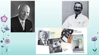
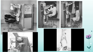
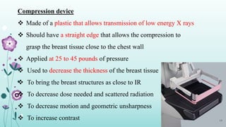
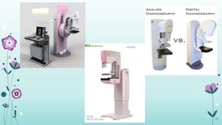
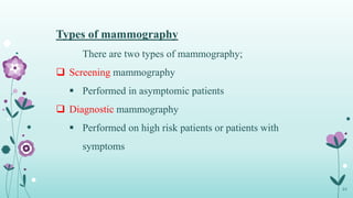
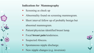
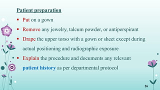
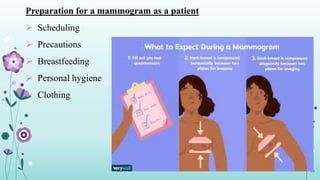
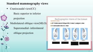
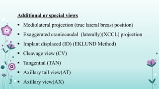
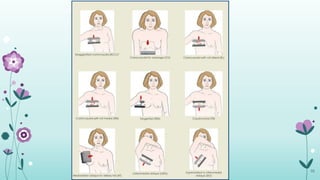
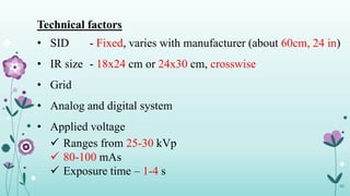
Mammography
Presented by Akari Kyaw
Fourth Year
B. Med Tech (MIT)
24.6.2020
Contents
• Definition
• History
• Anatomy
• Mammography equipments
• Types of mammography
• Indications
• Patient preparation
• Preparation as a patient
• mammography views
• Radiation safety
• Benefits
2
Mammography
 X ray imaging of breast organ
 Also called mastography
 Radiographic procedure that uses special equipment to produce images
of high contrast and high resolution for the diagnosis of breast leisions
 Using low energy X rays(usually around 30 kVp)
 goal of mammography is the early detection of breast cancer, typically
through detection of characteristic masses or microcalcifications.
3
History of mammography
 1913 (Dr. Albert Solomon)
First attempt at mammography
 1950 (Robert Egan)
First utilized low kVp and high mAs techniques
 1960
Development of xero-mammography
 1990
Screen-film mammography was accepted
4
5
6
Anatomy of breast
 Mammary gland
 A conic or hemispheric eminence
 Located on the anterior and lateral wall
 Average breast extends from the anterior portion of the 2nd
rib(clavicle) down to the 6th or 7th (mid sternum)
 Comprises 15 to 20 lobes which are covered by adipose tissue that
primarily accounts for its size and shape
7
8
Breast classifications
• Fibroglandular breast
• Fibrofatty breast
• Fatty breast
9
Fibroglandular breast
• Common age group- 15 to 30 years(and childless women> 30
years old)
• Pregnant or lactating women
• Radiographically dense
• Very little fat
10
Fibrofatty breast
• 30 to 50 years
• Young women with three or more pregnancies
• Average radiographic density
• 50% fat and 50% fibroglandular
11
Fatty Breast
• >50 years
• Postmenopausal
• Minimal radiographic density
• Breasts of children and men
12
Methods of localization
Two methods are
commonly used;
1. Quadrant system
• Upper outer quadrant
• Upper inner quadrant
• Lower outer quadrant
• Lower inner quadrant
2. Clock system
13
Mammography Equipments
Mammography equipment comprise of;
 High frequency generator
 Molybdenum target
 Filter
 Grid
 Compression device
 Automatic exposure control system
14
High frequency generator
 Works on single phase
 Provides rectified smoothed voltage supply to the tube
 Small in size with good reproducibility
 Providing up to 600 mAs
 Employ small focal spots, low kVp technique, low
grid ratio and special screen films
15
Target and filter
Most of the mammography X ray tubes are
designed with;
• Mo target with Mo Filter
• Mo target with Rh Filter
• Rh target with Rh Filter
16
Grid
 Placed between the breast and
cassette
 To reduce scatter radiation
 Improve contrast sensitivity
 Lower grid ratio than that of general
X ray(4:1 or 5:1)
17
Compression device
 Made of a plastic that allows transmission of low energy X rays
 Should have a straight edge that allows the compression to
grasp the breast tissue close to the chest wall
 Applied at 25 to 45 pounds of pressure
 Used to decrease the thickness of the breast tissue
 To bring the breast structures as close to IR
 To decrease dose needed and scattered radiation
 To decrease motion and geometric unsharpness
 To increase contrast 18
Automatic Exposure control (AEC)
 Employs phototimers to measure the X ray intensity and
quality
 Kept closer to the image receptor
 To minimize the object to image distance(OID)
 Improving spatial resolution
There are two types of AEC available,
 Ionization chamber type
 Solid state diode type
19
20
21
22
23
Types of mammography
There are two types of mammography;
 Screening mammography
 Performed in asymptomic patients
 Diagnostic mammography
 Performed on high risk patients or patients with
symptoms
24
Indications for Mammography
 Screening as check up
 Abnormality found on screening mammogram.
 Short interval follow up of probably benign but
abnormal mammogram.
 Patient/physician identified breast lump.
 Focal breast pain/tenderness.
 Suspected Abscess.
 Spontaneous nipple discharge.
 New nipple changes (e.g. inversion)
25
Patient preparation
 Put on a gown
 Remove any jewelry, talcum powder, or antiperspirant
 Drape the upper torso with a gown or sheet except during
actual positioning and radiographic exposure
 Explain the procedure and documents any relevant
patient history as per departmental protocol
26
• Patient history include the following
 Number of pregnancies
 Family history of cancer including breast cancer
 Medications (eg; hormone therapy) currently taken
 Previous breast surgery
 Previous mammograms when and where performed
 Reason for current visit such as screening mammogram,
lumps, pain, or discharge
27
Preparation for a mammogram as a patient
 Scheduling
 Precautions
 Breastfeeding
 Personal hygiene
 Clothing
28
What Not To Do Before a Mammogram
 DON'T apply deodorant before your mammogram
 DON'T wear a dress or one-piece outfit
 DON'T go right before or during your period
 DON'T consume caffeine products (coffee, chocolate)
several days up to two weeks before the appointment
 DON'T worry if you've had prior mammograms at other
facilities
29
Standard mammography views
 Craniocaudal view(CC)
Basic superior to inferior
projection
 Mediolateral oblique view(MLO)
Superomedial- inferolateral
oblique projection
30
31
Additional or special views
 Mediolateral projection (true lateral breast position)
 Exaggerated craniocaudal (laterally)(XCCL) projection
 Implant displaced (ID) (EKLUND Method)
 Cleavage view (CV)
 Tangential (TAN)
 Axillary tail view(AT)
 Axillary view(AX)
32
33
Technical factors
• SID - Fixed, varies with manufacturer (about 60cm, 24 in)
• IR size - 18x24 cm or 24x30 cm, crosswise
• Grid
• Analog and digital system
• Applied voltage
 Ranges from 25-30 kVp
 80-100 mAs
 Exposure time – 1-4 s
34
Radiation safety
We must obey cardinal rules;
• Time
• distance
• shielding
35
36
Benefits of mammography
•Reduces the risk of dying from breast cancer
•Reduces the risk of having to undergo chemotherapy; Screening often
allows for the detection of cancers at an early stage of development
•Allows women to know the health of their breasts.
37
38
References
 K. L. Bontrager and J. P. Lampignano (2014) Text Book Of
Radiographic Positioning And Related Anatomy, 8th Edition
 Department Of Radiology, Dartmouth Geisel School Of
Medicine 2020 Indications For Diagnostic Mammography
 Quebec; Breast cancer screening

Mammography

  • 1.
    Mammography Presented by AkariKyaw Fourth Year B. Med Tech (MIT) 24.6.2020
  • 2.
    Contents • Definition • History •Anatomy • Mammography equipments • Types of mammography • Indications • Patient preparation • Preparation as a patient • mammography views • Radiation safety • Benefits 2
  • 3.
    Mammography  X rayimaging of breast organ  Also called mastography  Radiographic procedure that uses special equipment to produce images of high contrast and high resolution for the diagnosis of breast leisions  Using low energy X rays(usually around 30 kVp)  goal of mammography is the early detection of breast cancer, typically through detection of characteristic masses or microcalcifications. 3
  • 4.
    History of mammography 1913 (Dr. Albert Solomon) First attempt at mammography  1950 (Robert Egan) First utilized low kVp and high mAs techniques  1960 Development of xero-mammography  1990 Screen-film mammography was accepted 4
  • 5.
  • 6.
  • 7.
    Anatomy of breast Mammary gland  A conic or hemispheric eminence  Located on the anterior and lateral wall  Average breast extends from the anterior portion of the 2nd rib(clavicle) down to the 6th or 7th (mid sternum)  Comprises 15 to 20 lobes which are covered by adipose tissue that primarily accounts for its size and shape 7
  • 8.
  • 9.
    Breast classifications • Fibroglandularbreast • Fibrofatty breast • Fatty breast 9
  • 10.
    Fibroglandular breast • Commonage group- 15 to 30 years(and childless women> 30 years old) • Pregnant or lactating women • Radiographically dense • Very little fat 10
  • 11.
    Fibrofatty breast • 30to 50 years • Young women with three or more pregnancies • Average radiographic density • 50% fat and 50% fibroglandular 11
  • 12.
    Fatty Breast • >50years • Postmenopausal • Minimal radiographic density • Breasts of children and men 12
  • 13.
    Methods of localization Twomethods are commonly used; 1. Quadrant system • Upper outer quadrant • Upper inner quadrant • Lower outer quadrant • Lower inner quadrant 2. Clock system 13
  • 14.
    Mammography Equipments Mammography equipmentcomprise of;  High frequency generator  Molybdenum target  Filter  Grid  Compression device  Automatic exposure control system 14
  • 15.
    High frequency generator Works on single phase  Provides rectified smoothed voltage supply to the tube  Small in size with good reproducibility  Providing up to 600 mAs  Employ small focal spots, low kVp technique, low grid ratio and special screen films 15
  • 16.
    Target and filter Mostof the mammography X ray tubes are designed with; • Mo target with Mo Filter • Mo target with Rh Filter • Rh target with Rh Filter 16
  • 17.
    Grid  Placed betweenthe breast and cassette  To reduce scatter radiation  Improve contrast sensitivity  Lower grid ratio than that of general X ray(4:1 or 5:1) 17
  • 18.
    Compression device  Madeof a plastic that allows transmission of low energy X rays  Should have a straight edge that allows the compression to grasp the breast tissue close to the chest wall  Applied at 25 to 45 pounds of pressure  Used to decrease the thickness of the breast tissue  To bring the breast structures as close to IR  To decrease dose needed and scattered radiation  To decrease motion and geometric unsharpness  To increase contrast 18
  • 19.
    Automatic Exposure control(AEC)  Employs phototimers to measure the X ray intensity and quality  Kept closer to the image receptor  To minimize the object to image distance(OID)  Improving spatial resolution There are two types of AEC available,  Ionization chamber type  Solid state diode type 19
  • 20.
  • 21.
  • 22.
  • 23.
  • 24.
    Types of mammography Thereare two types of mammography;  Screening mammography  Performed in asymptomic patients  Diagnostic mammography  Performed on high risk patients or patients with symptoms 24
  • 25.
    Indications for Mammography Screening as check up  Abnormality found on screening mammogram.  Short interval follow up of probably benign but abnormal mammogram.  Patient/physician identified breast lump.  Focal breast pain/tenderness.  Suspected Abscess.  Spontaneous nipple discharge.  New nipple changes (e.g. inversion) 25
  • 26.
    Patient preparation  Puton a gown  Remove any jewelry, talcum powder, or antiperspirant  Drape the upper torso with a gown or sheet except during actual positioning and radiographic exposure  Explain the procedure and documents any relevant patient history as per departmental protocol 26
  • 27.
    • Patient historyinclude the following  Number of pregnancies  Family history of cancer including breast cancer  Medications (eg; hormone therapy) currently taken  Previous breast surgery  Previous mammograms when and where performed  Reason for current visit such as screening mammogram, lumps, pain, or discharge 27
  • 28.
    Preparation for amammogram as a patient  Scheduling  Precautions  Breastfeeding  Personal hygiene  Clothing 28
  • 29.
    What Not ToDo Before a Mammogram  DON'T apply deodorant before your mammogram  DON'T wear a dress or one-piece outfit  DON'T go right before or during your period  DON'T consume caffeine products (coffee, chocolate) several days up to two weeks before the appointment  DON'T worry if you've had prior mammograms at other facilities 29
  • 30.
    Standard mammography views Craniocaudal view(CC) Basic superior to inferior projection  Mediolateral oblique view(MLO) Superomedial- inferolateral oblique projection 30
  • 31.
    31 Additional or specialviews  Mediolateral projection (true lateral breast position)  Exaggerated craniocaudal (laterally)(XCCL) projection  Implant displaced (ID) (EKLUND Method)  Cleavage view (CV)  Tangential (TAN)  Axillary tail view(AT)  Axillary view(AX)
  • 32.
  • 33.
    33 Technical factors • SID- Fixed, varies with manufacturer (about 60cm, 24 in) • IR size - 18x24 cm or 24x30 cm, crosswise • Grid • Analog and digital system • Applied voltage  Ranges from 25-30 kVp  80-100 mAs  Exposure time – 1-4 s
  • 34.
    34 Radiation safety We mustobey cardinal rules; • Time • distance • shielding
  • 35.
  • 36.
    36 Benefits of mammography •Reducesthe risk of dying from breast cancer •Reduces the risk of having to undergo chemotherapy; Screening often allows for the detection of cancers at an early stage of development •Allows women to know the health of their breasts.
  • 37.
  • 38.
    38 References  K. L.Bontrager and J. P. Lampignano (2014) Text Book Of Radiographic Positioning And Related Anatomy, 8th Edition  Department Of Radiology, Dartmouth Geisel School Of Medicine 2020 Indications For Diagnostic Mammography  Quebec; Breast cancer screening