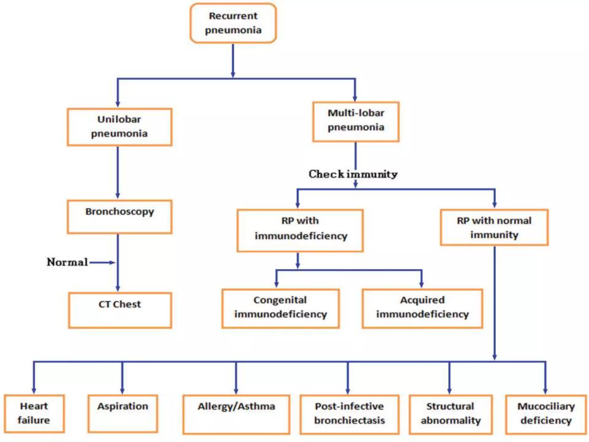
Dr seema rai
Assistant professor
Guru Gobind singh medical college Faridkot
 Pneumonia is an acute
infection of the pulmonary parenchyma
 Two episodes of pneumonia within the same
year or 3 or more episodes over any period of
time but with complete resolution of clinical
and radiological findings between acute
episodes.
 When there is clinical and radiological
evidence of pneumonia for more than a
month despite a course of adequate and
appropriate antibiotic therapy for 10 days.
 Congenital Malformations
a. Airways Cleft Palate, Pierre Robin syndrome
Tracheoesophageal fistulae,Tracheomalacia
b. Lungs Pulmonary hypoplasia, Pulmonary
sequestration, Congenital adenomatoid
malformation of the lung,Bronchogenic cyst
c. Cardiovascular Congenital heart disease,
especially L-R shunts, Vascular ring
 Aspirations Gastro-esophageal reflux
Foreign body, Anomalies of the upper airways
Swallowing abnormalities
 Defects in the clearance of airway secretions
Cystic fibrosis, Abnormalities of the ciliary
structure function, Abnormal clearance
secondary to infections, repair of congenital
defects, Airway compression
(intrinsic/extrinsic) e.g., mediastinal
tubercular lymphadenopathy
 Disorders of local/systemic immunity
 Primary immunodeficiency
 Acquired immunodeficiency
- HIV infection
- Immunosuppressive therapy
- Malnutrition
 Intraluminal obstruction
Foreign body
Bronchial tumor,
Adenoma, lipoma, papilloma
 Extraluminal obstruction
1 INFECTIOUS LAP(lymphadenopathy)
Tuberculosis, histoplasmosis.
2 Non-infectious lymphadenopathy
Tumors, sarcoidosis.
Vascular rings and slings
 Structural abnormalities
Bronchial stenosis or atresia,
Bronchomalacia, localized bronchiectasis,
pulmonary sequestration or congnital cystic adenoid
malformation, bronchogenic cyst
 Recurrent microaspiration
 Asthma
 Immunodeficiency
 Mucociliary dysfunction
 Structural Abnormality
 Congenital heart disease
 Bronchopulmonary dysplasia
 Age of onset: Onset of symptoms soon after
birth or in early infancy indicates hereditary/
congenital disorder. History of possible foreign
body aspiration followed by recurrent
episodes of pneumonia tends to occur in the
1-3 year age.
 Details of the episodes: Details of first
episode and subsequent episodes should be
obtained.
 Onset, nature and duration of cough,
 History of fever, and documentation of signs of
pneumonia and radiological evaluation
 Type and duration of antimicrobial therapy
(adequate/appropriate), response to
therapy and need for hospitalization also should
be recorded. It is important to differentiate these
episodes from recurrent wheezing episodes
 Ask about the timing of the symptoms in
relation to feeding and the change in
position.
 vomiting, irritability, and nocturnal symptoms
of coughing and wheezing.
 Sleep disturbances may be seen in gastro-
esophageal reflux and obstructive lesions,
especially of upper respiratory tract.
 Past history/associated complaints:
 History suggestive of systemic
immunodeficiency.
 Past history of tuberculosis and history of foreign
body inhalation is important.
 Symptoms of malabsorption, recurrent otitis
media and sinusitis and failure to thrive suggest
cystic fibrosis.
 Presence or absence of symptoms when the child
is well eg. intercurrent wheeze may suggest
poorly controlled asthma.
 Patient with congenital heart disease with
failure exhibit easy fatiguability, sweating
over the forehead on feeding.
 Swallowing dysfunction can present as
recurrent regurgitation of feeds and child
may develop cyanosis during these episodes
 Perinatal history:
 Prematurity, history of prolonged exposure to
oxygen and blood transfusions should be
looked for.
 History of ileus or delayed passage of
meconium should arouse suspicion of cystic
fibrosis.
 History of maternal infections such as HIV,
Chlamydia or other viral illnesses should be
sought
 history of allergic disorders, asthma, cystic
fibrosis, and congenital anomalies.
Occurrence of early or unexplained deaths or
recurrent infections in other family members
may suggest immunologic disorder.
 High risk behavior or history of blood
transfusion in parents is essential, to rule out
exposure to maternal HIV infection, in a child
where immunodeficiency is suspected.
 Crowded and polluted living environment
predispose to recurrent respiratory
infections.
 Daycare attendance, exposure to inhaled
pollutants, irritants and passive tobacco
smoking may provide important clues to the
aetiology, as well as the presence of siblings
with similar problems.
 Many medications also may cause pulmonary
problems (ACE inhibitors cough, aspirin-
induced wheeze, leukotriene antagonist
associated infiltrates) and hence, a
comprehensive drug history should be also
be taken
 Vaccination history as pertussis can present
with prolonged cough
 Obstructive upper respiratory tract lesions
present with sleep disturbances or even sleep
apnoea.
 Gastro-esophageal reflux also can have
similar presentation.
 If two or more of the following warning signs are present,
there is possibility of an underlying primary
immunodeficiency.
1. Four or more new ear infections within 1 year.
2. Two or more serious sinus infection within 1 year.
3. Two or more months on antibiotics with little effect.
4. Two or more pneumonia within 1 year.
5. Failure of an infant to gain weight or grow normally.
6. Recurrent, deep skin or organ abscesses.
7. Persistent thrush in mouth or fungal infection on skin.
8. Need for intravenous antibiotics to clear infection.
9. Two or more deep seated infection including septicemia.
10. A family history of primary immunodeficiency.
 Head and neck
 Signs of chronic otitis media or sinusitis. Chronic
otitis media is associated with ciliary dysmotility
syndromes or in boys with Wiscott-Aldrich
syndrome.
 Conjunctivitis, allergic shiners, Dennie morgan’s
lines and transverse creases over nose may
accompany dermatitits in atopic patients, who
have an increased frequency of asthma and
associated pneumonia.
 Presence of purulent conjunctivitis may suggest
a B cell immune deficiency.
 Phylectenular conjunctivitis may be seen in
several conditions having hyperactive immune
response including tuberculosis.
 Look for presence/ absence of
tonsillar/adenoidal tissue. Tonsils may be absent
in hypo/agammaglobulinemia.
2. Periodontal disease: indicates phagocytic
cell dysfunction, while oral candidiasis may
suggest T-cell abnormalities. Abnormal dentition
has been reported as a finding in hyper IgE
Syndrome.
 GROWTH AND DEVELOPMENT
 Normal growth is a reassuring sign, though
children may grow normally during the early
stages of serious systemic diseases.
 Malnutrition impose delay in clearing
infection
 Clubbing may be present in chronic disorders
like bronchiectasis, cystic fibrosis, and
bronchiolitis obliterans.
 Lymphadenopathy: Generalized
lymphadenopathy may be present
intuberculosis, HIV infection and histiocytosis
 Morphologic features: May point towards
specific disorders e.g., presence of “fish
mouth” with hypertelorism in DiGeorge’s
syndrome, telangiectasia of eyes/ears in
ataxia telangiectasia.
 Skin: Examined for evidence of infective foci
rash or for features of atopic dermatitis. Fine
and sparse hair is seen in severe combined
immunedeficiency. Multiple recurrent
pustules indicate immunodeficiency
 Observation during feeding: Nasopharyngeal
regurgitation, difficulty in sucking/swallowing
and associated coughing/choking should be
looked for. The palate, tongue and oro-
pharynx should be inspected for any
anomalies.
 Respiratory system: A meticulous
examination of respiratroy sytem including
assessment of :
 respiratory rate, evidence of distress, thoracic
deformities, wheezing, stridor,
 The dimensions of the chest
 Auscultation of the chest to localize the
infection should be done.
 1. A complete blood count: Anemia may
suggest chronic disease. Thrombocytopenia
can be seen in Wiskott-Aldrich syndrome.
 2. Xray chest: It helps to make the distinction
between recurrent and persistent pneumonia
and whether consolidation is localized to a
single lobe or multiple lobes.
 Mantoux test: Should be done on any child
with focal changes in chest x-ray and
consideration should be given to tests for
fungal infection in endemic areas.
 Computed tomography: Useful in diagnosing
structural anomalies and also helps in defining the
extent of involvement of the lung, especially in
diseases like bronchiectasis, cystic fibrosis and
interstitial lung disease.
 CT is the preferred method for investigating
perilaryngeal or mediastinal compressive masses
affecting the airway and has largely replaced
conventional angiography for investigation of
suspected vascular rings.
 High resolution CT(HRCT) needs to be done
when interstial lung disease is suspected.
 Pulmonary function tests (PFT), commonly
performed using spirometer, usually feasible
only in children >5 years age. It helps to
evaluate the airway hyper-reactivity.
 Bronchoscopy is indicated if abnormality of
bronchial anatomy or foreign body aspiration is
suspected. In addition, bronchoalveolar lavage
(BAL) can be performed to identify the etiologic
agent. Isolation of mucoid Pseudomonas
aeruginosa is a strong pointer to the diagnosis
of cystic fibrosis. Demonstration of
Pneumocystis jiroveci suggests underlying
immunodeficiency.
 Barium study: Barium swallow or cine
esophagogram may help in identifying the
disorders of swallowing. Radionuclide scans,
esophageal pH monitoring may be done to
confirm GER.
Presence of lipid laden macrophages in
bronchial washings has been
found to be of value in confirming recurrent or
chronic aspiration.
 Sweat chloride estimation should be
performed in all children with recurrent
pneumonia even in patients without evidence
of malabsorption or growth failure, because
20% of children with CF may have adequate
pancreatic function.
 Electron microscopy: Morphologic studies
are performed on nasal mucosal
scrapping/biopsy when ciliary abnormalities
are suspected in patients with bronchiectasis
or chronic sinobronchial disease.
i) Complete and differential blood counts
ii) Quantitative serum immunoglobulin G/M/A
including IgE.
iii) HIV screen by ELISA.
iv) T and B cell subset quantification.
If phagocytic defects are suspected,
screening tests include neutrophil count and
nitro blue tetrazolium test.
 Therapy for specific illness associated with
recurrent pulmonary infections
including asthma, chronic aspiration, GERD
and immunodeficiency should follow the
standard guidelines.
 Nutritional support chest physiotherapy
allergens are all important. Those with
congenital anatomic abnormalities or
acquired focal disease involving a single lobe
or segment may benefit from surgical
resection of that part.
Approach to recurrent pneumonia

Approach to recurrent pneumonia

  • 1.
    Dr seema rai Assistantprofessor Guru Gobind singh medical college Faridkot
  • 2.
     Pneumonia isan acute infection of the pulmonary parenchyma
  • 4.
     Two episodesof pneumonia within the same year or 3 or more episodes over any period of time but with complete resolution of clinical and radiological findings between acute episodes.
  • 5.
     When thereis clinical and radiological evidence of pneumonia for more than a month despite a course of adequate and appropriate antibiotic therapy for 10 days.
  • 6.
     Congenital Malformations a.Airways Cleft Palate, Pierre Robin syndrome Tracheoesophageal fistulae,Tracheomalacia b. Lungs Pulmonary hypoplasia, Pulmonary sequestration, Congenital adenomatoid malformation of the lung,Bronchogenic cyst c. Cardiovascular Congenital heart disease, especially L-R shunts, Vascular ring
  • 7.
     Aspirations Gastro-esophagealreflux Foreign body, Anomalies of the upper airways Swallowing abnormalities  Defects in the clearance of airway secretions Cystic fibrosis, Abnormalities of the ciliary structure function, Abnormal clearance secondary to infections, repair of congenital defects, Airway compression (intrinsic/extrinsic) e.g., mediastinal tubercular lymphadenopathy
  • 8.
     Disorders oflocal/systemic immunity  Primary immunodeficiency  Acquired immunodeficiency - HIV infection - Immunosuppressive therapy - Malnutrition
  • 9.
     Intraluminal obstruction Foreignbody Bronchial tumor, Adenoma, lipoma, papilloma  Extraluminal obstruction 1 INFECTIOUS LAP(lymphadenopathy) Tuberculosis, histoplasmosis. 2 Non-infectious lymphadenopathy Tumors, sarcoidosis. Vascular rings and slings  Structural abnormalities Bronchial stenosis or atresia, Bronchomalacia, localized bronchiectasis, pulmonary sequestration or congnital cystic adenoid malformation, bronchogenic cyst
  • 11.
     Recurrent microaspiration Asthma  Immunodeficiency  Mucociliary dysfunction  Structural Abnormality  Congenital heart disease  Bronchopulmonary dysplasia
  • 13.
     Age ofonset: Onset of symptoms soon after birth or in early infancy indicates hereditary/ congenital disorder. History of possible foreign body aspiration followed by recurrent episodes of pneumonia tends to occur in the 1-3 year age.
  • 14.
     Details ofthe episodes: Details of first episode and subsequent episodes should be obtained.  Onset, nature and duration of cough,  History of fever, and documentation of signs of pneumonia and radiological evaluation  Type and duration of antimicrobial therapy (adequate/appropriate), response to therapy and need for hospitalization also should be recorded. It is important to differentiate these episodes from recurrent wheezing episodes
  • 15.
     Ask aboutthe timing of the symptoms in relation to feeding and the change in position.  vomiting, irritability, and nocturnal symptoms of coughing and wheezing.  Sleep disturbances may be seen in gastro- esophageal reflux and obstructive lesions, especially of upper respiratory tract.
  • 16.
     Past history/associatedcomplaints:  History suggestive of systemic immunodeficiency.  Past history of tuberculosis and history of foreign body inhalation is important.  Symptoms of malabsorption, recurrent otitis media and sinusitis and failure to thrive suggest cystic fibrosis.  Presence or absence of symptoms when the child is well eg. intercurrent wheeze may suggest poorly controlled asthma.
  • 17.
     Patient withcongenital heart disease with failure exhibit easy fatiguability, sweating over the forehead on feeding.  Swallowing dysfunction can present as recurrent regurgitation of feeds and child may develop cyanosis during these episodes
  • 18.
     Perinatal history: Prematurity, history of prolonged exposure to oxygen and blood transfusions should be looked for.  History of ileus or delayed passage of meconium should arouse suspicion of cystic fibrosis.  History of maternal infections such as HIV, Chlamydia or other viral illnesses should be sought
  • 19.
     history ofallergic disorders, asthma, cystic fibrosis, and congenital anomalies. Occurrence of early or unexplained deaths or recurrent infections in other family members may suggest immunologic disorder.  High risk behavior or history of blood transfusion in parents is essential, to rule out exposure to maternal HIV infection, in a child where immunodeficiency is suspected.
  • 20.
     Crowded andpolluted living environment predispose to recurrent respiratory infections.  Daycare attendance, exposure to inhaled pollutants, irritants and passive tobacco smoking may provide important clues to the aetiology, as well as the presence of siblings with similar problems.
  • 21.
     Many medicationsalso may cause pulmonary problems (ACE inhibitors cough, aspirin- induced wheeze, leukotriene antagonist associated infiltrates) and hence, a comprehensive drug history should be also be taken  Vaccination history as pertussis can present with prolonged cough
  • 22.
     Obstructive upperrespiratory tract lesions present with sleep disturbances or even sleep apnoea.  Gastro-esophageal reflux also can have similar presentation.
  • 23.
     If twoor more of the following warning signs are present, there is possibility of an underlying primary immunodeficiency. 1. Four or more new ear infections within 1 year. 2. Two or more serious sinus infection within 1 year. 3. Two or more months on antibiotics with little effect. 4. Two or more pneumonia within 1 year. 5. Failure of an infant to gain weight or grow normally. 6. Recurrent, deep skin or organ abscesses. 7. Persistent thrush in mouth or fungal infection on skin. 8. Need for intravenous antibiotics to clear infection. 9. Two or more deep seated infection including septicemia. 10. A family history of primary immunodeficiency.
  • 24.
     Head andneck  Signs of chronic otitis media or sinusitis. Chronic otitis media is associated with ciliary dysmotility syndromes or in boys with Wiscott-Aldrich syndrome.  Conjunctivitis, allergic shiners, Dennie morgan’s lines and transverse creases over nose may accompany dermatitits in atopic patients, who have an increased frequency of asthma and associated pneumonia.  Presence of purulent conjunctivitis may suggest a B cell immune deficiency.
  • 25.
     Phylectenular conjunctivitismay be seen in several conditions having hyperactive immune response including tuberculosis.  Look for presence/ absence of tonsillar/adenoidal tissue. Tonsils may be absent in hypo/agammaglobulinemia. 2. Periodontal disease: indicates phagocytic cell dysfunction, while oral candidiasis may suggest T-cell abnormalities. Abnormal dentition has been reported as a finding in hyper IgE Syndrome.
  • 26.
     GROWTH ANDDEVELOPMENT  Normal growth is a reassuring sign, though children may grow normally during the early stages of serious systemic diseases.  Malnutrition impose delay in clearing infection
  • 27.
     Clubbing maybe present in chronic disorders like bronchiectasis, cystic fibrosis, and bronchiolitis obliterans.  Lymphadenopathy: Generalized lymphadenopathy may be present intuberculosis, HIV infection and histiocytosis
  • 28.
     Morphologic features:May point towards specific disorders e.g., presence of “fish mouth” with hypertelorism in DiGeorge’s syndrome, telangiectasia of eyes/ears in ataxia telangiectasia.  Skin: Examined for evidence of infective foci rash or for features of atopic dermatitis. Fine and sparse hair is seen in severe combined immunedeficiency. Multiple recurrent pustules indicate immunodeficiency
  • 29.
     Observation duringfeeding: Nasopharyngeal regurgitation, difficulty in sucking/swallowing and associated coughing/choking should be looked for. The palate, tongue and oro- pharynx should be inspected for any anomalies.
  • 30.
     Respiratory system:A meticulous examination of respiratroy sytem including assessment of :  respiratory rate, evidence of distress, thoracic deformities, wheezing, stridor,  The dimensions of the chest  Auscultation of the chest to localize the infection should be done.
  • 31.
     1. Acomplete blood count: Anemia may suggest chronic disease. Thrombocytopenia can be seen in Wiskott-Aldrich syndrome.  2. Xray chest: It helps to make the distinction between recurrent and persistent pneumonia and whether consolidation is localized to a single lobe or multiple lobes.
  • 32.
     Mantoux test:Should be done on any child with focal changes in chest x-ray and consideration should be given to tests for fungal infection in endemic areas.
  • 33.
     Computed tomography:Useful in diagnosing structural anomalies and also helps in defining the extent of involvement of the lung, especially in diseases like bronchiectasis, cystic fibrosis and interstitial lung disease.  CT is the preferred method for investigating perilaryngeal or mediastinal compressive masses affecting the airway and has largely replaced conventional angiography for investigation of suspected vascular rings.  High resolution CT(HRCT) needs to be done when interstial lung disease is suspected.
  • 34.
     Pulmonary functiontests (PFT), commonly performed using spirometer, usually feasible only in children >5 years age. It helps to evaluate the airway hyper-reactivity.
  • 35.
     Bronchoscopy isindicated if abnormality of bronchial anatomy or foreign body aspiration is suspected. In addition, bronchoalveolar lavage (BAL) can be performed to identify the etiologic agent. Isolation of mucoid Pseudomonas aeruginosa is a strong pointer to the diagnosis of cystic fibrosis. Demonstration of Pneumocystis jiroveci suggests underlying immunodeficiency.
  • 36.
     Barium study:Barium swallow or cine esophagogram may help in identifying the disorders of swallowing. Radionuclide scans, esophageal pH monitoring may be done to confirm GER. Presence of lipid laden macrophages in bronchial washings has been found to be of value in confirming recurrent or chronic aspiration.
  • 37.
     Sweat chlorideestimation should be performed in all children with recurrent pneumonia even in patients without evidence of malabsorption or growth failure, because 20% of children with CF may have adequate pancreatic function.
  • 38.
     Electron microscopy:Morphologic studies are performed on nasal mucosal scrapping/biopsy when ciliary abnormalities are suspected in patients with bronchiectasis or chronic sinobronchial disease.
  • 39.
    i) Complete anddifferential blood counts ii) Quantitative serum immunoglobulin G/M/A including IgE. iii) HIV screen by ELISA. iv) T and B cell subset quantification. If phagocytic defects are suspected, screening tests include neutrophil count and nitro blue tetrazolium test.
  • 40.
     Therapy forspecific illness associated with recurrent pulmonary infections including asthma, chronic aspiration, GERD and immunodeficiency should follow the standard guidelines.  Nutritional support chest physiotherapy allergens are all important. Those with congenital anatomic abnormalities or acquired focal disease involving a single lobe or segment may benefit from surgical resection of that part.