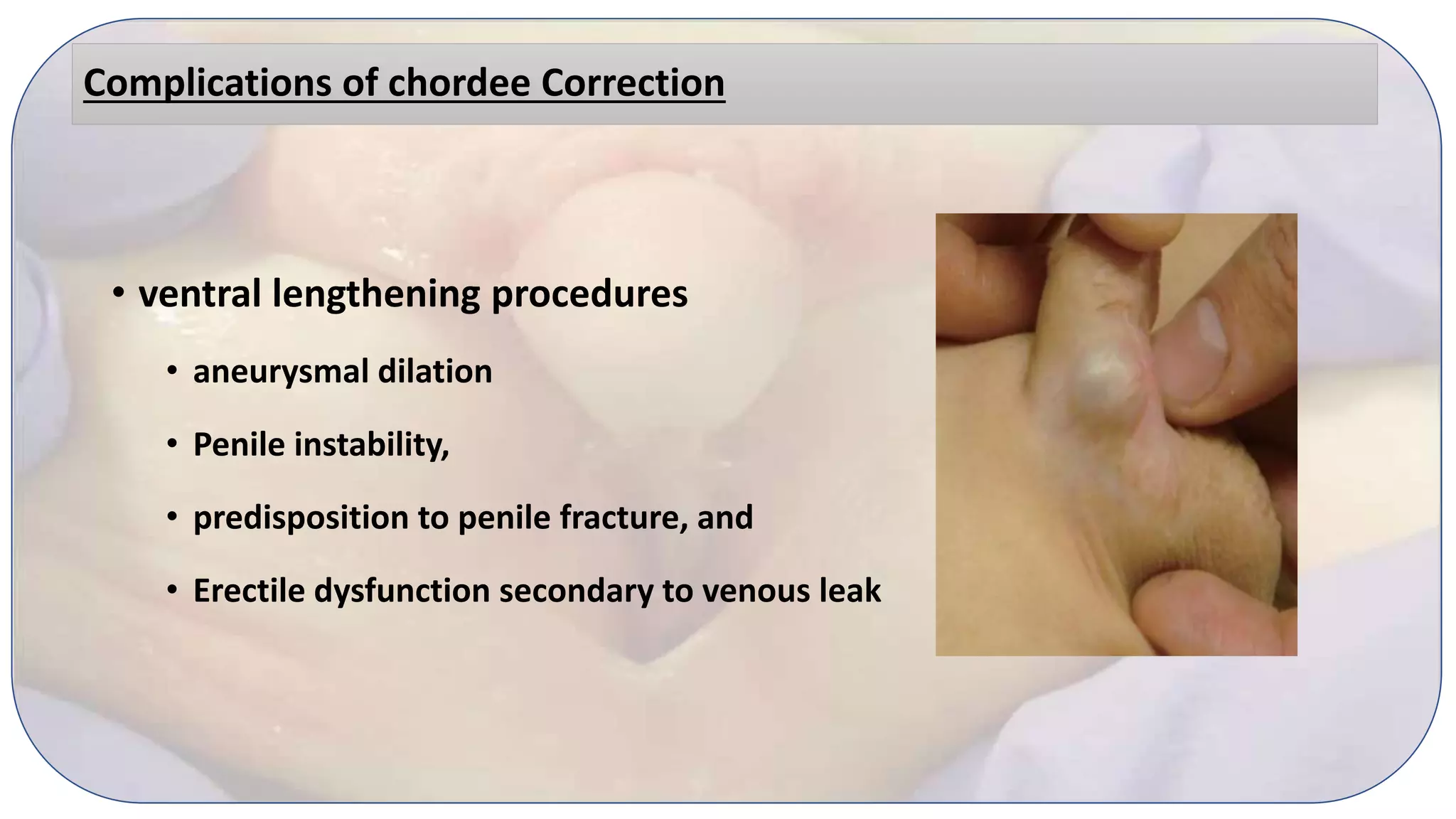
Staged Urethroplasty: approach to decision making
• Dr Faheem Ul Hassan Andrabi
Fellow Pediatric Urology
• Dr. Narender Babu
Associate Professor
Pediatric and Neonatal Surgery
IGICH Banglore
Severe Hypospadias
ventral triangular defect
• Its summit is formed by the proximal division of
the corpus spongiosum,
• the lateral sides by the two atretic pillars of
spongiosum, and
• the base by the widely open glans.
Severe Hypospadias
• The more proximally the spongiosum divides,
the more severe the hypospadias is.
• The position of meatus does not always reflect
severity
• Quite often, the urethra proximal to the meatus
is poorly developed.
Severe Hypospadias
• All tissues sitting inside this triangle are
• under-developed,
• hypoplastic or
• dysplastic.
Other Issues of severity
• Severe Chordee (≥ 30)
• Quality of Urethral Plate (tethered and inelastic)
• Urethral Plate ( Narrow ≤ 1cm)
• Proximal location of meatus > 2 cm
• Morphology of Glans (≤ 14mm)
• Shallow and narrow glanular groove
• Amount of dorsal skin
• Penoscrotal transposition
Chordee
• Devine and Horton classified chordee into
Type I – skin tethering
Type II- fibrotic dartos and buck’s fascia
Type III- corporal disproportion
Type IV congenital short urethra
Chordee
Chordee is graded as:
Mild 10-20 degree
Moderate 30-40 degree
Severe chordee 50 degree
Etiology of chordee
• abnormal development of the urethral plate
• presence of abnormal fibrotic mesenchymal tissue at the
urethral meatus,
• ventral-dorsal corporal disproportion
• Congenital short urethra
• (Montag and Palmer 2011)
Assesment of Chordee: Gittes test
Degloving Injection of Saline Assessment of Chordee
Chordee
• Chordee should be assessed after degloving and by gittes test
• The meatal position should be assessed after chordee correction
Chordee
• Distal hypospadias
• Chordee 25-30% (Campbell 10%)
• Tugtepe H, Thomas DT, Kandirici A, Yener S, Dagli T. Should we routinely test for chordee in patients
with distal hypospadias?. European Journal of Pediatric Surgery. 2015 Dec;3(02):195-8.
• Proximal Hypospadias ( Campbell 50%)
Management of Chordee
Glanular tilt Skin Bridge and Frenular Release
Cutaneous Chordee
Degloving upto Penopubic angle dorsally and PS
angle ventrally
Fibrous Tissue
Excision of fibrous tissue, lateral urethral plate
(Chordectomy)
Corporocavernosal Chordee Plication, ventral grafting
Management of Chordee
Urethral tethering and hypoplastic urethra Division and excision of urethral plate
Congenital short uretha Urethral reconstruction
Complex Chordee
Degloving+ Chordectomy+ Corporoplasty+ urethral
excision and reconstruction
Procedures
Nesbit: Excision of a wedge at maximum convexity and Closure
Procedures
Baskin: Dorsal midline Plication avoiding NVB
Procedures
Braga: Ventral corporal lenghthening: Incision in tunica albuginea of corpora
cavernosa extends from 3 to 9 o’clock positions.
Procedures
Braga: Ventral corporal lenghthening: Incision in tunica albuginea of corpora
cavernosa extends from 3 to 9 o’clock positions.
Urethral mobilization from corpora for chordee correction of >30 degrees
Management of chordee
• Surgical repair of minor VC includes urethral
plate preservation and dorsal shortening
techniques.
• Dorsal shortening techniques include
different types of dorsal plications, such as
the Nesbit and Baskin procedures.
Management of chordee
• In the survey by Cook et al. 83% of surgeons
reported using a dorsal plication technique for
VC of up to 30-40 degrees.
• With minor VC, urethral plate transection is
performed less than 25% of the time.
Management of chordee
• DP is not used when more than two rows of
plication sutures or four permanent sutures
are required
• Dorsal plication is reported to have a 7%
short-term recurrence
Management of chordee
• With higher degrees of VC (40 degrees or more),
urethral plate transection, ventral lengthening
techniques, or both employed.
• severe VC after extensive degloving down to the
bulbar urethra will require urethral plate
transection.
Management of chordee
• Similarly, most practitioners do not preserve
the urethral plate at 50 degree curvature
Complications of chordee Correction
• dorsal plication procedures may lead to
• penile shortening,
• erectile dysfunction, and
• dissatisfaction with
• reduced penile sensation
Complications of chordee Correction
• ventral lengthening procedures
• aneurysmal dilation
• Penile instability,
• predisposition to penile fracture, and
• Erectile dysfunction secondary to venous leak
Chordee Algorithm
Consensus
• With higher degrees of VC (40 degrees or more),
urethral plate transection, ventral lengthening
techniques, or both, are more frequently employed
Demarcation
Proximal
Hypospadias
UP supple elastic &
wide
Proximal TIP
Narrow Inelastic
Prepucial
graft
available
2 Stage Byars or
Bracka
2 stage Bracka
Rare
No
Dason S, Wong N, Braga LH. The contemporary role of 1 vs. 2-stage repair for proximal hypospadias. Translational andrology and urology. 2014 Dec;3(4):347.
Algorithm
Preserved UP
• Options
• Long TIP
• Onlay Island Flap
Long TIP
• fistula/dehiscence rate of 20%
• 17% of patients who had extensive urethral
plate mobilization developed symptomatic
strictures
• Very low Surgeon acceptability (10-15%)
Dason S, Wong N, Braga LH. The contemporary role of 1 vs. 2-stage repair for proximal hypospadias. Translational andrology and urology. 2014 Dec;3(4):347.
Onlay Island Flap
• Described Elder & Duckett 1987
• Modification of TTIF (Ducket)
• complication rate is 27.5%,
• fistula/dehiscence rate was 17.2%, and the stricture/stenosis rate was 4.4%
Excised UP
• Options
• TTIF ( Transverse Tubular Island Flap)
• Staged Urethroplasties
• Byar’s ( Flap)
• Bracka ( Graft)
TTIF
• Island Flap of preputial skin is tubularized and brought ventrally to serve as a
neourethra
• mean complication rate 38%,
• mean fistula/dehiscence rate 22.4%
• Mean stricture/stenosis rate 12.5%
• Diverticuli, megalourethra, and recurrent VC have also been reported
2 stage Bracka
• first stage
• correction of curvature,
• Excision of the urethral plate
• harvesting a graft or flap to create
a neourethral plate
Graft- 2 stage Bracka
• Second Stage
• involves tubularization of the graft or flap that was
placed during the first stage
Graft- 2 stage Bracka
• Indications
• Penoscrotal Hypospadias
• VC > 30 degrees
• Balanitis Xerotica
• Hypospadias Cripples
Graft- 2 stage Bracka
• In a survey by Steven et al. 50% of surgeons
preferred two-stage repairs overall
Bracka
Bracka
Group I___Single Stage Group 2 ___Bracka
Onlay Repair 72%, Tube Repair 28%
Success Rate 55% 80%
Complication rate 45%
infection in (2.5%), partial dehiscence in
(10%), urethrocutaneous fistula in (15%)
4 fistulas (16%), hematoma and complete
disruption in a redo case (4%).
meatal stenosis in (12.5%), urethral
diverticulum in (5%)
Two Stage Byars
• In the First Stage
• the penis is degloved,
• VC corrected,
• urethral plate is excised
• glans cleft is created, and the
• Prepucial flaps are sutured to the ventral penile shaft.
Two Stage Byars
• Byars flaps are tubularized in 2-layers to
bring the meatus to the glans.
Byar’s Vs Bracka
Advantages Disadvantages
Flap Suture line, Hair and sebaceous glands inside urethra
Better Take Redundant skin (inferior Cosmesis than Bracka)
Easy Poor fixation to corpora due to intervening dartos
Low Complication rate 21%
Advantages of Bracka
• Good take
• Easy
• Can be used when preputial skin is deficient
• Salvage procedure in Hypospadias cripples
• Better cosmesis
• Low complication rates
Thankyou
drfaheemandrabi@gmail.com

Staged urethroplasty decision making

  • 1.
    Staged Urethroplasty: approachto decision making • Dr Faheem Ul Hassan Andrabi Fellow Pediatric Urology • Dr. Narender Babu Associate Professor Pediatric and Neonatal Surgery IGICH Banglore
  • 2.
    Severe Hypospadias ventral triangulardefect • Its summit is formed by the proximal division of the corpus spongiosum, • the lateral sides by the two atretic pillars of spongiosum, and • the base by the widely open glans.
  • 3.
    Severe Hypospadias • Themore proximally the spongiosum divides, the more severe the hypospadias is. • The position of meatus does not always reflect severity • Quite often, the urethra proximal to the meatus is poorly developed.
  • 4.
    Severe Hypospadias • Alltissues sitting inside this triangle are • under-developed, • hypoplastic or • dysplastic.
  • 5.
    Other Issues ofseverity • Severe Chordee (≥ 30) • Quality of Urethral Plate (tethered and inelastic) • Urethral Plate ( Narrow ≤ 1cm) • Proximal location of meatus > 2 cm • Morphology of Glans (≤ 14mm) • Shallow and narrow glanular groove • Amount of dorsal skin • Penoscrotal transposition
  • 6.
    Chordee • Devine andHorton classified chordee into Type I – skin tethering Type II- fibrotic dartos and buck’s fascia Type III- corporal disproportion Type IV congenital short urethra
  • 7.
    Chordee Chordee is gradedas: Mild 10-20 degree Moderate 30-40 degree Severe chordee 50 degree
  • 8.
    Etiology of chordee •abnormal development of the urethral plate • presence of abnormal fibrotic mesenchymal tissue at the urethral meatus, • ventral-dorsal corporal disproportion • Congenital short urethra • (Montag and Palmer 2011)
  • 9.
    Assesment of Chordee:Gittes test Degloving Injection of Saline Assessment of Chordee
  • 10.
    Chordee • Chordee shouldbe assessed after degloving and by gittes test • The meatal position should be assessed after chordee correction
  • 11.
    Chordee • Distal hypospadias •Chordee 25-30% (Campbell 10%) • Tugtepe H, Thomas DT, Kandirici A, Yener S, Dagli T. Should we routinely test for chordee in patients with distal hypospadias?. European Journal of Pediatric Surgery. 2015 Dec;3(02):195-8. • Proximal Hypospadias ( Campbell 50%)
  • 12.
    Management of Chordee Glanulartilt Skin Bridge and Frenular Release Cutaneous Chordee Degloving upto Penopubic angle dorsally and PS angle ventrally Fibrous Tissue Excision of fibrous tissue, lateral urethral plate (Chordectomy) Corporocavernosal Chordee Plication, ventral grafting
  • 13.
    Management of Chordee Urethraltethering and hypoplastic urethra Division and excision of urethral plate Congenital short uretha Urethral reconstruction Complex Chordee Degloving+ Chordectomy+ Corporoplasty+ urethral excision and reconstruction
  • 14.
    Procedures Nesbit: Excision ofa wedge at maximum convexity and Closure
  • 15.
    Procedures Baskin: Dorsal midlinePlication avoiding NVB
  • 16.
    Procedures Braga: Ventral corporallenghthening: Incision in tunica albuginea of corpora cavernosa extends from 3 to 9 o’clock positions.
  • 17.
    Procedures Braga: Ventral corporallenghthening: Incision in tunica albuginea of corpora cavernosa extends from 3 to 9 o’clock positions.
  • 19.
    Urethral mobilization fromcorpora for chordee correction of >30 degrees
  • 21.
  • 22.
    Management of chordee •Surgical repair of minor VC includes urethral plate preservation and dorsal shortening techniques. • Dorsal shortening techniques include different types of dorsal plications, such as the Nesbit and Baskin procedures.
  • 23.
    Management of chordee •In the survey by Cook et al. 83% of surgeons reported using a dorsal plication technique for VC of up to 30-40 degrees. • With minor VC, urethral plate transection is performed less than 25% of the time.
  • 24.
    Management of chordee •DP is not used when more than two rows of plication sutures or four permanent sutures are required • Dorsal plication is reported to have a 7% short-term recurrence
  • 25.
    Management of chordee •With higher degrees of VC (40 degrees or more), urethral plate transection, ventral lengthening techniques, or both employed. • severe VC after extensive degloving down to the bulbar urethra will require urethral plate transection.
  • 26.
    Management of chordee •Similarly, most practitioners do not preserve the urethral plate at 50 degree curvature
  • 27.
    Complications of chordeeCorrection • dorsal plication procedures may lead to • penile shortening, • erectile dysfunction, and • dissatisfaction with • reduced penile sensation
  • 28.
    Complications of chordeeCorrection • ventral lengthening procedures • aneurysmal dilation • Penile instability, • predisposition to penile fracture, and • Erectile dysfunction secondary to venous leak
  • 29.
  • 30.
    Consensus • With higherdegrees of VC (40 degrees or more), urethral plate transection, ventral lengthening techniques, or both, are more frequently employed
  • 31.
  • 35.
    Proximal Hypospadias UP supple elastic& wide Proximal TIP Narrow Inelastic Prepucial graft available 2 Stage Byars or Bracka 2 stage Bracka Rare No Dason S, Wong N, Braga LH. The contemporary role of 1 vs. 2-stage repair for proximal hypospadias. Translational andrology and urology. 2014 Dec;3(4):347.
  • 36.
  • 37.
    Preserved UP • Options •Long TIP • Onlay Island Flap
  • 38.
    Long TIP • fistula/dehiscencerate of 20% • 17% of patients who had extensive urethral plate mobilization developed symptomatic strictures • Very low Surgeon acceptability (10-15%) Dason S, Wong N, Braga LH. The contemporary role of 1 vs. 2-stage repair for proximal hypospadias. Translational andrology and urology. 2014 Dec;3(4):347.
  • 39.
    Onlay Island Flap •Described Elder & Duckett 1987 • Modification of TTIF (Ducket) • complication rate is 27.5%, • fistula/dehiscence rate was 17.2%, and the stricture/stenosis rate was 4.4%
  • 40.
    Excised UP • Options •TTIF ( Transverse Tubular Island Flap) • Staged Urethroplasties • Byar’s ( Flap) • Bracka ( Graft)
  • 41.
    TTIF • Island Flapof preputial skin is tubularized and brought ventrally to serve as a neourethra • mean complication rate 38%, • mean fistula/dehiscence rate 22.4% • Mean stricture/stenosis rate 12.5% • Diverticuli, megalourethra, and recurrent VC have also been reported
  • 42.
    2 stage Bracka •first stage • correction of curvature, • Excision of the urethral plate • harvesting a graft or flap to create a neourethral plate
  • 43.
    Graft- 2 stageBracka • Second Stage • involves tubularization of the graft or flap that was placed during the first stage
  • 44.
    Graft- 2 stageBracka • Indications • Penoscrotal Hypospadias • VC > 30 degrees • Balanitis Xerotica • Hypospadias Cripples
  • 45.
    Graft- 2 stageBracka • In a survey by Steven et al. 50% of surgeons preferred two-stage repairs overall
  • 46.
  • 47.
    Bracka Group I___Single StageGroup 2 ___Bracka Onlay Repair 72%, Tube Repair 28% Success Rate 55% 80% Complication rate 45% infection in (2.5%), partial dehiscence in (10%), urethrocutaneous fistula in (15%) 4 fistulas (16%), hematoma and complete disruption in a redo case (4%). meatal stenosis in (12.5%), urethral diverticulum in (5%)
  • 48.
    Two Stage Byars •In the First Stage • the penis is degloved, • VC corrected, • urethral plate is excised • glans cleft is created, and the • Prepucial flaps are sutured to the ventral penile shaft.
  • 49.
    Two Stage Byars •Byars flaps are tubularized in 2-layers to bring the meatus to the glans.
  • 50.
    Byar’s Vs Bracka AdvantagesDisadvantages Flap Suture line, Hair and sebaceous glands inside urethra Better Take Redundant skin (inferior Cosmesis than Bracka) Easy Poor fixation to corpora due to intervening dartos Low Complication rate 21%
  • 51.
    Advantages of Bracka •Good take • Easy • Can be used when preputial skin is deficient • Salvage procedure in Hypospadias cripples • Better cosmesis • Low complication rates
  • 52.
  • 53.