Downloaded 276 times


![• A 16-year-old person, reared as female presented with complaints of genital ambiguity and
primary amenorrhoea along with lack of secondary sexual characters, Karyotype analysis revealed
46XY karyotype. There was no evidence of hypocortisolemia (cortisol 9.08 μg/dl,
adrenocorticotropic hormone [ACTH] 82.5 pg/ml) or elevated level of 17-OH-progesterone (0.16
ng/ml). Pooled luteinizing hormone (LH) was 11.79 mIU/ml and follicle-stimulating hormone (FSH)
was 66.37 mIU/ml. Serum estradiol level was 25 pg/ml (21-251). Basal and 72 h post beta-human
chorionic gonadotropin (hCG) levels of androstenedione and testosterone levels were done (basal
testosterone of 652 ng/dl and basal androstenedione of 1.17 ng/ml; 72 h post hCG testosterone of
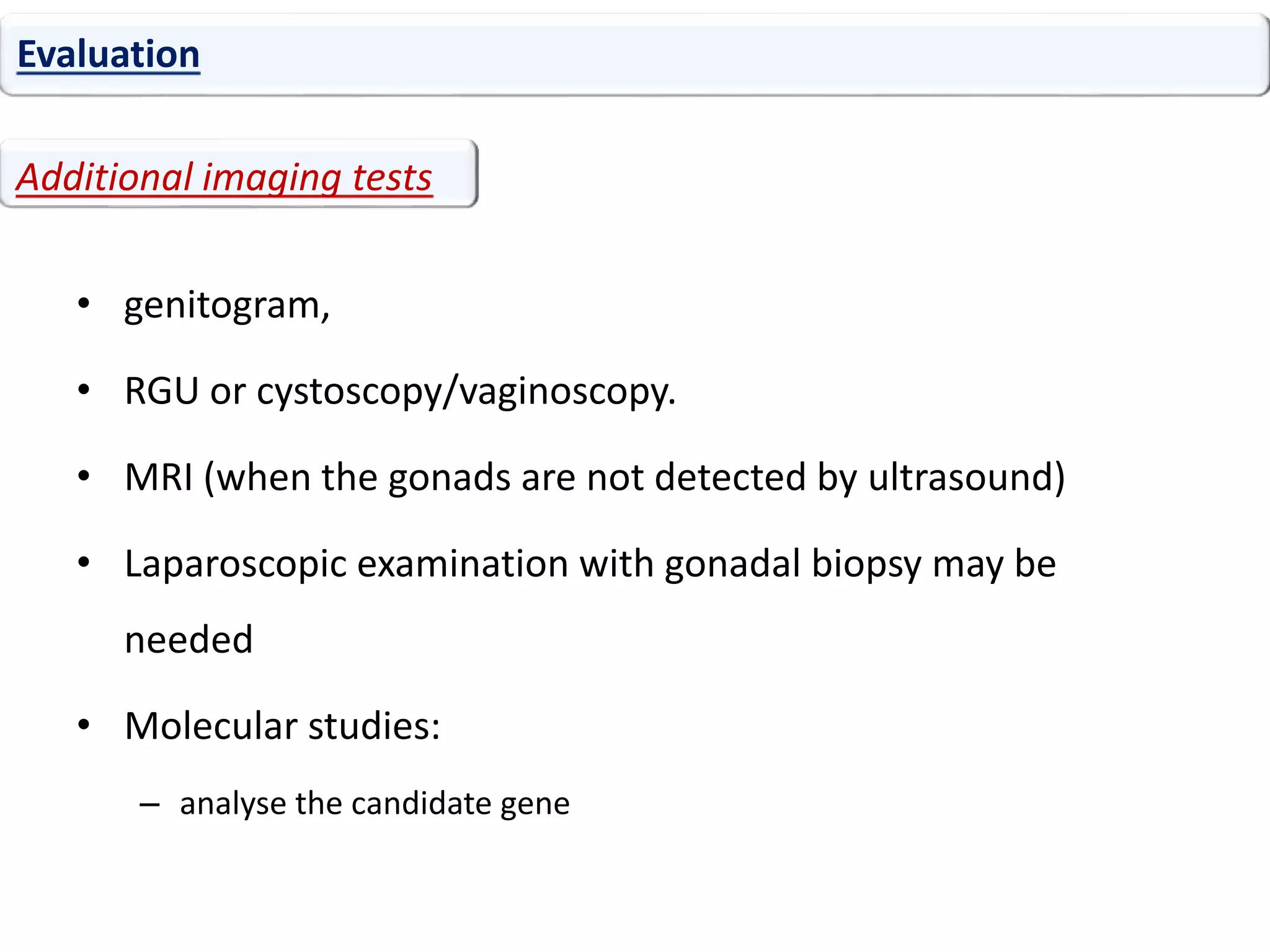
896 ng/dl and androstenedione of 1.34 ng/ml). Magnetic resonance imaging (MRI) pelvis (with
ultrasonogrphy [USG] correlation) revealed uterus didelphys and bilateral ovarian-like structures.
Right sided gonads and adjacent tubal structures were visualized laparoscopically and removed.
Left sided gonads were not visualized and Mullerian remnants were adhered to sigmoid colon.
Case study
Case No;4
MGD](https://image.slidesharecdn.com/dsdpresenstation-190717150409/75/Disorders-of-sexual-differentiation-59-2048.jpg)


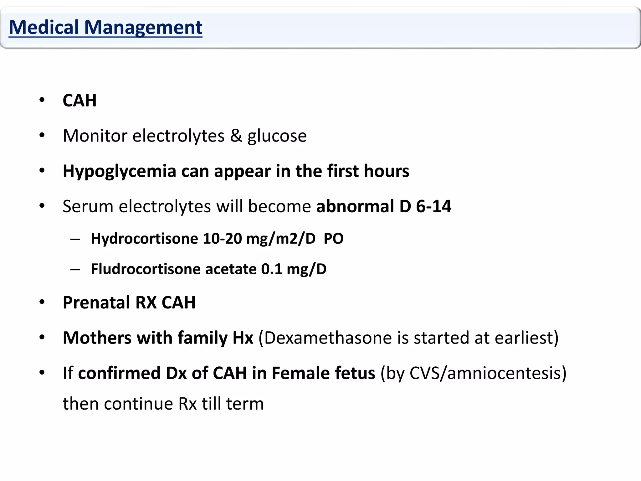


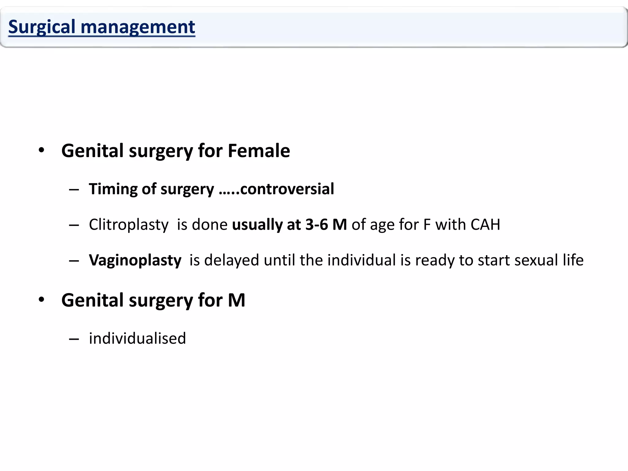
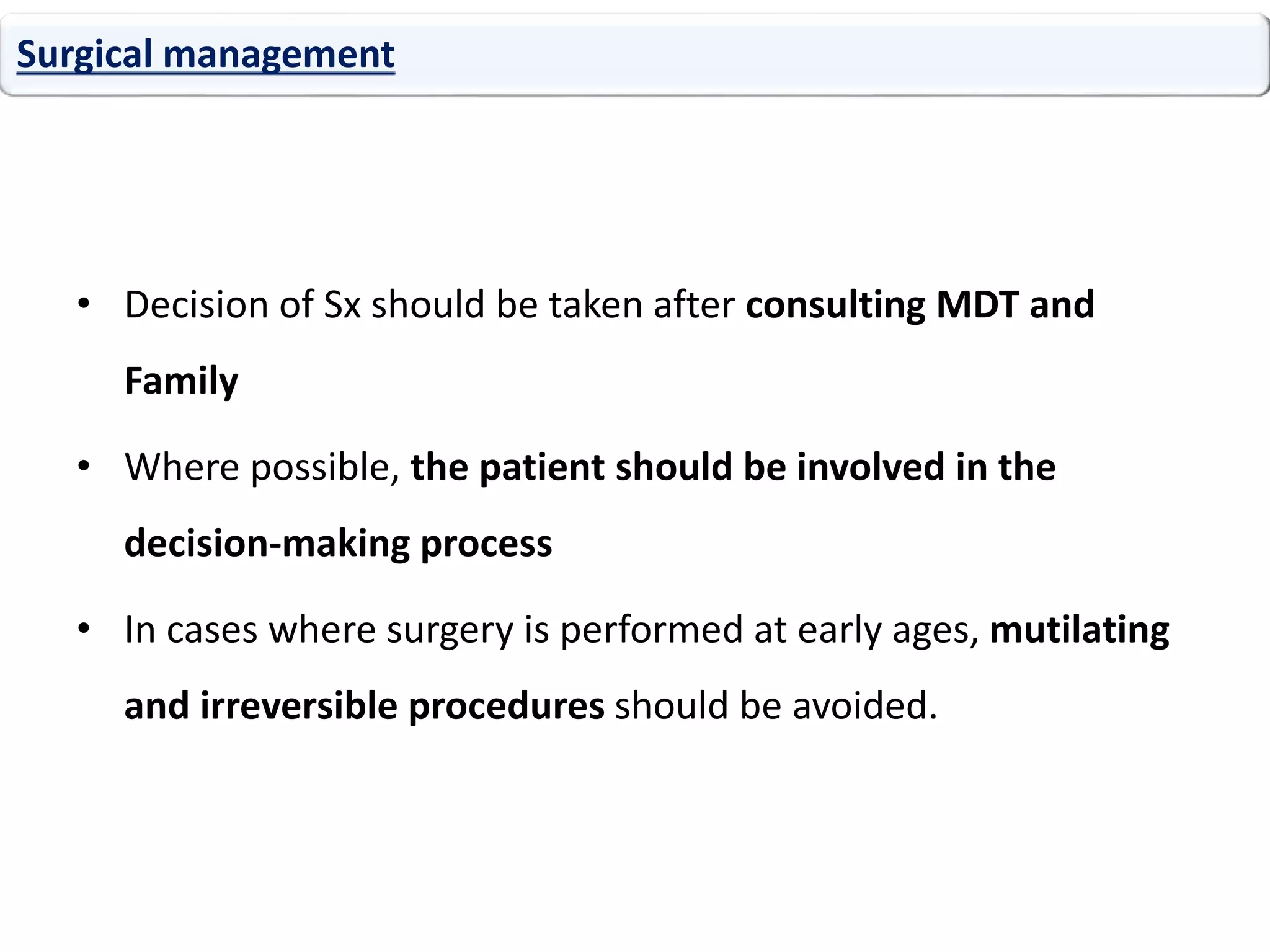


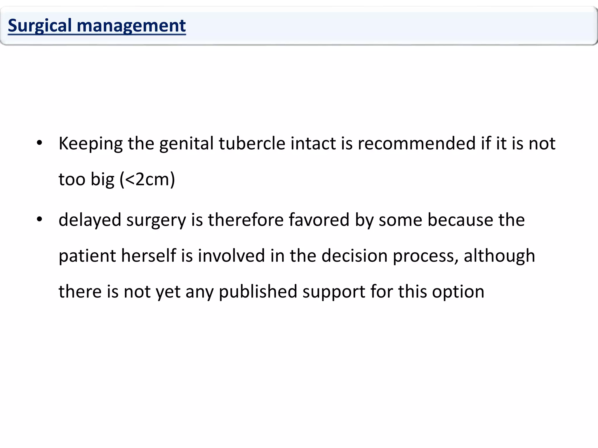


The document discusses disorders of sexual differentiation (DSD). It begins by describing typical embryonic development of male and female genitalia. Common causes of DSD are then discussed, including congenital adrenal hyperplasia (CAH), which can cause virilization of 46,XX individuals. Other conditions mentioned are ovotesticular DSD, complete and partial androgen insensitivity syndrome, 5-alpha reductase deficiency, persistent Müllerian duct syndrome, mixed gonadal dysgenesis, and complete gonadal dysgenesis. The roles of various genes in sexual development are also summarized. Clinical features, investigations, and management considerations are provided for different DSD conditions.