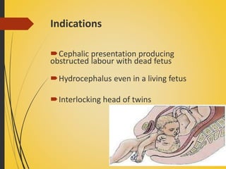
This document provides information about Caesarean section (C-section), including its definition, indications, types, and procedures. Some key points:
- A C-section is a surgical procedure to deliver babies through incisions in the abdominal and uterine walls after 28 weeks of pregnancy. Its use has steadily increased to around 25% currently due to various medical and safety factors.
- Indications can be maternal, fetal, or both and include conditions like cephalopelvic disproportion, breech presentation, fetal distress, and previous C-section.
- The two main types are lower segment (transverse incision below the bladder) and upper segment (vertical incision through the upper uterus), with
Call Girls Service Chennai Jiya 7001305949 Independent Escort Service Chennai
Caesarean Section Guide
1. Caesarean Section
Mrs. U SREEVIDYA Msc.
NURSING,
Associate Professor,
Apollo college of nursing,
CHITTOOR
2. Caesarean Section
Definition:
It is an operative
procedure whereby the
fetus or foetuses after
the end of 28th week are
delivered through an
incision on the
abdominal and uterine
walls.
3. Incidence
* It is steadily raising about 10%
* Increased from 5% in 1970 to 25% in 1990 due to:
* Procedures as high forceps and difficult mid forceps
are abandoned in favour of Caesarean Section (C.S.)
* Increased C.S delivery in breech presentation.
* Destructive operations are abandoned in favour of
C.S.
* Decreased morbidity and mortality due to C.S
encourages its use.
4. Incidence
* Increased safety of the operation
* Improved anaesthesia
* Availability of blood transfusion and antibiotics
* Early identification of at risk mothers
* Wider use of c/s in post c/s pregnancies and Mal -
presentations.
6. Indications
Maternal indications:
1. Severe degree of Contracted pelvis and cephalopelvic disproportion
2. Pelvic tumours especially if impacted in the pelvis or cancer
cervix.
3. Vaginal atresia
The above are considered as absolute indications of c/s,
whereby the vaginal delivery is impossible.
The relative indications are as follows -
1.Minor degree of contracted pelvis and CPD
2.Antepartum hemorrhage
3.4.Hypertensive disorders with pregnancy
4.Abnormal uterine action
5.Previous uterine scar as previous c/s, hysterotomy or
metroplasty.
6.Previous successful repair of vesico-vaginal fistula.
7. Indications cont..
7.Previous caesarean section if,
a. the cause of the previous section is permanent e.g.
contracted pelvis.
b.previous section was upper segment.
c.suspected weak scar as evidencedby:
> History of puerperal infection after the previous
section.
> Hysterosalpingography or hysteroscopy done after the
previous section reveals a defect in the scar.
> Vaginal bleeding during current labour.
> Marked tenderness over the scar during current labour.
>Associated conditions as antepartumhaemorrhage or
malpresentations.
8. FETAL INDICATION
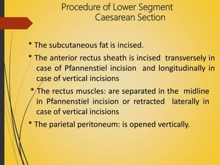
• Fetal distress and cord prolapse
• Breech presentation –[footling, knee presentation,
complicated breech]
• Malpresentation [ brow, transverse lie persistent
mentoposterior ]
• Sever IUGR
• Macrosomia
• Multiple pregnancy[first twin non -vertex and
monoamniotic twin]
• HIV complicating
9. Indications cont..
Foetal indications:
1. Malpresentations and malposition
2.Prolapsed pulsating cord or foetal distress
before full cervical dilatation.
3.Diabetes mellitus
4.Bad obstetric history as recurrent intrauterine
foetal death in last weeks of pregnancy or
repeated intranatal foetal death.
5.Post-mortem C.S. done within 10 minutes of
maternal death to save a living baby.
10. Contraindications
1.Dead foetus: except in;
a. Extreme degree of pelvic contraction.
b. Neglected shoulder presentation
c. Severe accidental haemorrhage.
2. Disseminated intravascular coagulation (blood
coagulation disorder): to minimise blood loss.
3. Extensive scar or pyogenic infection in the
abdominal wall e.g. in burns.
4. Too premature baby.
11. Time of operation
1.Elective
2. Emergency
• Elective: when the operation is done at a pre-planned
time during pregnancy.
Conditions to be fulfilled – Maturity of the
foetus and lungs maturity should be complete.
The operation is done at a pre-selected time
before onset of labour, usually at completed 39
weeks
• Emergency: when the operation is performed due to
sudden complication arising either during pregnancy or
during labour.
12. Types of Caesarean Section
According to the site of uterine incision
• a. Upper segment caesarean section (classical C.S.):
The incision is done in the upper uterine segment
and it is always vertical.
• b. Lower segment caesarean section (LSCS): It is the
common type. The incision is done in the lower uterine
segment and may be transverse (the usual) or vertical in
the following conditions:
> Presence of lateral varicosities.
> Constriction ring to cut through it.
> Deeply engaged head.
13. Classical CS - Disadvantages
• Chance of scar rupture more
• General peritonitis ,if infection occurs
14. Abdominal Incisions
1.Vertical Incision
• Vertical incisions are very rare.
• quickest to make
• greater chance of dehiscence
2. The horizontal or Pfannenstiel Incision
• It is placed at the top of to pubic hair or just over the hair
line as the c-section is started.
• cosmetically better & stronger
• less chance of dehiscence
15. Types of Caesarean Section
According to number of the operation
a. Primary caesarean section: for the first time.
b.Repeated caesarean section: with previous
caesarean section(s).
16. Types of Caesarean Section
According to opening the peritoneal cavity
a. Transperitoneal: The ordinary operation where
the peritoneal cavity is opened before incising the
uterus.
b. Extraperitoneal: The peritoneal cavity is not opened
and the lower uterine segment is reached either
laterally or inferiorly by reflecting the peritoneum of
the vesico-uterine pouch . It is indicated in case of
infected uterine contents as chorioamnionitis.
17. Advantages of elective C.S.
* Pre - operative good preparation as regard
sterilisation and antiseptic measures, fasting
and bowel preparation.
* The risk of puerperal sepsis is minimised.
* The operation is scheduled and working is in
ease.
18. Disadvantages of elective C.S.
* The risk of immaturity of the foetus or its lung is
present.
* Higher incidence of respiratory distress syndrome.
* The lower segment may be not well formed.
* Postpartum haemorrhage is more liable to occur.
* Imperfect drainage of lochia as the cervix is closed so
it should be dilated by the index finger introduced
abdominally through the uterine incision.
19. Pre operative preparation
• Skin preparation
• Antacid: Rantidine (H2 blocker)- 150mg is given orally
before night and repeated 50mg, IM/IV Injection one hour
before surgery.
• Administer Metoclopramide – 10mg, IV
• Bowel and bladder should be emptied.
• FHS should be recorded.
• Anaesthesia: General inhalation anaesthesia with nitrous
oxide + oxygen (the most commonly used), epidural, spinal
or rarely local infiltration anaesthesia.
* Position: Tilting the patient 15 degree to the left in the dorsal
position to minimise the aorto-caval compression.
20. Procedure of Lower Segment Caesarean
Section
• Skin incision: Pfannenstiel (transverse suprapubic) incision is
the most commonly used, but midline or paramedian,
vertical suprapubic incisions may be used. If the patient had
a previous C.S, incise in the same incision with trimming of
the fibrosed edges of the wound to help good healing.
Pfannenstiel incision has a better cosmetic
appearance, better healing and less incidence of incisional
hernia but it is more time consuming associated with more
blood loss and gives less exposure.
21.
22.
23. Procedure of Lower Segment
Caesarean Section
* The subcutaneous fat is incised.
* The anterior rectus sheath is incised transversely in
case of Pfannenstiel incision and longitudinally in
case of vertical incisions
* The rectus muscles: are separated in the midline
in Pfannenstiel incision or retracted laterally in
case of vertical incisions
* The parietal peritoneum: is opened vertically.
24. *The uterus is centralised, the bowel and omentum are
packed off with moist laparotomy pads, however this is
usually unnecessary.
*The loose peritoneum over the lower uterine segment is
held and incised transversely, for about 10 cm in a
semilunar fashion with its edges directed upwards.
* The bladder is compressed downward and is retained
behind a Doyne retractor placed over the symphysis.
25.
26. * The uterus is incised: in the same semilunar fashion by one
of the following methods:
> A semilunar mark is made by the scalpel cutting partially
through the myometrium for 10 cm. A short (3cm) cut is made
in the middle of this incision mark reaching up to but not
through the membranes. The incision is completed by the 2
index fingers along the incision mark. If the lower uterine
segment is very thin, injury of the foetus can be avoided by
using the handle of the scalpel or a haemostat (an artery
forceps) to open the uterus.
> The short (3cm) middle incision may be enlarged by a
bandage scissors over 2 fingers introduced into the uterus
to protect the foetus.
27. *Membranes are ruptured by toothed or Kocher’s
forceps.
* The head is delivered by:
> introducing the right hand gently below it and lifting it
up helped by fundal pressure done by the assistant,
> using one blade of the forceps or,
> using Wrigley’s forceps.
>If the head is deep in the pelvis it can be pushed up
vaginally by an assistant.
> The Doyen’s retractor is removed after the hand or
forceps blade is applied and before head extraction.
28.
29. * Suction for the foetus is carried out before
delivery of the head.
* In breech or transverse lie the foetus is
extracted as breech.
* The placenta is removed.
30. * Closure of the uterine incision is done in 3 layers.
>The first is a continuous locking suture taking most of
the myometrium but not passing through the decidua
to guard against endometriosis and weakness of the
scar.
>The second is a continuous or interrupted one inverting
the first layer.
>The third is a continuous or interrupted layer to close
the visceral peritoneum of the uterus. Closure of
visceral and/or parietal peritoneum is omitted by some
surgeons.
* The abdomen is then closed in layers .
• Clean the vagina and place the sterile pad.
31.
32. Upper Segment Caesarean Section
Indications:
* Dense adhesions, extensive varicosity or myoma in the lower
uterine segment making its exposure or incising through it
difficult.
* Impacted shoulder presentation.
* Anterior placenta praevia.
* Defective scar in the lower segment.
* Cancer cervix.
* Rapid delivery is indicated.
* If an associated tubal sterilisation will be done.
* Previous successful repair of high vesico-vaginal or cervico-
vaginal fistula.
* Post-mortem hysterectomy.
33. • Procedure:
* Abdominal incision: is vertical.
* Uterine incision: 10 cm vertical incision is made in
the midline of upper uterine segment without
incising the peritoneal coat separately as it is
adherent in the upper segment.
* Extraction of the foetus: as a breech in cephalic
presentation.
* The last layer of the uterine incision closure
includes the superficial part of the myometrium
with the peritoneal covering.
* The remainder of the procedure is as lower
segment C.S.
34. Post operative care
• Patient is observed for at least 6-8 hours
• Vital signs should be recorded.
• I.V fluids should be administered
• Blood transfusion if necessary
• Inj. Methergin – 0.2mg, IM, should be given. Repeat if
necessary.
• Prophylactic antibiotics for 48 hours.
• Analgesics – Pethidine-75 to 100 mg
• Ambulation: sit on the bed with in 24 hrs
• Breast feeding
• Diet: second day- liquid diet like electrolyte water or tea,
third day – light solid diet
• Mild laxative – milk of magnesia – 4-6 spoons
• Removal of stiches – 6th or 7th day.
• Discharge: on the day following removal of the stiches.
35. Advantages of the lower segment over the upper
segment operation
* Less blood loss: due to less vascularity and the placental
bed is away from the incision.
* Easier to repair.
*The resultant uterine scar is stronger
*Less subsequent adhesions to the bowel and omentum.
*Less liability to acute gastric dilatation and paralytic
ileus.
* Less liability to peritonitis due to better
peritonization and healing.
36. Caesarean Hysterectomy
Hysterectomy is carried out after caesarean section in the
same sitting for one of the following reasons:
* Uncontrollable postpartum haemorrhage.
* Unrepairable rupture uterus.
* Operable cancer cervix.
* Couvelaire uterus.
* Placenta accreta cannot be separated.
* Severe uterine infection particularly that caused by Cl.
welchii.
*Multiple uterine myomas in a woman not desiring
future pregnancy.
38. Complications of Caesarean Section
1. Operative:
a. Primary maternal mortality is 4 times that of vaginal
delivery which may be due to:
> shock .
>Anaesthetic complications particularly Mendelson’s
syndrome
>Haemorrhage usually due to extension of the uterine
incision to the uterine vessels, atony of the uterus or
DIC.
b. Injuries to the bladder or ureter.
c. Fetal injuries, RDS- Respiratory distress syndrome to
foetus.
39. Complications of Caesarean Section
2.Post-operative:
b. Early:
> Thrombosis and pulmonary embolism.
> Acute dilatation of the stomach and paralytic ileus.
> Wound infection, puerperal sepsis and burst abdomen.
>Chest infection.
c. Late:
> Rupture of the uterine scar.
> Incisional hernia.
> Chronic pelvic pain or backache
> Menstrual irregularities.
41. DESTRUCTIVE OPERATIONS
DEFINITION:
The destructive operations are designed to diminish the
bulk of the fetus so as to facilitate easy delivery through
the birth canal.
These procedures are difficult and may be dangerous
too unless the operator is sufficiently skilled.
42. Types of destructive operations
Some commonly performed operations are
Craniotomy
Evisceration
Decapitation
Cleidotomy
43. CRANIOTOMY
Definition
It is an operation to make a perforation
on the fetal head , to evacuate the contents
followed by extraction of the fetus.
46. Contraindications
The operation should not be done when the
pelvis is severly contracted. So as to shortened the
true conjugate to less than 7.5cm (3``).
Rupture of the uterus
Condition to be full filled
The cervix must be fully dilated
Baby must be dead
47. Procedure
Step1: Two fingers are introduced into the vagina and the fingertips are to be planned on
proposed site of perforation. However when the suture line cannot be defined because of
big caput , the perforation should be done through the dependent part.
Step 2: The Oldham’s perforator , with the blades closed , is introduced protecting the
anterior vaginal wall and the adjacent bladder until the tip reaches the proposed site of
perforation.
Step 3 :- By rotating movements the skull is perforated . After the skull is perforated , the
instrument is thrust up to the shoulder and the handles approximated ,so as to allow
separation of the sharp blades for about 2.5 cm .
Step 4 :- With the fingers brain matter is evacuated. The idea is to make the skull collapsed
as much as possible.
Step 5 :-when the skull is found sufficiently compressed, the extraction of the fetus is
achieved either by using cranioclast or by two Gaint Vulsella are used to hold the incised
skull and scalp margins.
Step 6 :- the traction is now excreted in the same direction is like that mentioned in forceps
operations.
Step 7 :- after the delivery of the placenta, the uterovaginal canal must be explored as a
routine for evidence of rupture uterus or any tear.
Inj. Methergin 0.2mg is to be given intravenously with the delivery of the anterior
shoulder. The rest of the delivery is completed as in normal delivery.
48.
49. EVISCERATION
The operation consists in removal of thoracic and abdominal
contents piecemeal through an opening on the thoracic or
abdominal cavity at the most accessible site.
The objective is to diminish the bulk of the fetus which
facilitates its extraction.
If difficulty arises , the spine may have to be divided
( spondylotomy) with embryotomy scissors.
Indications
Neglected shoulder presentation with dead fetus , the neck
is not easily assessable.
Fetal malformation such as fetal ascites or hugely distended
bladder or monsters.
50.
51. DECAPITATION
Definition
It is a destructive operation whereby the fetal
head is severed from the trunk and the delivery is
completed with the extraction of the trunk and that
of the decapitated head per vaginam.
Indications
Neglected shoulder presentation with dead fetus
where neck is easily assessable
Interlocking head of the twins
52. Procedure
The operation should be done at general anesthesia
Step 1 :- if the fetal hand is not prolapsed bring down the hand. A roller gauze is tied on the
fetal wrist and assistant is asked to give the traction towards the side away from the fetal
head to make the neck more assessable and fixed
Step 2 :- two fingers of the left hand are introduced with the palmar surface downwards
and the fingertips are to be placed on the superior surface of the neck –the prolapsed site of
decapitation.
Step 3 :- the decapitation hook with knife is to be introduced flushed under the guidance of
the fingers placed into the vagina, with knob pointing toward the fetal head. The hook is
pushed above the neck and rotated to 90 ̊ , to placed the knife firmly against the neck.
Step 4 :- by upward and downward movement of the hook with knife the vertebral column
is severed.
Step 5:- delivery of the decapitation head – the methods are
By hooking the index fingers into the mouth
By holding the severed head with Giant Vulsellum and delivery of he head as that of
aftercoming head in breech.
Using forceps
Step 6 :- routine exploration of the utero vaginal canal to exclude rupture of the uterus or any
other injury.
53.
54. CLEIDOTOMY
The operation consists of reduction in the bulk of the
shoulder girdle by division of one or both the clavicles.
The operation is done only in dead fetus ( anencephaly
excluded ) with shoulder dystocia.
The clavicles are divided by the embryotomy scissors or
long straight scissors introduced under the guidance of
left two fingers placed inside the vagina.
55. Post operative care following destructive
operation
Exploration of the utero-vaginal canal must be done to
exclude rupture of the uterus or lacerations on the vagina or
any genital injury.
A self retaining catheter is put inside specially following
craniotomy for a period of 3-5 days or until the bladder tone
is regained.
Dextrose saline drip is to be continued till dehydration is
corrected.
Blood transfusion may be given if required .
Ceftriaxone 1gm IV infusion is given twice daily.
56. Complications
Injury to the utero-vaginal canal
Rupture of the uterus
Post partum haemorrhage-atonic or traumatic
Shock due to blood loss and or dehydration
Puerperal sepsis
Subinvolution
Injury to the adjacent viscera
Prolonged ill health
57. Nursing diagnosis
1. Alteration in comfort due to pain related to delivery process
Assess the types of pain ,types , duration and intensity
Provide comfortable left lateral position to the mother
Provide psychological reassurance to the mother
2. Potential for complication related to destructive operation
Assess for any types of laceration
Maintain aseptic technique
Handle the case carefully
Case should handle by experts
Clean the perineal area with betadine
58. Nursing diagnosis
3. Potential for infection related to destructive operation
Assess for any scar
Maintain strict aseptic technique
Clean the surrounding
Avoid many visitors
4. Fear and anxiety of parents related to delivery process
Provide proper explanation about baby’s condition
Give information about progress of delivery frequently
Provide psychological reassurance to the mother
Clarify the mother doubts.
60. DEFINITION
Version is the turning out of fetus from one
presentation to another and may be done either
externally or internally by the physician.
If the aim is to make the head the presenting part is
called cephalic version and if the breech will be the
presenting part it is called podalic version.
61. TYPES OF VERSION
According to the methods employed.
1. External cephalic version
2. Internal podalic version
3. Bipolar version
62. External cephalic version
It is a procedure used to turn a fetus from a breech
position or transverse position into a cephalic pole of
the uterus.
64. PRELIMINARIES
The patient is asked to empty bladder.
She is to lie on her back with the sholders slightly
raised and the thighs slightly flexed.
abdomen is fully exposed and FHR is auscultated.
The most commonly used tocolytic medication
(terbutaline-0.25mg sc) to relax the uterus.
66. PROCEDURE
(a) (a)
Step-1. The breech mobilised
using both hands On the
surface of the abdomen
at first. Then one by the
fetus'head and the other
by the buttocks the fetus
is turned and rolled to the
vertex position.
(b) (b)
Step-2. Generally podalic pole is
grasped by right hand and
head is grasped by left hand
till the lie becomes transverse.
67. CONTINUE:
Step-3. The hand is now changed one after the
other hold the fetal poles to prevent
crossing of the hand.
(c) ( (d)
68. INSTRUCTIONS
The patient is advised for follow up to check the
corrected position.
To report to the physician if there is vaginal bleeding
or liquor amnii.
Rh-negative non immunised women must be
protected by intramuscular administration of 100.mug
anti-D gamma globulin.
69. CONTRAINDICATION
Fetal distress.
The amniotic sac has ruptured.
A mother has a condition(such a heart problem).
A caesarean delivery is needed,such as when there
is placenta praevia or abruptio placentae.
70. Advantages of ECV
1. Reduces the number of caesarean delivery.
2. Reduces maternal morbidity due to caesarean or
vaginal breech delivery .
3. Reduces the fetal hazards of vaginal breech
delivery.
71. EXTERNAL PODALIC VERSION
The external podalic version may be done
in cases when the external cephalic version
fails in transverse lie in case of the second
baby of twins.
72. INTERNAL PODALIC VERSION
Internal version is always a podalic version
and is almost always completed with the
extraction of the fetus.
73.
74. INDICATIONS
Its only indication being the transverse lie in case of
the second baby of twins.
However, it may be employed in singleton pregnancy
to expedite delivery in adverse condition where the
caesarean section facilities are lacking. Such condition
are:
1. Transverse lie with cervix fully dilated.
2.Cord prolapse with cervix fully dilated with
transverse lie or head high up and the baby is alive.
76. PROCEDURE
Step-1: If the podalic pole of the fetus is on left side of
the mother, the right hand isto be introduced and vice
versa.
Step-2: The identification of the foot is done by
palpation of theheel.
Step-3: While the leg is brought down by a steady
traction the cephalic pole is pushed up using the
external hand.
Step-4: After one leg is brought down,there is no
difficulty todelivertheother leg.
79. COMPLICATIONS
Maternal risk include -
1. Placental abruption.
2. Rupture of theuterus.
Fetal risk include -
1. Asphyxia.
2. Cord prolapse.
3. Intra cranial hemorrhage.
80. BIPOLAR
VERSION
It also known as braxton-hicks.
The version is done introducing
one or two finger through the
cervix and the other hand on the
abdomen.
83. PROCEDURE
Under the generalanethesia.
At least two finger are passed through the partially
dialated cervix, the foot is grasped as in internal
podalicversion pulled through the cervix while the
other hand is assisting theversion externally .
84. BIBLIOGRAPHY
BOOK REFERENCE
1. Annama Jocab, text book of comprehensive
text book of ‘MIDWIFY and GYNECOLOGY
nursing ‘ JAYPEE publication 3rd edition page
no.285-287.
2. D.C. DUTTA text book of obsterical including
perinatary and contraception central publication
7th edition page no. 582-585.
3. NETREFERENCE
www.wikipedia.c
om
www.pubmed.co
m