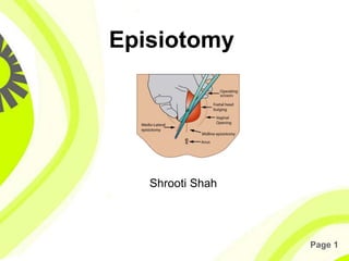
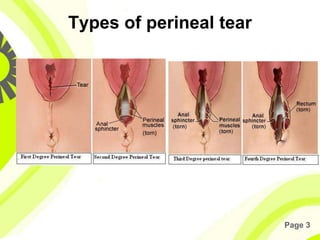
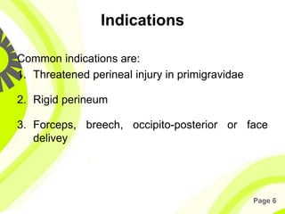
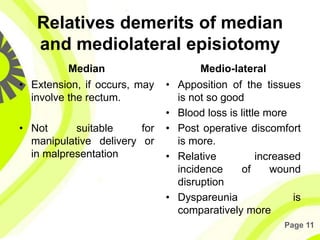
Page 1
Episiotomy
Shrooti Shah
Page 2
Definition
• A surgically planned incision on the
perineum and the posterior vaginal wall
during the second stage of labour is called
episiotomy.
Page 3
Types of perineal tear
Page 4
Objectives
• To enlarge the vaginal introitus so as to
facilitate easy and safe delivery of the
fetus- spontaneous or manipulative.
• To minimise overstretching and rupture of
the perineal muscles and fascia; to reduce
the stress and strain on the fetal head.
Page 5
Indications
• In elastic rigid perineum
• Anticipating perineal tear: Big baby, face to
pubis delivery, Breech delivery, Shoulder
dystocia
• Operative delivery: Forceps delivery, Ventouse
delivery
• Previous perineal surgery: Pelvic floor repair,
Perineal reconstructive surgery
Page 6
Indications
Common indications are:
1. Threatened perineal injury in primigravidae
2. Rigid perineum
3. Forceps, breech, occipito-posterior or face
delivey
Page 7
Timing of the episiotomy
Bulging thinned perineum during
contraction just prior to crowning is the
ideal time.
Page 8
Advantages
• Maternal:
– A clear and controlled incision is easy to repair and
heals better than a lacerated wound that might occur
otherwise.
– Reduction in the duration of second stage
– Reduction of trauma to the pelvic floor muscles
• Fetal:
– It minimises intracranial injuries specially in premature
babies or after coming head of breech
Page 9
Types
• Medio-lateral
• Median
• Lateral
• T-shaped
Page 10
Relatives merit of median and
mediolateral episiotomy
Median
• The muscles are not cut.
• Blood loss is least.
• Repair is easy
• Post operative comfort is
maximum
• Healing is superior
• Wound disruption is rare
• Dyspareunia is rare
Medio-lateral
• Relatively safety from
rectal involvement from
extension.
• If necessary, the incision
can be extennded
Page 11
Relatives demerits of median
and mediolateral episiotomy
Median
• Extension, if occurs, may
involve the rectum.
• Not suitable for
manipulative delivery or
in malpresentation
Medio-lateral
• Apposition of the tissues
is not so good
• Blood loss is little more
• Post operative discomfort
is more.
• Relative increased
incidence of wound
disruption
• Dyspareunia is
comparatively more
Page 12
Structure cut are:
• Posterior vaginal wall
• Superior and deep transverse perineal muscles,
bulbospongiosus and part of levator ani.
• Fascia covering those muscles
• Transverse perineal branches of pudendal
vessels and nerves
• Subcutaneous tissue and skin
Page 13
Steps of episiotomy
• Provide emotional support and encouragement.
• Use local infiltration with lignocaine.
• Make sure there are no known allergies to
lignocaine or related drugs.
• Infiltrate beneath the vaginal mucosa, beneath
the skin of the perineum and deeply into the
perineal muscle.
• Note: Aspirate (pull back on the plunger) to
be sure that no vessel has been penetrated
Page 14
Steps of episiotomy
Page 15
Steps of episiotomy
• Wait 2 minutes and then pinch the incision
site with forceps.
• Wait to perform episiotomy until:
- the perineum is thinned out; and
- 3–4 cm of the baby’s head is visible during a
contraction.
Page 16
Steps of episiotomy
• Wearing high-level disinfected gloves,
place two fingers between the baby’s head
and the perineum.
• Use scissors to cut the perineum about 3–
4 cm in the mediolateral direction
Page 17
Steps of episiotomy
• Use scissors to cut 2–3 cm up the middle
of the posterior vagina.
• Control the baby’s head and shoulders as
they deliver.
• Carefully examine for extensions and
other tears and repair
Page 18
Repair of episiotomy
• Apply antiseptic solution to the area around the
episiotomy.
• If the episiotomy is extended through the anal
sphincter or rectalmucosa, manage as third or
fourth degree tears, respectively
• Close the vaginal mucosa using continuous 1-0
suture
Page 19
Repair of episiotomy
• Start the repair about 1 cm above the apex (top)
of the episiotomy. Continue the suture to the
level of the vaginal opening.
• At the opening of the vagina, bring together the
cut edges of the vaginal opening
• - Bring the needle under the vaginal opening
and out through the incision and tie.
Page 20
Repair of episiotomy
• Close the perineal muscle using
interrupted 1-0 sutures
• Close the skin using interrupted (or
subcuticular) 1-0 sutures
Page 21
Post operative care
• Dressing
• Comfort
• Ambulation
• Removal of stitches
Page 22
Complications
• Immediate
• Extension of the incision to involve the rectum
• Vulval haematoma
• Infection
• Wound dehiscence
• Injury to anal sphincter causing incontinence of
flatus or faeces
• Rectovaginal fistula (Rarely)
• Necrotising fascitis
Page 23
Complications
• Remote
• Dyspareunia
• Chance of perineal lacerations
• Scar endometriosis (rare)

Episiotomy

  • 1.
  • 2.
    Page 2 Definition • Asurgically planned incision on the perineum and the posterior vaginal wall during the second stage of labour is called episiotomy.
  • 3.
    Page 3 Types ofperineal tear
  • 4.
    Page 4 Objectives • Toenlarge the vaginal introitus so as to facilitate easy and safe delivery of the fetus- spontaneous or manipulative. • To minimise overstretching and rupture of the perineal muscles and fascia; to reduce the stress and strain on the fetal head.
  • 5.
    Page 5 Indications • Inelastic rigid perineum • Anticipating perineal tear: Big baby, face to pubis delivery, Breech delivery, Shoulder dystocia • Operative delivery: Forceps delivery, Ventouse delivery • Previous perineal surgery: Pelvic floor repair, Perineal reconstructive surgery
  • 6.
    Page 6 Indications Common indicationsare: 1. Threatened perineal injury in primigravidae 2. Rigid perineum 3. Forceps, breech, occipito-posterior or face delivey
  • 7.
    Page 7 Timing ofthe episiotomy Bulging thinned perineum during contraction just prior to crowning is the ideal time.
  • 8.
    Page 8 Advantages • Maternal: –A clear and controlled incision is easy to repair and heals better than a lacerated wound that might occur otherwise. – Reduction in the duration of second stage – Reduction of trauma to the pelvic floor muscles • Fetal: – It minimises intracranial injuries specially in premature babies or after coming head of breech
  • 9.
    Page 9 Types • Medio-lateral •Median • Lateral • T-shaped
  • 10.
    Page 10 Relatives meritof median and mediolateral episiotomy Median • The muscles are not cut. • Blood loss is least. • Repair is easy • Post operative comfort is maximum • Healing is superior • Wound disruption is rare • Dyspareunia is rare Medio-lateral • Relatively safety from rectal involvement from extension. • If necessary, the incision can be extennded
  • 11.
    Page 11 Relatives demeritsof median and mediolateral episiotomy Median • Extension, if occurs, may involve the rectum. • Not suitable for manipulative delivery or in malpresentation Medio-lateral • Apposition of the tissues is not so good • Blood loss is little more • Post operative discomfort is more. • Relative increased incidence of wound disruption • Dyspareunia is comparatively more
  • 12.
    Page 12 Structure cutare: • Posterior vaginal wall • Superior and deep transverse perineal muscles, bulbospongiosus and part of levator ani. • Fascia covering those muscles • Transverse perineal branches of pudendal vessels and nerves • Subcutaneous tissue and skin
  • 13.
    Page 13 Steps ofepisiotomy • Provide emotional support and encouragement. • Use local infiltration with lignocaine. • Make sure there are no known allergies to lignocaine or related drugs. • Infiltrate beneath the vaginal mucosa, beneath the skin of the perineum and deeply into the perineal muscle. • Note: Aspirate (pull back on the plunger) to be sure that no vessel has been penetrated
  • 14.
    Page 14 Steps ofepisiotomy
  • 15.
    Page 15 Steps ofepisiotomy • Wait 2 minutes and then pinch the incision site with forceps. • Wait to perform episiotomy until: - the perineum is thinned out; and - 3–4 cm of the baby’s head is visible during a contraction.
  • 16.
    Page 16 Steps ofepisiotomy • Wearing high-level disinfected gloves, place two fingers between the baby’s head and the perineum. • Use scissors to cut the perineum about 3– 4 cm in the mediolateral direction
  • 17.
    Page 17 Steps ofepisiotomy • Use scissors to cut 2–3 cm up the middle of the posterior vagina. • Control the baby’s head and shoulders as they deliver. • Carefully examine for extensions and other tears and repair
  • 18.
    Page 18 Repair ofepisiotomy • Apply antiseptic solution to the area around the episiotomy. • If the episiotomy is extended through the anal sphincter or rectalmucosa, manage as third or fourth degree tears, respectively • Close the vaginal mucosa using continuous 1-0 suture
  • 19.
    Page 19 Repair ofepisiotomy • Start the repair about 1 cm above the apex (top) of the episiotomy. Continue the suture to the level of the vaginal opening. • At the opening of the vagina, bring together the cut edges of the vaginal opening • - Bring the needle under the vaginal opening and out through the incision and tie.
  • 20.
    Page 20 Repair ofepisiotomy • Close the perineal muscle using interrupted 1-0 sutures • Close the skin using interrupted (or subcuticular) 1-0 sutures
  • 21.
    Page 21 Post operativecare • Dressing • Comfort • Ambulation • Removal of stitches
  • 22.
    Page 22 Complications • Immediate •Extension of the incision to involve the rectum • Vulval haematoma • Infection • Wound dehiscence • Injury to anal sphincter causing incontinence of flatus or faeces • Rectovaginal fistula (Rarely) • Necrotising fascitis
  • 23.
    Page 23 Complications • Remote •Dyspareunia • Chance of perineal lacerations • Scar endometriosis (rare)