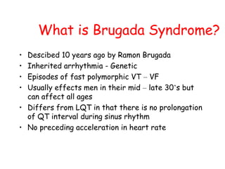
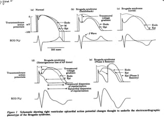
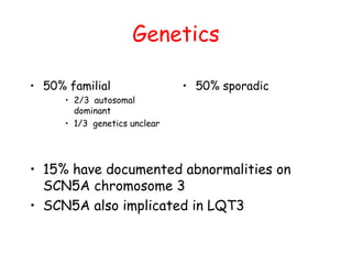
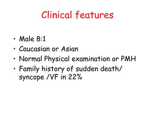
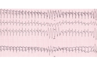
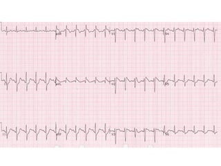
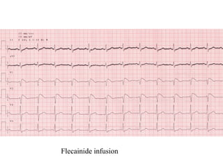
Brugada Syndrome
Definition
“ Syncopal episodes and/or sudden death in patients
with a structurally normal heart and a
characteristic electrocardiogram displaying a
pattern resembling right bundle branch block
with ST segment elevation in leads V1 to V3 ”
Brugada et al. Circulation
What is Brugada Syndrome?
• Descibed 10 years ago by Ramon Brugada
• Inherited arrhythmia - Genetic
• Episodes of fast polymorphic VT – VF
• Usually effects men in their mid – late 30’s but
can affect all ages
• Differs from LQT in that there is no prolongation
of QT interval during sinus rhythm
• No preceding acceleration in heart rate
Other Forms of
Presentation
• Symptomatic with normal ECG during control
• Asymptomatic with abnormal ECG
• Asymptomatic family members with an abnormal
ECG either during, or after flecainide challenge
Epidemiology
• First reported in Poland
• Incidence & distribution difficult to ascertain
• Geographic distribution – worldwide but common in
S.E. Asia
– Philippines - Bangungut (Scream followed by
sudden death at night)
– Japan - Pokkuri (Unexpected sudden death at
night)
– Thailand - Lai Tai (Death during sleep)
Presentation
• Sudden cardiac death survivor (SCD)
• Approximately 10% of patients experience
paroxysmal AF
• Unexplained syncope
Genetic Considerations
• Link to gene SCN5A located in chromosome 3
• Approximately 60% of patients with aborted SCD
with the typical ECG, have family members with
the same ECG abnormalities or a family history of
SCD.
• The defect may be due to anomalies in the cellular
conduction channels, which may be inherited.
• Predominance of affected males
• Different from LQT and ARVD
Mechanism of
arrhythmogenesis
• Loss of AP dome in epicardial tissue
• Dispersion of refractories
• Phase II re-entry
• Rapid polymorphic VT
• VF
• Changes enhanced by Sodium channel
blockers (class Ia/Ic agents)
Genetics
• 50% familial
• 2/3 autosomal
dominant
• 1/3 genetics unclear
• 50% sporadic
• 15% have documented abnormalities on
SCN5A chromosome 3
• SCN5A also implicated in LQT3
Clinical features
• Male 8:1
• Caucasian or Asian
• Normal Physical examination or PMH
• Family history of sudden death/
syncope /VF in 22%
Diagnosis
• FH of sudden cardiac death or PMVT
• Brugada Shift pattern on ECG
• Normal structural heart
• Accentuation of ECG changes with
class Ia/Ic agents
• Demonstration of defects on SCN5A
Differential diagnosis 1
• Arrhythmogenic right ventricular
dysplasia
• Idiopathic ventricular fibrillation
• Polymorphic ventricular tachycardia
(inherited or acquired prolonged QT)
• Asymptomatic variants
Differential diagnosis 2
• Brugada vs idiopathic VF
40-60 % Chen et al
3-24% Remmne et al
• Brugada vs ARVD
No structural or histopathological similarities
Different genetic loci
Morphological abnormalities on MRI
Differential diagnosis 3
• Brugada vs MI
Similar to Right ventricular infarction
Younger age group
Asmyptomatic individuals
with Brugada ECG
• 0.05-0.14% general population
• No symptoms over 2-3 year follow up
• Variable response to Na blockers
• Brugada reported 27% event rate in
patients picked up with family
screening over a 3 year follow up
Treatment
• Brugada recommends/ESC advisory
board
• ICD for symptomatic individuals
• Extensive investigation of asymptomatic
individuals to exclude structural heart
disease
• ICD in patients with inducible VT/VF on EPS
Unanswered Questions
• Risk to asymtomatic individuals ?
• Interplay of pathological,
physiological and pharmacological
factors ?
• The predictive role of
electrophysiological studies
Prognosis & Treatment
• Poor prognosis when left untreated
• Antiarrhythmic therapy does not protect against
SCD
• Only available treatment is the Implantable
Cardioverter Defibrillator (ICD)
Considerations
• Genetic screening
• Exclude conditions where the ECG may mimic
Brugada
– Pectus excavatum
– Mediastinal tumours
– Steinert’s disease
The Flecainide Challenge
• IV flecainide is the standard test used to identify
individuals with concealed forms of the disease
• May reveal the characteristic ECG changes
• Conducted at the request of the attending
consultant cardiologist and with the approval of
the consultant electrophysiologist
N.B.The flecainide challenge is a significant moment
for the patient & their family, as it may represent the
confirmation of a lifelong and potentially life threatening
condition
Patient Education
• Discuss why the flecainide challenge is necessary,
the side effects & the need for continuous cardiac
monitoring
• The treatment options available must be discussed
should the flecainide challenge prove positive
• Full informed consent must be obtained by the
doctor administrating the dose.
• If the patient declines the flecainide challenge
then the associated risks must be discussed with
them
Medical Considerations
• Only medical personnel trained to administer
flecainide for this study may instigate therapy
• Raise awareness of the potential for ventricular
arrhythmias as in 0.5% of cases Ventricular
Fibrillation may be induced
• Additional counselling and support may be required
to help the patient cope with the associated
psychological, social and medical problems
Pre-Procedural Considerations
• Full explanation must be given and treatment
options outlined
• Informed consent must be obtained and
documented in the patients’ notes
• Serum electrolytes should be checked & corrected
• Record baseline observations
• Record a 12 lead ECG on admission
• Ensure cannula is sited and patent
Pre-Procedural Considerations
2
• The patient should be nil by mouth for at least 4
hours preceding the test
• The use of a pre-med sedation (eg. Midazolam)
may be required
• Ensure full resuscitation facilities, equipment and
drugs are available
N.B. - For paediatric cases the correct doses of
resuscitation drugs should be calculated and
prepared beforehand
Prescribed Drug Dosage
• The intravenous flecainide must be prescribed and
signed by the doctor administrating the dose,
clearly marked “For Flecainide challenge only”
• Confirm the correct dose of Flecainide is
prescribed according to the patients weight
• The recommended IV dose is 2mg/kg
N.B.The maximum dose licensed for Flecainide is 150mg = 75kg
The Clinical Setting
• The flecainide challenge should only be carried out
in a high dependency setting
• For paediatric cases:
- PICU should be informed with the bed space
booked and agreed in advance
- The medical team must liaise with the consultant
intensivist & PICU registrar
- A senior paediatric nurse to accompany & assist
- PICU staff will not be directly involved other
than in the event of a resuscitation
The Procedure
• Establish continuous 12 lead ECG monitoring for
duration of the challenge
• Monitor and record the patients BP, HR & Oxygen
saturations at 5 minute intervals
• The prescribing doctor must administer the IV
flecainide slowly over 10 minutes
• During administration, 12 lead ECGs should be
recorded at 5 minute intervals or with any
observed ECG changes on the monitor
• If, at any time, there are changes suggestive of
Brugada the remaining infusion must be
discontinued
• Twelve lead ECG monitoring should continue for 30
minutes on completion of the infusion
Study Interpretation
• The flecainide challenge is considered positive if
there is a 1mm ST elevation in leads V1, V2 & V3
Follow up Care
• The patient should be closely monitored until the
results of the test are known & the effects of
sedation have worn off
• If the test is positive the doctor should confirm
this with the consultant electrophysiologist, prior
to informing the patient and/or parents
• The discharging doctor should check with the
consultant electrophysiologist & cardiologist
regarding follow-up & arrange this prior to
discharge
• A discharge summary should be completed prior to
the patient leaving the ward
Case Report
• PC - 52 yr male
• 3 yr history of palpitations
• Usually on exertion
• 15 times in last 3 yrs
• Associated with pre syncope
• Previously fit and well
• Examination normal
• ECG
• Echo normal
Flecainide infusion
Investigations
• Multiple ambulatory monitoring
normal
• ETT
• Coronary angio normal
• EPS – VF with S4
Outcome
• Dual chamber ICD
• Currently well
– NSVT
– No therapy

Brugada syndrome

  • 1.
  • 2.
    Definition “ Syncopal episodesand/or sudden death in patients with a structurally normal heart and a characteristic electrocardiogram displaying a pattern resembling right bundle branch block with ST segment elevation in leads V1 to V3 ” Brugada et al. Circulation
  • 3.
    What is BrugadaSyndrome? • Descibed 10 years ago by Ramon Brugada • Inherited arrhythmia - Genetic • Episodes of fast polymorphic VT – VF • Usually effects men in their mid – late 30’s but can affect all ages • Differs from LQT in that there is no prolongation of QT interval during sinus rhythm • No preceding acceleration in heart rate
  • 4.
    Other Forms of Presentation •Symptomatic with normal ECG during control • Asymptomatic with abnormal ECG • Asymptomatic family members with an abnormal ECG either during, or after flecainide challenge
  • 5.
    Epidemiology • First reportedin Poland • Incidence & distribution difficult to ascertain • Geographic distribution – worldwide but common in S.E. Asia – Philippines - Bangungut (Scream followed by sudden death at night) – Japan - Pokkuri (Unexpected sudden death at night) – Thailand - Lai Tai (Death during sleep)
  • 6.
    Presentation • Sudden cardiacdeath survivor (SCD) • Approximately 10% of patients experience paroxysmal AF • Unexplained syncope
  • 7.
    Genetic Considerations • Linkto gene SCN5A located in chromosome 3 • Approximately 60% of patients with aborted SCD with the typical ECG, have family members with the same ECG abnormalities or a family history of SCD. • The defect may be due to anomalies in the cellular conduction channels, which may be inherited. • Predominance of affected males • Different from LQT and ARVD
  • 9.
    Mechanism of arrhythmogenesis • Lossof AP dome in epicardial tissue • Dispersion of refractories • Phase II re-entry • Rapid polymorphic VT • VF • Changes enhanced by Sodium channel blockers (class Ia/Ic agents)
  • 10.
    Genetics • 50% familial •2/3 autosomal dominant • 1/3 genetics unclear • 50% sporadic • 15% have documented abnormalities on SCN5A chromosome 3 • SCN5A also implicated in LQT3
  • 11.
    Clinical features • Male8:1 • Caucasian or Asian • Normal Physical examination or PMH • Family history of sudden death/ syncope /VF in 22%
  • 12.
    Diagnosis • FH ofsudden cardiac death or PMVT • Brugada Shift pattern on ECG • Normal structural heart • Accentuation of ECG changes with class Ia/Ic agents • Demonstration of defects on SCN5A
  • 13.
    Differential diagnosis 1 •Arrhythmogenic right ventricular dysplasia • Idiopathic ventricular fibrillation • Polymorphic ventricular tachycardia (inherited or acquired prolonged QT) • Asymptomatic variants
  • 14.
    Differential diagnosis 2 •Brugada vs idiopathic VF 40-60 % Chen et al 3-24% Remmne et al • Brugada vs ARVD No structural or histopathological similarities Different genetic loci Morphological abnormalities on MRI
  • 15.
    Differential diagnosis 3 •Brugada vs MI Similar to Right ventricular infarction Younger age group
  • 16.
    Asmyptomatic individuals with BrugadaECG • 0.05-0.14% general population • No symptoms over 2-3 year follow up • Variable response to Na blockers • Brugada reported 27% event rate in patients picked up with family screening over a 3 year follow up
  • 17.
    Treatment • Brugada recommends/ESCadvisory board • ICD for symptomatic individuals • Extensive investigation of asymptomatic individuals to exclude structural heart disease • ICD in patients with inducible VT/VF on EPS
  • 18.
    Unanswered Questions • Riskto asymtomatic individuals ? • Interplay of pathological, physiological and pharmacological factors ? • The predictive role of electrophysiological studies
  • 19.
    Prognosis & Treatment •Poor prognosis when left untreated • Antiarrhythmic therapy does not protect against SCD • Only available treatment is the Implantable Cardioverter Defibrillator (ICD)
  • 20.
    Considerations • Genetic screening •Exclude conditions where the ECG may mimic Brugada – Pectus excavatum – Mediastinal tumours – Steinert’s disease
  • 21.
    The Flecainide Challenge •IV flecainide is the standard test used to identify individuals with concealed forms of the disease • May reveal the characteristic ECG changes • Conducted at the request of the attending consultant cardiologist and with the approval of the consultant electrophysiologist N.B.The flecainide challenge is a significant moment for the patient & their family, as it may represent the confirmation of a lifelong and potentially life threatening condition
  • 22.
    Patient Education • Discusswhy the flecainide challenge is necessary, the side effects & the need for continuous cardiac monitoring • The treatment options available must be discussed should the flecainide challenge prove positive • Full informed consent must be obtained by the doctor administrating the dose. • If the patient declines the flecainide challenge then the associated risks must be discussed with them
  • 23.
    Medical Considerations • Onlymedical personnel trained to administer flecainide for this study may instigate therapy • Raise awareness of the potential for ventricular arrhythmias as in 0.5% of cases Ventricular Fibrillation may be induced • Additional counselling and support may be required to help the patient cope with the associated psychological, social and medical problems
  • 24.
    Pre-Procedural Considerations • Fullexplanation must be given and treatment options outlined • Informed consent must be obtained and documented in the patients’ notes • Serum electrolytes should be checked & corrected • Record baseline observations • Record a 12 lead ECG on admission • Ensure cannula is sited and patent
  • 25.
    Pre-Procedural Considerations 2 • Thepatient should be nil by mouth for at least 4 hours preceding the test • The use of a pre-med sedation (eg. Midazolam) may be required • Ensure full resuscitation facilities, equipment and drugs are available N.B. - For paediatric cases the correct doses of resuscitation drugs should be calculated and prepared beforehand
  • 26.
    Prescribed Drug Dosage •The intravenous flecainide must be prescribed and signed by the doctor administrating the dose, clearly marked “For Flecainide challenge only” • Confirm the correct dose of Flecainide is prescribed according to the patients weight • The recommended IV dose is 2mg/kg N.B.The maximum dose licensed for Flecainide is 150mg = 75kg
  • 27.
    The Clinical Setting •The flecainide challenge should only be carried out in a high dependency setting • For paediatric cases: - PICU should be informed with the bed space booked and agreed in advance - The medical team must liaise with the consultant intensivist & PICU registrar - A senior paediatric nurse to accompany & assist - PICU staff will not be directly involved other than in the event of a resuscitation
  • 28.
    The Procedure • Establishcontinuous 12 lead ECG monitoring for duration of the challenge • Monitor and record the patients BP, HR & Oxygen saturations at 5 minute intervals • The prescribing doctor must administer the IV flecainide slowly over 10 minutes • During administration, 12 lead ECGs should be recorded at 5 minute intervals or with any observed ECG changes on the monitor • If, at any time, there are changes suggestive of Brugada the remaining infusion must be discontinued • Twelve lead ECG monitoring should continue for 30 minutes on completion of the infusion
  • 29.
    Study Interpretation • Theflecainide challenge is considered positive if there is a 1mm ST elevation in leads V1, V2 & V3
  • 30.
    Follow up Care •The patient should be closely monitored until the results of the test are known & the effects of sedation have worn off • If the test is positive the doctor should confirm this with the consultant electrophysiologist, prior to informing the patient and/or parents • The discharging doctor should check with the consultant electrophysiologist & cardiologist regarding follow-up & arrange this prior to discharge • A discharge summary should be completed prior to the patient leaving the ward
  • 31.
    Case Report • PC- 52 yr male • 3 yr history of palpitations • Usually on exertion • 15 times in last 3 yrs • Associated with pre syncope • Previously fit and well • Examination normal • ECG • Echo normal
  • 36.
  • 37.
    Investigations • Multiple ambulatorymonitoring normal • ETT • Coronary angio normal • EPS – VF with S4
  • 38.
    Outcome • Dual chamberICD • Currently well – NSVT – No therapy