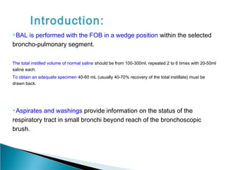
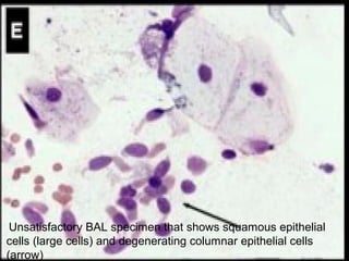
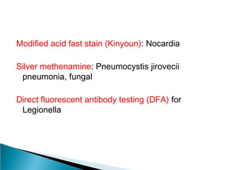
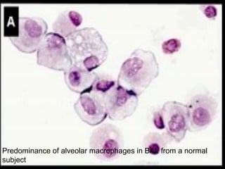
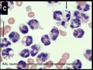
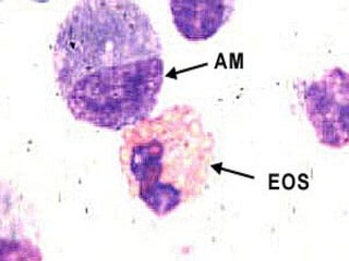
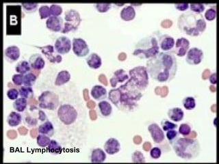
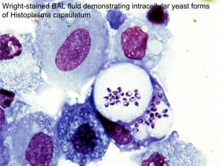
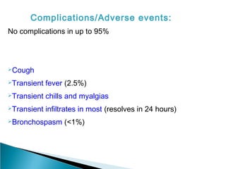
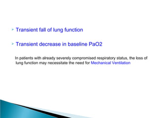
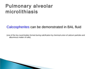
Broncho-AlveolarBroncho-Alveolar
Lavage Fluid AnalysisLavage Fluid Analysis
By Dr Uttam Kumar DasBy Dr Uttam Kumar Das
PGT Dept of Pathology
BSMC Bankura
05.03.2014
Introduction:
BAL is performed with the FOB in a wedge position within the selected
broncho-pulmonary segment.
The total instilled volume of normal saline should be from 100-300ml, repeated 2 to 6 times with 20-50ml
saline each.
To obtain an adequate specimen 40-60 mL (usually 40-70% recovery of the total instillate) must be
drawn back.
Aspirates and washings provide information on the status of the
respiratory tract in small bronchi beyond reach of the bronchoscopic
brush.
Area That Is Lavaged
 Procedures were usually performed in the right
middle lobe or lingula
 But lavage can be done in the most affected areas
of the lung
(In evaluating BAL in patients with Pneumocystis jirovecii
pneumonia, it was found that lavage in the upper lobes
had a higher yield than the traditional right middle lobe
or lingula)
Handling of Aspirated Fluid
 At the time of the lavage cells should be stored in
silicone-coated or similar containers
 Cell counts should probably be made on
unfiltered, unwashed, and unconcentrated
samples (If concentration is performed, the method should be
specified)
 Centrifugation to concentrate proteins and cells
can lead to loss of cells
 Washing the cells can change the differential
count considerably
Satisfactory Sample
1. A total of 2×10⁶
cells is considered a minimum requirement
2. Furthermore, more than 10 macrophages should be
present in a high-powered microscopic field
3. Degenerative changes should cover less than 20% of the
specimen area on the slide
4. If the number of squamous epithelial cells, bronchial cells,
RBCs, or inflammatory cells exceeds that of macrophages,
the specimen is considered unsatisfactory
The Storage of Fluid
 Cells stored at 4̊ C can be analyzed up to 24
hours after the procedure without significant
changes in the count and differentials
 Certain proteins may be temperature sensitive
and the samples may need to be stored at -80̊ C
Correcting for Bronchoalveolar
Lavage Dilution
 Instilled fluid is mixed with the endogenous fluid in the
alveoli
 Alveolar space is also in contact with a vascular space-
So water and solutes can transfer into the alveolar space
This process leads to the uncertainty of any measurement
of the concentration of any material in the alveolar space
Solution
One method has been to report per mL of
aspirated fluid.
Using this correction method has allowed clinicians
to quantitate the number of bacteria in the alveolar
space and to therefore diagnose bacterial
pneumonia
Unsatisfactory BAL specimen that shows squamous epithelial
cells (large cells) and degenerating columnar epithelial cells
(arrow)
Steps in Handling Cellular Population
of Bronchoalveolar Lavage Fluid
Cellular Staining
Papanicolaou Stain:
-Detect Cancer & Infection
-Not good at differentiating between
inflammatory cells
Toluidine blue staining:
-Mast cells are better seen
Wright-Giemsa stain:
-Good at differentiating between inflammatory cells
Diff-Quik (modification of the Wright-Giemsa stain):
-Is a rapid method allowing staining of the slide within a
few minutes
Limitations:
-The cells must be adequately adhered to the slide prior
to fixation
-Some cells are underestimated by these techniques
Oil red O stain:
-In fat embolism
Fat and Lipid stain (e.g. Sudan III):
-Lipoid pneumonia (aspiration)
Lipid-laden alveolar macrophage index > 100
(Sensitivity of 100%, Specificity 57%)
Periodic acid-Schiff (PAS):
-Pulmonary alveolar proteinosis
Other stains
KOH preparation: Fungal
Auramine-rhodamine or Ziehl-Neelson:
Mycobacterial
Modified acid fast stain (Kinyoun): Nocardia
Silver methenamine: Pneumocystis jirovecii
pneumonia, fungal
Direct fluorescent antibody testing (DFA) for
Legionella
Number of Cells Counted
De Brauwer et al determined that between 300 and
500 cells counted provided a good representation
of the number of nucleated cells for a BAL sample
Different cell
types in respiratory tract
 Upper respiratory tract
Ciliated pseudostratified columnar cells
Squamous cells
 Trachea and bronchi
Peudostratified Ciliated columnar cells
Goblet cells
 Terminal bronchioles
Low columnar or cuboidal-may be ciliated
Club cells (Clara cells)-nonciliated, secretory
cuboidal cells
 Alveoli
Type I pneumocytes-simple squamous alveolar cells
Type-II pneumocytes-great alveolar cells
Dust Cell-in the alveoli
Alveolar macrophages- in the connective
tissue of alveolar walls or interalveolar septa
General indications for BAL:
-Non-resolving pneumonia
- Diffuse lung infiltrates (interstitial and/or
alveolar)
- Infiltrates in an immunocompromised host
- Suspected alveolar hemorrhage
- Quantitative cultures for VAP
- Exclusion of diagnosable conditions by BAL
- Research
Gross examination-
Pulmonary alveolar proteinosis 
-Opaque or translucent brownish or sandy colored fluid
-Sediments out into two layers if left to sit
 Alveolar hemorrhage 
-Sequentially more hemorrhagic with each aliquot
Amorphous, predominantly acellular debris (pulmonary alveolar
proteinosis)
Papanicolaou stain- foamy proteinaceous alveolar cast
Alveolar macrophages
Normal >80%
Decreased in
Sarcoidosis (to 55% or less)
Cell count and differential count
Predominance of alveolar macrophages in BAL from a normal
subject
This photomicrograph shows an asbestos body under higher
magnification, surrounded by alveolar macrophages
Neutrophils (Normal <3%):
Nonspecific, but suggests active alveolitis
Increased in:
ARDS
Connective tissue disorders
Idiopathic pulmonary fibrosis
Infection
Pneumoconiosis
Wegener's granulomatosis
BAL neutrophil predomnance with intracellular bacteria
Eosinophils (Normal <1-2%)
Low to Moderate Eosinophilia (5-20%):
Drug induced lung disease
Minocycline
Nitrofurantoin
Penicillin
Infections
Parasitic
Mycobacterial
Fungal
 Bronchial Asthma
 Malignancies (infrequently)
 Other interstitial pneumonias occasionally (BOOP
or COP, IPF/UIP, ILD associated with Connective
tissue disorders, Sarcoidosis)
BAL eosinophilia
Moderate to Marked Eosinophilia (>20%):
 Allergic bronchopulmonary aspergillosis
 Acute eosinophilic pneumonia
 Churg-Strauss syndrome
 Chronic eosinophilic pneumonia
 Idiopathic hypereosinophilic syndrome
Lymphocytes (Normal <15%)
Normal CD4/CD8 (0.9-2.5:1):
Tuberculosis
Malignancies
Low CD4/CD8:
Hypersensitivity Pneumonitis
Silicosis
Drug-induced lung disease
HIV infection
BOOP (COP)
Elevated CD4/CD8:
Active sarcoidosis (>4:1 up to 10:1)
Asbestosis
Berylliosis
Crohn's disease
Connective tissue disorders
Sometimes in normal persons (inc. with age)
BAL Lymphocytosis
Erythrocytes
◦ Elevated erythrocyte count - early sign of alveolar
hemorrhage (first several hours)
◦ Phagocytosed erythrocytes - alveolar hemorrhage
within 48 hrs
◦ Hemosiderin laden macrophages - alveolar
hemorrhage > 48hrs
Foamy macrophages:
Non specific finding
May be seen in amiodarone use
Malignancies (sensitivity ranges from 35% to 70%)
◦ Lymphangitic carcinomatosis
◦ Lymphoma
◦ Bronchoalveolar carcinoma and other primary lung malignancies
◦ Extrapulmonary malignancies
Hemosiderin Laden Macrophages:
20% is highly specific and sensitive for alveolar hemorrhage
 Langerhans cells
>5% suggestive of Pulmonary Langerhans cell histiocytosis
 Cytomegalic cells
Viral pneumonias (cytomegalovirus, herpes)
Sulfur granules: Actinomycetes
Microbiology
Cultures
Polymerase chain reaction (PCR)  TB and
others
Quantitative or semi-quantitative cultures 
VAP
GMS BAL fluid showing round to cup shaped cysts of Pneumocystis
Pap-stained BAL fluid demonstrating large, retractile yeast forms
of Blastomyces dermatiditis (400X)
Pap-stained BAL fluid showing variably-sized, round yeast forms
of Cryptococcus neoformans (1000X)
Wright-stained BAL fluid demonstrating intracellular yeast forms
of Histoplasma capsulatum
Wright-stained BAL fluid demonstrating oblong, budding yeast
forms with pseudohyphae (1000X)
Complications/Adverse events:
No complications in up to 95%
Cough
Transient fever (2.5%)
Transient chills and myalgias
Transient infiltrates in most (resolves in 24 hours)
Bronchospasm (<1%)
 Transient fall of lung function
 Transient decrease in baseline PaO2
In patients with already severely compromised respiratory status, the loss of
lung function may necessitate the need for Mechanical Ventilation
Pulmonary alveolar
microlithiasis
Calcospherites can be demonstrated in BAL fluid
(one of the tiny round bodies formed during calcification by chemical union of calcium particles and
albuminous matter of cells)
Thank You

Bal fluid analysis

  • 1.
    Broncho-AlveolarBroncho-Alveolar Lavage Fluid AnalysisLavageFluid Analysis By Dr Uttam Kumar DasBy Dr Uttam Kumar Das PGT Dept of Pathology BSMC Bankura 05.03.2014
  • 2.
    Introduction: BAL is performedwith the FOB in a wedge position within the selected broncho-pulmonary segment. The total instilled volume of normal saline should be from 100-300ml, repeated 2 to 6 times with 20-50ml saline each. To obtain an adequate specimen 40-60 mL (usually 40-70% recovery of the total instillate) must be drawn back. Aspirates and washings provide information on the status of the respiratory tract in small bronchi beyond reach of the bronchoscopic brush.
  • 4.
    Area That IsLavaged  Procedures were usually performed in the right middle lobe or lingula  But lavage can be done in the most affected areas of the lung (In evaluating BAL in patients with Pneumocystis jirovecii pneumonia, it was found that lavage in the upper lobes had a higher yield than the traditional right middle lobe or lingula)
  • 5.
    Handling of AspiratedFluid  At the time of the lavage cells should be stored in silicone-coated or similar containers  Cell counts should probably be made on unfiltered, unwashed, and unconcentrated samples (If concentration is performed, the method should be specified)
  • 6.
     Centrifugation toconcentrate proteins and cells can lead to loss of cells  Washing the cells can change the differential count considerably
  • 7.
    Satisfactory Sample 1. Atotal of 2×10⁶ cells is considered a minimum requirement 2. Furthermore, more than 10 macrophages should be present in a high-powered microscopic field 3. Degenerative changes should cover less than 20% of the specimen area on the slide 4. If the number of squamous epithelial cells, bronchial cells, RBCs, or inflammatory cells exceeds that of macrophages, the specimen is considered unsatisfactory
  • 8.
    The Storage ofFluid  Cells stored at 4̊ C can be analyzed up to 24 hours after the procedure without significant changes in the count and differentials  Certain proteins may be temperature sensitive and the samples may need to be stored at -80̊ C
  • 9.
    Correcting for Bronchoalveolar LavageDilution  Instilled fluid is mixed with the endogenous fluid in the alveoli  Alveolar space is also in contact with a vascular space- So water and solutes can transfer into the alveolar space This process leads to the uncertainty of any measurement of the concentration of any material in the alveolar space
  • 10.
    Solution One method hasbeen to report per mL of aspirated fluid. Using this correction method has allowed clinicians to quantitate the number of bacteria in the alveolar space and to therefore diagnose bacterial pneumonia
  • 11.
    Unsatisfactory BAL specimenthat shows squamous epithelial cells (large cells) and degenerating columnar epithelial cells (arrow)
  • 12.
    Steps in HandlingCellular Population of Bronchoalveolar Lavage Fluid
  • 13.
    Cellular Staining Papanicolaou Stain: -DetectCancer & Infection -Not good at differentiating between inflammatory cells Toluidine blue staining: -Mast cells are better seen
  • 14.
    Wright-Giemsa stain: -Good atdifferentiating between inflammatory cells Diff-Quik (modification of the Wright-Giemsa stain): -Is a rapid method allowing staining of the slide within a few minutes Limitations: -The cells must be adequately adhered to the slide prior to fixation -Some cells are underestimated by these techniques
  • 15.
    Oil red Ostain: -In fat embolism Fat and Lipid stain (e.g. Sudan III): -Lipoid pneumonia (aspiration) Lipid-laden alveolar macrophage index > 100 (Sensitivity of 100%, Specificity 57%) Periodic acid-Schiff (PAS): -Pulmonary alveolar proteinosis
  • 16.
    Other stains KOH preparation:Fungal Auramine-rhodamine or Ziehl-Neelson: Mycobacterial
  • 17.
    Modified acid faststain (Kinyoun): Nocardia Silver methenamine: Pneumocystis jirovecii pneumonia, fungal Direct fluorescent antibody testing (DFA) for Legionella
  • 18.
    Number of CellsCounted De Brauwer et al determined that between 300 and 500 cells counted provided a good representation of the number of nucleated cells for a BAL sample
  • 19.
    Different cell types inrespiratory tract  Upper respiratory tract Ciliated pseudostratified columnar cells Squamous cells  Trachea and bronchi Peudostratified Ciliated columnar cells Goblet cells
  • 20.
     Terminal bronchioles Lowcolumnar or cuboidal-may be ciliated Club cells (Clara cells)-nonciliated, secretory cuboidal cells  Alveoli Type I pneumocytes-simple squamous alveolar cells Type-II pneumocytes-great alveolar cells Dust Cell-in the alveoli Alveolar macrophages- in the connective tissue of alveolar walls or interalveolar septa
  • 22.
    General indications forBAL: -Non-resolving pneumonia - Diffuse lung infiltrates (interstitial and/or alveolar) - Infiltrates in an immunocompromised host - Suspected alveolar hemorrhage - Quantitative cultures for VAP - Exclusion of diagnosable conditions by BAL - Research
  • 23.
    Gross examination- Pulmonary alveolarproteinosis  -Opaque or translucent brownish or sandy colored fluid -Sediments out into two layers if left to sit  Alveolar hemorrhage  -Sequentially more hemorrhagic with each aliquot
  • 24.
    Amorphous, predominantly acellulardebris (pulmonary alveolar proteinosis)
  • 25.
    Papanicolaou stain- foamyproteinaceous alveolar cast
  • 26.
    Alveolar macrophages Normal >80% Decreasedin Sarcoidosis (to 55% or less) Cell count and differential count
  • 27.
    Predominance of alveolarmacrophages in BAL from a normal subject
  • 29.
    This photomicrograph showsan asbestos body under higher magnification, surrounded by alveolar macrophages
  • 30.
    Neutrophils (Normal <3%): Nonspecific,but suggests active alveolitis Increased in: ARDS Connective tissue disorders Idiopathic pulmonary fibrosis Infection Pneumoconiosis Wegener's granulomatosis
  • 31.
    BAL neutrophil predomnancewith intracellular bacteria
  • 32.
    Eosinophils (Normal <1-2%) Lowto Moderate Eosinophilia (5-20%): Drug induced lung disease Minocycline Nitrofurantoin Penicillin Infections Parasitic Mycobacterial Fungal
  • 33.
     Bronchial Asthma Malignancies (infrequently)  Other interstitial pneumonias occasionally (BOOP or COP, IPF/UIP, ILD associated with Connective tissue disorders, Sarcoidosis)
  • 34.
  • 36.
    Moderate to MarkedEosinophilia (>20%):  Allergic bronchopulmonary aspergillosis  Acute eosinophilic pneumonia  Churg-Strauss syndrome  Chronic eosinophilic pneumonia  Idiopathic hypereosinophilic syndrome
  • 37.
    Lymphocytes (Normal <15%) NormalCD4/CD8 (0.9-2.5:1): Tuberculosis Malignancies Low CD4/CD8: Hypersensitivity Pneumonitis Silicosis Drug-induced lung disease HIV infection BOOP (COP)
  • 38.
    Elevated CD4/CD8: Active sarcoidosis(>4:1 up to 10:1) Asbestosis Berylliosis Crohn's disease Connective tissue disorders Sometimes in normal persons (inc. with age)
  • 39.
  • 40.
    Erythrocytes ◦ Elevated erythrocytecount - early sign of alveolar hemorrhage (first several hours) ◦ Phagocytosed erythrocytes - alveolar hemorrhage within 48 hrs ◦ Hemosiderin laden macrophages - alveolar hemorrhage > 48hrs
  • 41.
    Foamy macrophages: Non specificfinding May be seen in amiodarone use Malignancies (sensitivity ranges from 35% to 70%) ◦ Lymphangitic carcinomatosis ◦ Lymphoma ◦ Bronchoalveolar carcinoma and other primary lung malignancies ◦ Extrapulmonary malignancies
  • 42.
    Hemosiderin Laden Macrophages: 20%is highly specific and sensitive for alveolar hemorrhage  Langerhans cells >5% suggestive of Pulmonary Langerhans cell histiocytosis  Cytomegalic cells Viral pneumonias (cytomegalovirus, herpes) Sulfur granules: Actinomycetes
  • 43.
    Microbiology Cultures Polymerase chain reaction(PCR)  TB and others Quantitative or semi-quantitative cultures  VAP
  • 44.
    GMS BAL fluidshowing round to cup shaped cysts of Pneumocystis
  • 45.
    Pap-stained BAL fluiddemonstrating large, retractile yeast forms of Blastomyces dermatiditis (400X)
  • 46.
    Pap-stained BAL fluidshowing variably-sized, round yeast forms of Cryptococcus neoformans (1000X)
  • 47.
    Wright-stained BAL fluiddemonstrating intracellular yeast forms of Histoplasma capsulatum
  • 48.
    Wright-stained BAL fluiddemonstrating oblong, budding yeast forms with pseudohyphae (1000X)
  • 49.
    Complications/Adverse events: No complicationsin up to 95% Cough Transient fever (2.5%) Transient chills and myalgias Transient infiltrates in most (resolves in 24 hours) Bronchospasm (<1%)
  • 50.
     Transient fallof lung function  Transient decrease in baseline PaO2 In patients with already severely compromised respiratory status, the loss of lung function may necessitate the need for Mechanical Ventilation
  • 51.
    Pulmonary alveolar microlithiasis Calcospherites canbe demonstrated in BAL fluid (one of the tiny round bodies formed during calcification by chemical union of calcium particles and albuminous matter of cells)
  • 52.