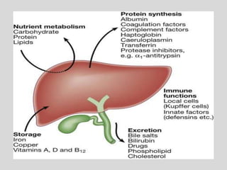
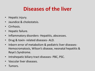
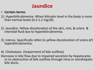
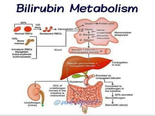
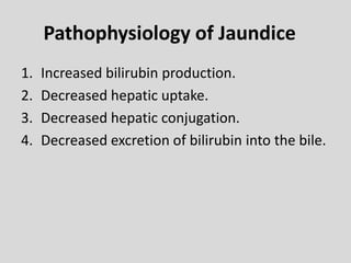
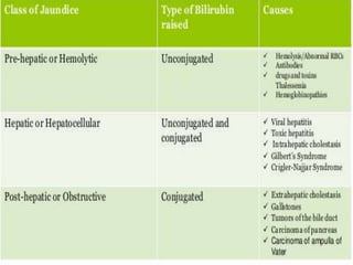
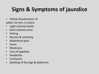
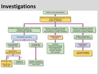
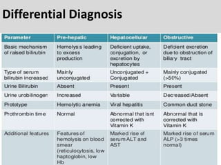
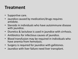
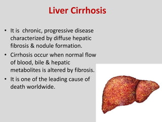
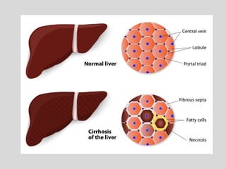
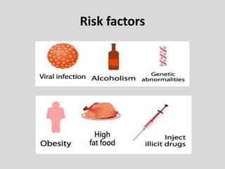
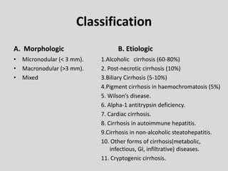
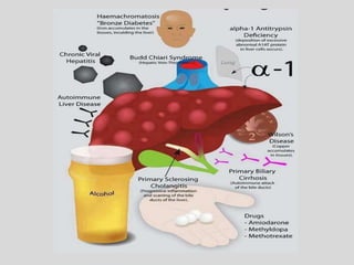
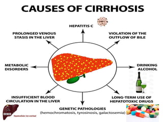
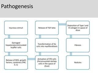
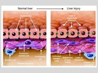
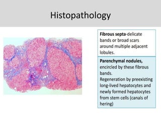
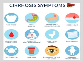
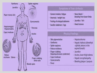
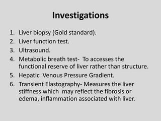
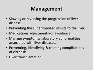
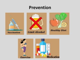
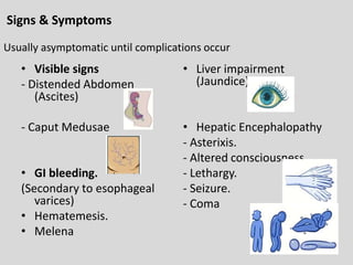
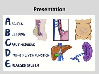
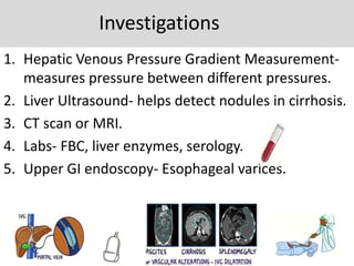
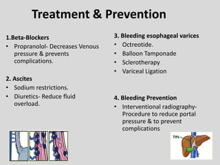
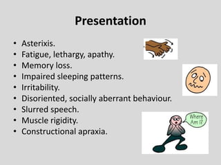
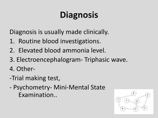
Liver Pathology & Disease
Condition
Dr. Deepika R. Kenjale
Liver
• Largest organ situated in the RUQ.
• Color- Reddish brown & has soft consistency.
• Weight- 1400-1600 gm in males & 1200-1400 gm in
females.
• External feature-
a) 2 lobes- Right & left. (right forms 5/6th part of liver.
b) 5 surfaces- Anterior, posterior, superior, inferior, & right.
c) 1 prominent border- Inferior.
• Blood supply- 20 % from hepatic artery & 80 % through
portal vein.
Function
Metabolism – Carbohydrate, Fat & Protein
Secretory – Bile, bile acids, salts & pigments.
Excretory – Bilirubin, drugs, toxins.
Synthesis – Albumin, coagulation factors.
Storage – Vitamins, carbohydrates etc.
Detoxification – Toxins, ammonia etc.
Diseases of the liver
• Hepatic injury.
• Jaundice & cholestatsis.
• Cirrhosis.
• Hepatic failure.
• Inflammatory disorders- Hepatitis, abscesses.
• Drug & toxin- related diseases- ALD.
• Inborn error of metabolism & pediatric liver diseases-
Hemocromatosis, Wilson’s disease, neonatal hepatitis &
Reye’s Syndrome.
• Intrahepatic biliary tract diseases- PBC, PSC.
• Vascular liver diseases.
• Tumors.
Jaundice
• Certain terms:
1). Hyperbilirubinemia- When bilirubin level in the body is more
than normal levels (0.2-1.2 mg/dl).
2). Jaundice- Yellow discoloration of the skin, mm, & sclera &
interstial fluid due to hyperbilirubinemia.
3). Icterus- Specifically refers to yellow discoloration of sclera d/t
hyperbilirubinemia.
4). Cholestasis- (Impairment of bile outflow)
Decrease in bile flow due to impaired secretion by hepatocytes
or to obstruction of bile outflow through intra-or extrahepatic
bile ducts.
Pathophysiology of Jaundice
1. Increased bilirubin production.
2. Decreased hepatic uptake.
3. Decreased hepatic conjugation.
4. Decreased excretion of bilirubin into the bile.
Signs & Symptoms of jaundice
• Yellow discoloration of-
a)Skin, b) mm, c) sclera.
• Light-colored stools.
• Dark-colored urine.
• Itching.
• Nausea & vomiting.
• Abdominal pain.
• Fever.
• Weakness.
• Loss of appetite.
• Headache.
• Confusion.
• Swelling of the legs & abdomen.
Investigations
Differential Diagnosis
Treatment
• Supportive care.
• Jaundice caused by medication/drugs requires
antidote.
• Steroids in individuals who have autoimmune disease
with jaundice.
• Diuretics & lactulose is used in jaundice with cirrhosis.
• Antibiotics for infectious causes of jaundice.
• Blood transfusion may be required in individuals who
have anemia from hemolysis.
• Surgery is required for jaundice with gallstones.
• Jaundice with liver failure need liver transplant.
Liver Cirrhosis
• It is chronic, progressive disease
characterized by diffuse hepatic
fibrosis & nodule formation.
• Cirrhosis occur when normal flow
of blood, bile & hepatic
metabolites is altered by fibrosis.
• It is one of the leading cause of
death worldwide.
Risk factors
Classification
A. Morphologic
• Micronodular (< 3 mm).
• Macronodular (>3 mm).
• Mixed
B. Etiologic
1.Alcoholic cirrhosis (60-80%)
2. Post-necrotic cirrhosis (10%)
3.Biliary Cirrhosis (5-10%)
4.Pigment cirrhosis in haemochromatosis (5%)
5. Wilson’s disease.
6. Alpha-1 antitrypsin deficiency.
7. Cardiac cirrhosis.
8. Cirrhosis in autoimmune hepatitis.
9.Cirrhosis in non-alcoholic steatohepatitis.
10. Other forms of cirrhosis(metabolic,
infectious, GI, infiltrative) diseases.
11. Cryptogenic cirrhosis.
Pathogenesis
Histopathology
Investigations
1. Liver biopsy (Gold standard).
2. Liver function test.
3. Ultrasound.
4. Metabolic breath test- To accesses the
functional reserve of liver rather than structure.
5. Hepatic Venous Pressure Gradient.
6. Transient Elastography- Measures the liver
stiffness which may reflect the fibrosis or
edema, inflammation associated with liver.
Management
• Slowing or reversing the progression of liver
disease.
• Preventing the superimposed insults to the liver.
• Medications adjustments/or avoidance.
• Manage symptoms/ laboratory abnormalities
associated with liver diseases.
• Preventing, identifying & treating complications
of cirrhosis.
• Liver transplantation.
Prevention
Complications
• Spontaneous bacterial peritonitis.
• Portal Hypertension.
• Hepatic Encephalopathy.
• Hepatorenal Syndrome.
• Portal vein Thrombosis.
• Pulmonary complications of portal hypertension
- Hepatopulmonary Syndrome
- Portopulmonary Hypertension.
• Tumors (late complications).
- Hepatocellular carcinoma. (HCC)
Portal Hypertension
• Increase in pressure in portal system usually
follows obstruction to the portal blood flow
anywhere along its course.
• Present- portal venous pressure is ≥ 6mmHg
(1-5 mmHg).
• >10 mmHg- Clinically significant
• >12 mmHg- Associated with complications.
Classification
Signs & Symptoms
• Visible signs
- Distended Abdomen
(Ascites)
- Caput Medusae
• GI bleeding.
(Secondary to esophageal
varices)
• Hematemesis.
• Melena
• Liver impairment
(Jaundice)
• Hepatic Encephalopathy
- Asterixis.
- Altered consciousness.
- Lethargy.
- Seizure.
- Coma
Usually asymptomatic until complications occur
Presentation
Investigations
1. Hepatic Venous Pressure Gradient Measurement-
measures pressure between different pressures.
2. Liver Ultrasound- helps detect nodules in cirrhosis.
3. CT scan or MRI.
4. Labs- FBC, liver enzymes, serology.
5. Upper GI endoscopy- Esophageal varices.
Treatment & Prevention
1.Beta-Blockers
• Propranolol- Decreases Venous
pressure & prevents
complications.
2. Ascites
• Sodium restrictions.
• Diuretics- Reduce fluid
overload.
3. Bleeding esophageal varices
• Octreotide.
• Balloon Tamponade
• Sclerotherapy
• Variceal Ligation
4. Bleeding Prevention
• Interventional radiography-
Procedure to reduce portal
pressure & to prevent
complications
Hepatic Encephalopathy
• HE is defined as fluctuation in mental status &
cognition function in presence of severe liver
disease.
• Hepatic dysfunction  inadequate
elimination of metabolic products with
subsequent accumulation of neurotoxic
metabolites (like ammonia).
Presentation
• Asterixis.
• Fatigue, lethargy, apathy.
• Memory loss.
• Impaired sleeping patterns.
• Irritability.
• Disoriented, socially aberrant behaviour.
• Slurred speech.
• Muscle rigidity.
• Constructional apraxia.
Diagnosis
Diagnosis is usually made clinically.
1. Routine blood investigations.
2. Elevated blood ammonia level.
3. Electroencephalogram- Triphasic wave.
4. Other-
-Trial making test,
- Psychometry- Mini-Mental State
Examination..
Management Goals
• General measures:
Avoidance of trigger substances.
Treatment of further complications which
might aggravate hepatic encephalopathy.
• Medications-
Lactulose- 1st line treatment for HE.
Liver pathology & Disease condition

Liver pathology & Disease condition

  • 1.
    Liver Pathology &Disease Condition Dr. Deepika R. Kenjale
  • 2.
    Liver • Largest organsituated in the RUQ. • Color- Reddish brown & has soft consistency. • Weight- 1400-1600 gm in males & 1200-1400 gm in females. • External feature- a) 2 lobes- Right & left. (right forms 5/6th part of liver. b) 5 surfaces- Anterior, posterior, superior, inferior, & right. c) 1 prominent border- Inferior. • Blood supply- 20 % from hepatic artery & 80 % through portal vein.
  • 4.
    Function Metabolism – Carbohydrate,Fat & Protein Secretory – Bile, bile acids, salts & pigments. Excretory – Bilirubin, drugs, toxins. Synthesis – Albumin, coagulation factors. Storage – Vitamins, carbohydrates etc. Detoxification – Toxins, ammonia etc.
  • 6.
    Diseases of theliver • Hepatic injury. • Jaundice & cholestatsis. • Cirrhosis. • Hepatic failure. • Inflammatory disorders- Hepatitis, abscesses. • Drug & toxin- related diseases- ALD. • Inborn error of metabolism & pediatric liver diseases- Hemocromatosis, Wilson’s disease, neonatal hepatitis & Reye’s Syndrome. • Intrahepatic biliary tract diseases- PBC, PSC. • Vascular liver diseases. • Tumors.
  • 7.
    Jaundice • Certain terms: 1).Hyperbilirubinemia- When bilirubin level in the body is more than normal levels (0.2-1.2 mg/dl). 2). Jaundice- Yellow discoloration of the skin, mm, & sclera & interstial fluid due to hyperbilirubinemia. 3). Icterus- Specifically refers to yellow discoloration of sclera d/t hyperbilirubinemia. 4). Cholestasis- (Impairment of bile outflow) Decrease in bile flow due to impaired secretion by hepatocytes or to obstruction of bile outflow through intra-or extrahepatic bile ducts.
  • 9.
    Pathophysiology of Jaundice 1.Increased bilirubin production. 2. Decreased hepatic uptake. 3. Decreased hepatic conjugation. 4. Decreased excretion of bilirubin into the bile.
  • 11.
    Signs & Symptomsof jaundice • Yellow discoloration of- a)Skin, b) mm, c) sclera. • Light-colored stools. • Dark-colored urine. • Itching. • Nausea & vomiting. • Abdominal pain. • Fever. • Weakness. • Loss of appetite. • Headache. • Confusion. • Swelling of the legs & abdomen.
  • 13.
  • 14.
  • 15.
    Treatment • Supportive care. •Jaundice caused by medication/drugs requires antidote. • Steroids in individuals who have autoimmune disease with jaundice. • Diuretics & lactulose is used in jaundice with cirrhosis. • Antibiotics for infectious causes of jaundice. • Blood transfusion may be required in individuals who have anemia from hemolysis. • Surgery is required for jaundice with gallstones. • Jaundice with liver failure need liver transplant.
  • 16.
    Liver Cirrhosis • Itis chronic, progressive disease characterized by diffuse hepatic fibrosis & nodule formation. • Cirrhosis occur when normal flow of blood, bile & hepatic metabolites is altered by fibrosis. • It is one of the leading cause of death worldwide.
  • 18.
  • 19.
    Classification A. Morphologic • Micronodular(< 3 mm). • Macronodular (>3 mm). • Mixed B. Etiologic 1.Alcoholic cirrhosis (60-80%) 2. Post-necrotic cirrhosis (10%) 3.Biliary Cirrhosis (5-10%) 4.Pigment cirrhosis in haemochromatosis (5%) 5. Wilson’s disease. 6. Alpha-1 antitrypsin deficiency. 7. Cardiac cirrhosis. 8. Cirrhosis in autoimmune hepatitis. 9.Cirrhosis in non-alcoholic steatohepatitis. 10. Other forms of cirrhosis(metabolic, infectious, GI, infiltrative) diseases. 11. Cryptogenic cirrhosis.
  • 22.
  • 24.
  • 27.
    Investigations 1. Liver biopsy(Gold standard). 2. Liver function test. 3. Ultrasound. 4. Metabolic breath test- To accesses the functional reserve of liver rather than structure. 5. Hepatic Venous Pressure Gradient. 6. Transient Elastography- Measures the liver stiffness which may reflect the fibrosis or edema, inflammation associated with liver.
  • 28.
    Management • Slowing orreversing the progression of liver disease. • Preventing the superimposed insults to the liver. • Medications adjustments/or avoidance. • Manage symptoms/ laboratory abnormalities associated with liver diseases. • Preventing, identifying & treating complications of cirrhosis. • Liver transplantation.
  • 29.
  • 30.
    Complications • Spontaneous bacterialperitonitis. • Portal Hypertension. • Hepatic Encephalopathy. • Hepatorenal Syndrome. • Portal vein Thrombosis. • Pulmonary complications of portal hypertension - Hepatopulmonary Syndrome - Portopulmonary Hypertension. • Tumors (late complications). - Hepatocellular carcinoma. (HCC)
  • 31.
    Portal Hypertension • Increasein pressure in portal system usually follows obstruction to the portal blood flow anywhere along its course. • Present- portal venous pressure is ≥ 6mmHg (1-5 mmHg). • >10 mmHg- Clinically significant • >12 mmHg- Associated with complications.
  • 32.
  • 34.
    Signs & Symptoms •Visible signs - Distended Abdomen (Ascites) - Caput Medusae • GI bleeding. (Secondary to esophageal varices) • Hematemesis. • Melena • Liver impairment (Jaundice) • Hepatic Encephalopathy - Asterixis. - Altered consciousness. - Lethargy. - Seizure. - Coma Usually asymptomatic until complications occur
  • 35.
  • 36.
    Investigations 1. Hepatic VenousPressure Gradient Measurement- measures pressure between different pressures. 2. Liver Ultrasound- helps detect nodules in cirrhosis. 3. CT scan or MRI. 4. Labs- FBC, liver enzymes, serology. 5. Upper GI endoscopy- Esophageal varices.
  • 37.
    Treatment & Prevention 1.Beta-Blockers •Propranolol- Decreases Venous pressure & prevents complications. 2. Ascites • Sodium restrictions. • Diuretics- Reduce fluid overload. 3. Bleeding esophageal varices • Octreotide. • Balloon Tamponade • Sclerotherapy • Variceal Ligation 4. Bleeding Prevention • Interventional radiography- Procedure to reduce portal pressure & to prevent complications
  • 38.
    Hepatic Encephalopathy • HEis defined as fluctuation in mental status & cognition function in presence of severe liver disease. • Hepatic dysfunction  inadequate elimination of metabolic products with subsequent accumulation of neurotoxic metabolites (like ammonia).
  • 42.
    Presentation • Asterixis. • Fatigue,lethargy, apathy. • Memory loss. • Impaired sleeping patterns. • Irritability. • Disoriented, socially aberrant behaviour. • Slurred speech. • Muscle rigidity. • Constructional apraxia.
  • 43.
    Diagnosis Diagnosis is usuallymade clinically. 1. Routine blood investigations. 2. Elevated blood ammonia level. 3. Electroencephalogram- Triphasic wave. 4. Other- -Trial making test, - Psychometry- Mini-Mental State Examination..
  • 44.
    Management Goals • Generalmeasures: Avoidance of trigger substances. Treatment of further complications which might aggravate hepatic encephalopathy. • Medications- Lactulose- 1st line treatment for HE.