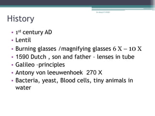
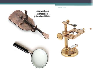
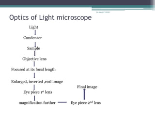
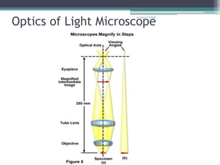
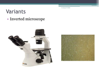
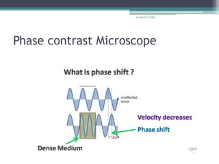
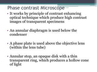
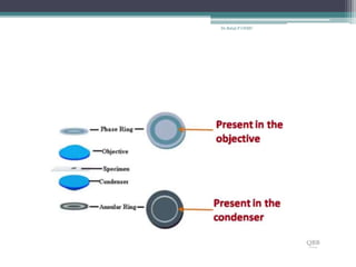
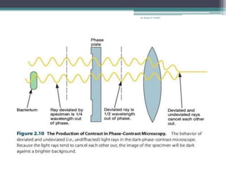
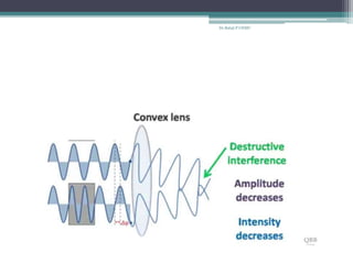
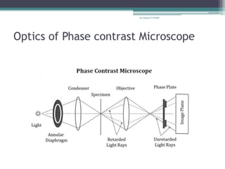
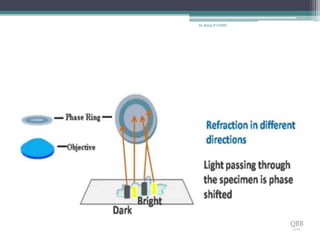
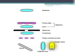
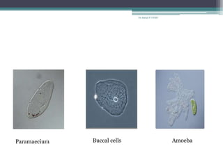
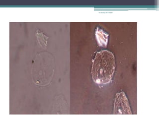
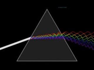
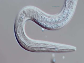
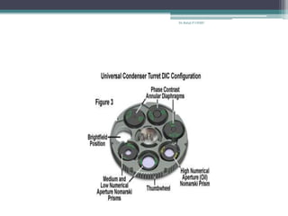
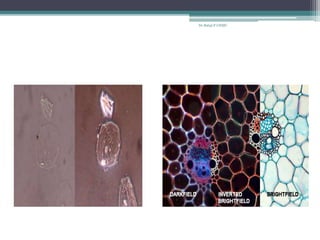
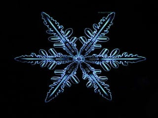
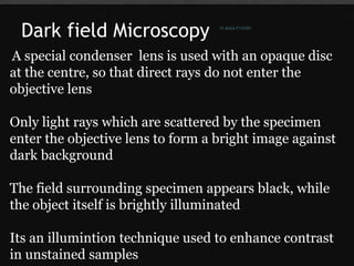
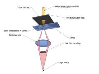
LIGHT MICROSCOPY
Dr.Balaji P,
First year Post Graduate,
M.D., Microbiology,
Dept. of Microbiology,
Chengalpattu Medical college.
Dr.Balaji P CHMC
Dr.Balaji P CHMC
Dr.Balaji P CHMC
History
• 1st century AD
• Lentil
• Burning glasses /magnifying glasses 6 X – 10 X
• 1590 Dutch , son and father – lenses in tube
• Galileo –principles
• Antony von leeuwenhoek 270 X
• Bacteria, yeast, Blood cells, tiny animals in
water
Dr.Balaji P CHMC
OPTICAL/LIGHT MICROSCOPE
• Microscopes which uses visible light
and lens(es) to magnify small objects
• Two types :
a) Simple
b) Compound
Dr.Balaji P CHMC
Simple Microscope
• This is the type of microscope which was
invented first
• Original design of Light microscope
• Uses single lens
Dr.Balaji P CHMC
Dr.Balaji P CHMC
Compound Microscope:
• Uses multiple lenses to get image from specimen
and uses another set of lenses to magnify it
before seeing it as a final image
• Advantages:
magnification is high
resolution is high
changeable objective lens
Dr.Balaji P CHMC
Dr.Balaji P CHMC
Lens
collection of
prisms as a unit
Dr.Balaji P CHMC
Magnification
degree of
enlargement of the
specimen’s image
by means of no.of
times of
length,breadth
Dr.Balaji P CHMC
Refraction
Bending of light when it
enters from one media
to another
Refractive Index
Ratio of velocity of light
in vacuum and in any
other medium
Dr.Balaji P CHMC
Resolution
Ability of a lens to
separate or
distinguish between
small objects that
are close together
Dr.Balaji P CHMC
Focal point & Focal length
Dr.Balaji P CHMC
Focal point
convergence of
light rays at a
point by lens
Focal length
Distance between
focal point and
centre of lens
Dr.Balaji P CHMC
Working distance
Distance between
the the front surface
of lens and the
surface of the cover
glass
Dr.Balaji P CHMC
Abbe’s formula:
• Described by a German physicist Ernst Abbe in 1870
• The resolution of a microscope depends upon the
numerical aperture of its condenser, objective lens,
and wave length of the light
• Goes by formula
d = 0.5λ
nsinθ
Dr.Balaji P CHMC
d- distance
λ - wave length of light
nsinθ - Numerical aperture
θ- angular aperture ( ½ the angle of cone of light enters
objective lens from specimen)
n – refractive index
Dr.Balaji P CHMC
NUMERICAL APERTURE
• Applies for condenser and objective lens
• Light gathering (converging) ability of a lens
• Depends on angular aperture(θ)
• Higher the numerical aperture lesser the
working distance and vice versa
• Cone of light depends on refractive index (n)
Dr.Balaji P CHMC
Dr.Balaji P CHMC
NA of various objectives:
4 X 0.1
10 X 0.25
40 X 0.65
100 X 1.25
Working Distance:
4 X 17-20 mm
10 X 4-8 mm
40 X 0.5-0.7 mm
100 X 0.1 mm
Dr.Balaji P CHMC
• Most microscopes posses NA 1.2 to 1.4
(objective lens)
• Condenser NA 0.9
• Refractive index (n) of air is 1
• Hence , lens working in air couldn’t give much
resolution, for which we are using immersion oil
which has more refractive index than air, which
in turn increases NA (max 1.25)
• Spectrum of light (blue green) used in
microscope is 380 nm -530 nm
Dr.Balaji P CHMC
• The maximum theoretical resolving power of a
microscope with an oil immersion objective with
blue-green light is approximately 0.2 mcm
• d= (0.5)(530 nm) = 0.212 / 0.2 mcm
1.25
• At best, a bright-field microscope can
distinguish between two dots around 0.2 mcm
apart
Dr.Balaji P CHMC
TYPES
• Bright field
• Dark field
• Phase contrast
• Differential interference contrast microscope
• Fluorescence microscopes
Dr.Balaji P CHMC
BRIGHT-FIELD MICROSCOPE
Dr.Balaji P CHMC
Dr.Balaji P CHMC
Optics of Light microscope
Light
Condenser
Sample
Objective lens
Focused at its focal length
Enlarged, inverted ,real image
Final image
Eye piece 1st lens
magnification further Eye piece 2nd lens
Dr.Balaji P CHMC
Optics of Light Microscope
Dr.Balaji P CHMC
Uses and advantages:
• Simple set up
• Used to view live / stained cells and organisms
• Little preparation is required
• Adaptable with new technology
Disadvantages:
• Biological specimens are of low contrast and
needs to be stained
• Needs stronger light source for high
magnification
• Artefacts of staining
Dr.Balaji P CHMC
Variants
• Inverted microscope
Dr.Balaji P CHMC
• Uses:
Live cell culture
Large specimens
Micromanipulation of sample
Advantages:
Large/ high weight specimens
More samples in shorter time
safety of objective
Works in same direction
Disdvantage:
Higher cost
Limited working distance
Thickness of container
Dr.Balaji P CHMC
• Stereo microscope
Dr.Balaji P CHMC
• Comparison microscope
Dr.Balaji P CHMC
• Petrographic microscope
Dr.Balaji P CHMC
Phase contrast Microscope
Dr.Balaji P CHMC
Phase contrast Microscope
• It works by principle of contrast enhancing
optical technique which produce high contrast
images of transparent specimens
• An annular diaphragm is used below the
condenser
• A phase plate is used above the objective lens
(within the lens tube)
• Annular stop, an opaque disk with a thin
transparent ring, which produces a hollow cone
of light
Dr.Balaji P CHMC
• As this cone passes through specimen ,some light
rays are bent and are retarded ¼ wave length
• This deviated light is focused to form an image of
the object
• Undeviated light rays strike phase ring in the phase
plate ,which is located in the objective
• after passing through phase plate ,undeviated light
is advanced by ¼ wavelength
• Finally , deviated and undeviated waves will be ½
wavelength out of phase and will cancel/merge each
other and forms final image
Dr.Balaji P CHMC
Dr.Balaji P CHMC
Dr.Balaji P CHMC
Dr.Balaji P CHMC
Dr.Balaji P CHMC
Dr.Balaji P CHMC
Optics of Phase contrast Microscope
Dr.Balaji P CHMC
Dr.Balaji P CHMC
Dr.Balaji P CHMC
Paramaecium Buccal cells Amoeba
Dr.Balaji P CHMC
Dr.Balaji P CHMC
Uses
• study of unstained living cells, microorganisms
such as bacteria,molds and their
shape,motility,subcellular particles bacterial
components such as endospores and inclusion
bodies etc.
Dr.Balaji P CHMC
Advantages:
• Observing of living cells in its natural state
• Specimen need not to be killed, fixed or stained
• High-contrast images
• Ideal for thin specimens
Disadvantages:
• Annuli or rings limit the aperture to some
extent, which decreases resolution
• Not ideal for thick specimens
• Images may appear grey or green, if white or
green lights are used, respectively, resulting in
poor photomicrography
• phase artifacts
Dr.Balaji P CHMC
Dr.Balaji P CHMC
Differential interference contrast
microscope
• Similar to the phase-contrast microscope
• But , a polarizer is used after light source
followed by a prism
• Two beams of plane polarized light at right
angles to each other generated by prism
• Object beam passes through the specimen, while
the reference beam passes through a clear area
of the slide
Dr.Balaji P CHMC
• After passing through specimen both waves will
combine and interfere with each others to form
image
• It creates an image by detecting differences in
refractive indices and thickness of specimen
• Finally a live, unstained specimen appears
brightly colored and three-dimensional
Dr.Balaji P CHMC
Optics of Differential-interference Phase
contrast Microscope
Dr.Balaji P CHMC
Dr.Balaji P CHMC
Uses
• Living cells and its structures such as cell walls,
endospores, granules, vacuoles,and eucaryotic
nuclei are clearly visible
• used to visualize living cells and for quantitative
studies
Dr.Balaji P CHMC
Advantages:
• It gives a better image than phase contrast
microscope
• 3D colorful images of specimen
• No Halo effect
• No shade off effect
• Disadvantages:
• three-dimensional image of a specimen
may not be accurate.
• The enhanced areas of light and shadow
might add distortion to the appearance of
the image.
Dr.Balaji P CHMC
Fungal spores
Paramecium
Dr.Balaji P CHMC
Egg of Trichuris trichiura Yeast cells
Dr.Balaji P CHMC
Dr.Balaji P CHMC
Dr.Balaji P CHMC
Dr.Balaji P CHMC
Dr.Balaji P CHMC
Dark field Microscopy
A special condenser lens is used with an opaque disc
at the centre, so that direct rays do not enter the
objective lens
Only light rays which are scattered by the specimen
enter the objective lens to form a bright image against
dark background
The field surrounding specimen appears black, while
the object itself is brightly illuminated
Its an illumintion technique used to enhance contrast
in unstained samples
Dr.Balaji P CHMC
Dr.Balaji P CHMC
Dr.Balaji P CHMC
Dr.Balaji P CHMC
• Variants
Treponema pallidum
Borrelia
Leptospira
Treponema Borrelia
Leptospira
Dr.Balaji P CHMC
Uses
Ideal for viewing objects that are unstained,
transparent and absorb little or no light
Live blood cells
Live bacteria
Other live organisms
Environmental water samples
Uses and advantages :
Dr.Balaji P CHMC
• Disadvantages: Prone to degradation,
distortion and inaccuracies
• A specimen that is not thin enough may appear
to have artifacts throughout the image
• Special care to be taken to prepare sample and
work place setup
• need to use oil on the condenser and/or slide , if
it contains liquid bubbles which will cause
images degradation, flare, distortion and even
decrease in contrast and details of the specimen.
• Dark field needs an intense amount of light to
work
Dr.Balaji P CHMC
References
• Prescot Microbiology 5th edition
• Bailey and Scott 14th edition
• Monica-Cheesbrough 2nd edition
Dr.Balaji P CHMC
Dr.Balaji P CHMC

Light and Dark field microscopy

  • 1.
    LIGHT MICROSCOPY Dr.Balaji P, Firstyear Post Graduate, M.D., Microbiology, Dept. of Microbiology, Chengalpattu Medical college. Dr.Balaji P CHMC
  • 2.
  • 3.
  • 4.
    History • 1st centuryAD • Lentil • Burning glasses /magnifying glasses 6 X – 10 X • 1590 Dutch , son and father – lenses in tube • Galileo –principles • Antony von leeuwenhoek 270 X • Bacteria, yeast, Blood cells, tiny animals in water Dr.Balaji P CHMC
  • 5.
    OPTICAL/LIGHT MICROSCOPE • Microscopeswhich uses visible light and lens(es) to magnify small objects • Two types : a) Simple b) Compound Dr.Balaji P CHMC
  • 6.
    Simple Microscope • Thisis the type of microscope which was invented first • Original design of Light microscope • Uses single lens Dr.Balaji P CHMC
  • 7.
  • 8.
    Compound Microscope: • Usesmultiple lenses to get image from specimen and uses another set of lenses to magnify it before seeing it as a final image • Advantages: magnification is high resolution is high changeable objective lens Dr.Balaji P CHMC
  • 9.
  • 10.
    Lens collection of prisms asa unit Dr.Balaji P CHMC
  • 11.
    Magnification degree of enlargement ofthe specimen’s image by means of no.of times of length,breadth Dr.Balaji P CHMC
  • 12.
    Refraction Bending of lightwhen it enters from one media to another Refractive Index Ratio of velocity of light in vacuum and in any other medium Dr.Balaji P CHMC
  • 13.
    Resolution Ability of alens to separate or distinguish between small objects that are close together Dr.Balaji P CHMC
  • 14.
    Focal point &Focal length Dr.Balaji P CHMC
  • 15.
    Focal point convergence of lightrays at a point by lens Focal length Distance between focal point and centre of lens Dr.Balaji P CHMC
  • 16.
    Working distance Distance between thethe front surface of lens and the surface of the cover glass Dr.Balaji P CHMC
  • 17.
    Abbe’s formula: • Describedby a German physicist Ernst Abbe in 1870 • The resolution of a microscope depends upon the numerical aperture of its condenser, objective lens, and wave length of the light • Goes by formula d = 0.5λ nsinθ Dr.Balaji P CHMC
  • 18.
    d- distance λ -wave length of light nsinθ - Numerical aperture θ- angular aperture ( ½ the angle of cone of light enters objective lens from specimen) n – refractive index Dr.Balaji P CHMC
  • 19.
    NUMERICAL APERTURE • Appliesfor condenser and objective lens • Light gathering (converging) ability of a lens • Depends on angular aperture(θ) • Higher the numerical aperture lesser the working distance and vice versa • Cone of light depends on refractive index (n) Dr.Balaji P CHMC
  • 20.
  • 21.
    NA of variousobjectives: 4 X 0.1 10 X 0.25 40 X 0.65 100 X 1.25 Working Distance: 4 X 17-20 mm 10 X 4-8 mm 40 X 0.5-0.7 mm 100 X 0.1 mm Dr.Balaji P CHMC
  • 22.
    • Most microscopesposses NA 1.2 to 1.4 (objective lens) • Condenser NA 0.9 • Refractive index (n) of air is 1 • Hence , lens working in air couldn’t give much resolution, for which we are using immersion oil which has more refractive index than air, which in turn increases NA (max 1.25) • Spectrum of light (blue green) used in microscope is 380 nm -530 nm Dr.Balaji P CHMC
  • 23.
    • The maximumtheoretical resolving power of a microscope with an oil immersion objective with blue-green light is approximately 0.2 mcm • d= (0.5)(530 nm) = 0.212 / 0.2 mcm 1.25 • At best, a bright-field microscope can distinguish between two dots around 0.2 mcm apart Dr.Balaji P CHMC
  • 24.
    TYPES • Bright field •Dark field • Phase contrast • Differential interference contrast microscope • Fluorescence microscopes Dr.Balaji P CHMC
  • 25.
  • 26.
  • 27.
    Optics of Lightmicroscope Light Condenser Sample Objective lens Focused at its focal length Enlarged, inverted ,real image Final image Eye piece 1st lens magnification further Eye piece 2nd lens Dr.Balaji P CHMC
  • 28.
    Optics of LightMicroscope Dr.Balaji P CHMC
  • 29.
    Uses and advantages: •Simple set up • Used to view live / stained cells and organisms • Little preparation is required • Adaptable with new technology Disadvantages: • Biological specimens are of low contrast and needs to be stained • Needs stronger light source for high magnification • Artefacts of staining Dr.Balaji P CHMC
  • 30.
  • 31.
    • Uses: Live cellculture Large specimens Micromanipulation of sample Advantages: Large/ high weight specimens More samples in shorter time safety of objective Works in same direction Disdvantage: Higher cost Limited working distance Thickness of container Dr.Balaji P CHMC
  • 32.
  • 33.
  • 34.
  • 35.
  • 36.
    Phase contrast Microscope •It works by principle of contrast enhancing optical technique which produce high contrast images of transparent specimens • An annular diaphragm is used below the condenser • A phase plate is used above the objective lens (within the lens tube) • Annular stop, an opaque disk with a thin transparent ring, which produces a hollow cone of light Dr.Balaji P CHMC
  • 37.
    • As thiscone passes through specimen ,some light rays are bent and are retarded ¼ wave length • This deviated light is focused to form an image of the object • Undeviated light rays strike phase ring in the phase plate ,which is located in the objective • after passing through phase plate ,undeviated light is advanced by ¼ wavelength • Finally , deviated and undeviated waves will be ½ wavelength out of phase and will cancel/merge each other and forms final image Dr.Balaji P CHMC
  • 38.
  • 39.
  • 40.
  • 41.
  • 42.
  • 43.
    Optics of Phasecontrast Microscope Dr.Balaji P CHMC
  • 44.
  • 45.
  • 46.
    Paramaecium Buccal cellsAmoeba Dr.Balaji P CHMC
  • 47.
  • 48.
    Uses • study ofunstained living cells, microorganisms such as bacteria,molds and their shape,motility,subcellular particles bacterial components such as endospores and inclusion bodies etc. Dr.Balaji P CHMC
  • 49.
    Advantages: • Observing ofliving cells in its natural state • Specimen need not to be killed, fixed or stained • High-contrast images • Ideal for thin specimens Disadvantages: • Annuli or rings limit the aperture to some extent, which decreases resolution • Not ideal for thick specimens • Images may appear grey or green, if white or green lights are used, respectively, resulting in poor photomicrography • phase artifacts Dr.Balaji P CHMC
  • 50.
  • 51.
    Differential interference contrast microscope •Similar to the phase-contrast microscope • But , a polarizer is used after light source followed by a prism • Two beams of plane polarized light at right angles to each other generated by prism • Object beam passes through the specimen, while the reference beam passes through a clear area of the slide Dr.Balaji P CHMC
  • 52.
    • After passingthrough specimen both waves will combine and interfere with each others to form image • It creates an image by detecting differences in refractive indices and thickness of specimen • Finally a live, unstained specimen appears brightly colored and three-dimensional Dr.Balaji P CHMC
  • 53.
    Optics of Differential-interferencePhase contrast Microscope Dr.Balaji P CHMC
  • 54.
  • 55.
    Uses • Living cellsand its structures such as cell walls, endospores, granules, vacuoles,and eucaryotic nuclei are clearly visible • used to visualize living cells and for quantitative studies Dr.Balaji P CHMC
  • 56.
    Advantages: • It givesa better image than phase contrast microscope • 3D colorful images of specimen • No Halo effect • No shade off effect • Disadvantages: • three-dimensional image of a specimen may not be accurate. • The enhanced areas of light and shadow might add distortion to the appearance of the image. Dr.Balaji P CHMC
  • 57.
  • 58.
    Egg of Trichuristrichiura Yeast cells Dr.Balaji P CHMC
  • 59.
  • 60.
  • 61.
  • 62.
  • 63.
    Dark field Microscopy Aspecial condenser lens is used with an opaque disc at the centre, so that direct rays do not enter the objective lens Only light rays which are scattered by the specimen enter the objective lens to form a bright image against dark background The field surrounding specimen appears black, while the object itself is brightly illuminated Its an illumintion technique used to enhance contrast in unstained samples Dr.Balaji P CHMC
  • 64.
  • 65.
  • 66.
  • 67.
  • 68.
    Uses Ideal for viewingobjects that are unstained, transparent and absorb little or no light Live blood cells Live bacteria Other live organisms Environmental water samples Uses and advantages : Dr.Balaji P CHMC
  • 69.
    • Disadvantages: Proneto degradation, distortion and inaccuracies • A specimen that is not thin enough may appear to have artifacts throughout the image • Special care to be taken to prepare sample and work place setup • need to use oil on the condenser and/or slide , if it contains liquid bubbles which will cause images degradation, flare, distortion and even decrease in contrast and details of the specimen. • Dark field needs an intense amount of light to work Dr.Balaji P CHMC
  • 70.
    References • Prescot Microbiology5th edition • Bailey and Scott 14th edition • Monica-Cheesbrough 2nd edition Dr.Balaji P CHMC
  • 71.