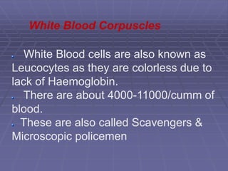
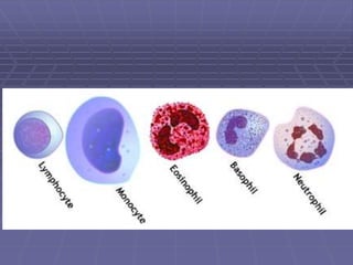
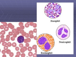
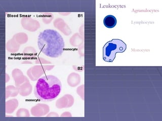
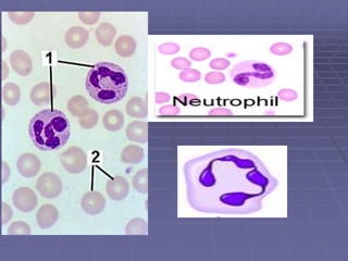
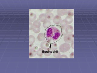
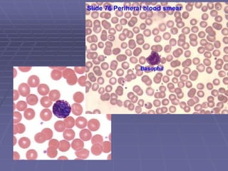
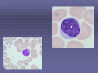
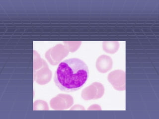
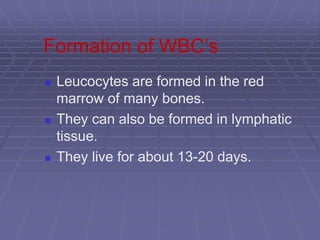
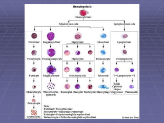
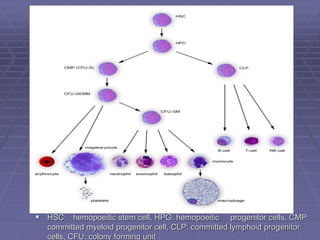
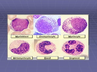
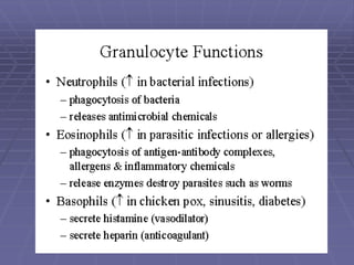
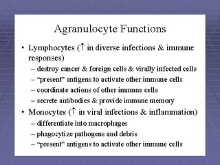
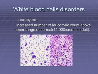
White blood cells
Dr. Sanjeev shrivastava
White Blood cells are also known as
Leucocytes as they are colorless due to
lack of Haemoglobin.
There are about 4000-11000/cumm of
blood.
These are also called Scavengers &
Microscopic policemen
White Blood Corpuscles
WBC’s
 Five Types
 Classified according to the presence or
absence of granules and the staining
characteristics of their cytoplasm.
 Leucocytes appear brightly colored in
stained preparations, they have a nuclei
and are generally larger in size than
RBC’s.
Type of WBC’s
 Granulocytes—have large
granules in their cytoplasm
 Neutrophils
 Eosinophils
 Basophils
Types of WBC’s
 Agranulocytes—do not have
granules in their cytoplasm
 Lymphocytes
 Monocytes
Granuloctyes
 Neutrophils
 Stain light purple with neutral dyes
 Granules are small and numerous—(fine)
 Several lobes in nucleus
 50-70% of WBC count
 Highly mobile/very active
 Diapedesis—Can leave blood vessels and
enter tissue space
 Phagocytosis (eater), contain several
lysosomes (janitor)
Granulocytes
 Eosinophils or Acidophils:
 Large, numerous granules
 Nuclei with two lobes
 1-4% of WBC count
 Found in lining of respiratory and digestive tracts
 Important functions involve protections against
infections caused by parasitic worms and
involvement in allergic reactions
 Secrete anti-inflammatory substances in allergic
reactions
 Basophils
 Least numerous-- 0-1%
 Diapedesis—Can leave blood vessels and
enter tissue space
 Contain histamine,serotonin,heparin—
inflammatory chemical
Agranulocytes
 Lymphocytes
 Smallest WBC
 Large nuclei/small amount of cytoplasm
 Account for 25-30% of WBC count
 Two types—T lymphocytes—attack an
infect or cancerous cell, B lymphocytes—
produce antibodies against specific
antigens (foreign body)
Agranulocytes
 Monocytes
 Largest of WBCs
 Dark kidney bean shaped nuclei
 Highly phagocytic
Formation of WBC’s
 Leucocytes are formed in the red
marrow of many bones.
 They can also be formed in lymphatic
tissue.
 They live for about 13-20 days.
Myelopoeisis
 HSC: hemopoeitic stem cell, HPG: hemopoeitic progenitor cells, CMP:
committed myeloid progenitor cell, CLP: committed lymphoid progenitor
cells, CFU: colony forming unit
White blood cells disorders
I. Leukocytosis
increased number of leucocytic count above
upper range of normal(11,000/cmm in adult).
Neutrophilia
 Neutrophils > 7500/cumm
 Pathological
1. Acute pyogenic infection
2. Acute hemorrhage or hemolysis
3. Tissue damage: Truma or infarction
4. Malignancy
5. Myeloproliferative disorders : CML
6. Drugs: digitalis, Cortisone
7. t/t with myeloid growth factors
8. Asplenia
NEUTROPHILIA
Eosinophilia
 Eosinphils >
500/mm3 .
 Causes:
1. Allergic diseases
2. parasitic diseases
3. certain skin
diseases
4. Drug sensitivity
Basophilia
 Basophils > 100/mm3.
 Causes:
1. CML,MF.
2. Reactive:
3 small box or chicken box
Monocytosis
 Monocytes > 800/mm3
 Causes:
1. Chronic bacterial
infection e.g. T.B,
brucllosis, typhoid.
2. Conective tissue disease
3. Chronic neutropenia
4. Malignancies
Lymphocytosis
 Relative Lymphocytosis:
PMN leucocytes are
decreased, so the
lymphocytes are
relatively increased
without change in their
absolute number.
 Absolute lymphocytosis
>3500/cmm in adult.
 Causes:
1. Viral infection
CMV
Measels
2. Bacterial infection
TB
Pertusis
Brucellosis
3. Chronic lymphocytic leukemia
4. Postsplenectomy
Leucopenia
 Decrease in leucocytic count below
4000/cmm
I. neutropenia:
<2000/cmm
 Causes:
1. Drugs:
anti inflammatory
anti bacterial
anti convalsant
2. Infection:
Viral: Hepatitis, HIV
Bacterial: TB, brucellosis
some protozoal and fungal infections
3. Immune:
systemic lupus erythematosis
Felty’s syndrome
4. Part of general pan cytopenia
Agranulocytosis
 Marked reduction of neutrophils below
500/cumm.
 Associated with acute febrile illness and
necrotic lesions in the mouth and the
throat.
Lymphopenia
 Lymphocytes <1500/cumm
 Causes:
1. Irradiation
2. Steroids and Immunosuppressive drugs
3. Hodgkin’s disease
4. Blood diseases: e.g. myelocytic leukemia
leukemias
 The leukemia are a group of disorders
characterized by the accumulation of
malignant white cells in the bone marrow
and blood.
Created by

White blood cells / Leucocytes

  • 1.
    White blood cells Dr.Sanjeev shrivastava
  • 2.
    White Blood cellsare also known as Leucocytes as they are colorless due to lack of Haemoglobin. There are about 4000-11000/cumm of blood. These are also called Scavengers & Microscopic policemen White Blood Corpuscles
  • 3.
    WBC’s  Five Types Classified according to the presence or absence of granules and the staining characteristics of their cytoplasm.  Leucocytes appear brightly colored in stained preparations, they have a nuclei and are generally larger in size than RBC’s.
  • 5.
    Type of WBC’s Granulocytes—have large granules in their cytoplasm  Neutrophils  Eosinophils  Basophils
  • 7.
    Types of WBC’s Agranulocytes—do not have granules in their cytoplasm  Lymphocytes  Monocytes
  • 9.
    Granuloctyes  Neutrophils  Stainlight purple with neutral dyes  Granules are small and numerous—(fine)  Several lobes in nucleus  50-70% of WBC count  Highly mobile/very active  Diapedesis—Can leave blood vessels and enter tissue space  Phagocytosis (eater), contain several lysosomes (janitor)
  • 11.
    Granulocytes  Eosinophils orAcidophils:  Large, numerous granules  Nuclei with two lobes  1-4% of WBC count  Found in lining of respiratory and digestive tracts  Important functions involve protections against infections caused by parasitic worms and involvement in allergic reactions  Secrete anti-inflammatory substances in allergic reactions
  • 13.
     Basophils  Leastnumerous-- 0-1%  Diapedesis—Can leave blood vessels and enter tissue space  Contain histamine,serotonin,heparin— inflammatory chemical
  • 15.
    Agranulocytes  Lymphocytes  SmallestWBC  Large nuclei/small amount of cytoplasm  Account for 25-30% of WBC count  Two types—T lymphocytes—attack an infect or cancerous cell, B lymphocytes— produce antibodies against specific antigens (foreign body)
  • 17.
    Agranulocytes  Monocytes  Largestof WBCs  Dark kidney bean shaped nuclei  Highly phagocytic
  • 19.
    Formation of WBC’s Leucocytes are formed in the red marrow of many bones.  They can also be formed in lymphatic tissue.  They live for about 13-20 days.
  • 21.
    Myelopoeisis  HSC: hemopoeiticstem cell, HPG: hemopoeitic progenitor cells, CMP: committed myeloid progenitor cell, CLP: committed lymphoid progenitor cells, CFU: colony forming unit
  • 25.
    White blood cellsdisorders I. Leukocytosis increased number of leucocytic count above upper range of normal(11,000/cmm in adult).
  • 26.
    Neutrophilia  Neutrophils >7500/cumm  Pathological 1. Acute pyogenic infection 2. Acute hemorrhage or hemolysis 3. Tissue damage: Truma or infarction 4. Malignancy 5. Myeloproliferative disorders : CML 6. Drugs: digitalis, Cortisone 7. t/t with myeloid growth factors 8. Asplenia
  • 27.
  • 28.
    Eosinophilia  Eosinphils > 500/mm3.  Causes: 1. Allergic diseases 2. parasitic diseases 3. certain skin diseases 4. Drug sensitivity
  • 29.
    Basophilia  Basophils >100/mm3.  Causes: 1. CML,MF. 2. Reactive: 3 small box or chicken box
  • 30.
    Monocytosis  Monocytes >800/mm3  Causes: 1. Chronic bacterial infection e.g. T.B, brucllosis, typhoid. 2. Conective tissue disease 3. Chronic neutropenia 4. Malignancies
  • 31.
    Lymphocytosis  Relative Lymphocytosis: PMNleucocytes are decreased, so the lymphocytes are relatively increased without change in their absolute number.  Absolute lymphocytosis >3500/cmm in adult.
  • 32.
     Causes: 1. Viralinfection CMV Measels 2. Bacterial infection TB Pertusis Brucellosis 3. Chronic lymphocytic leukemia 4. Postsplenectomy
  • 33.
    Leucopenia  Decrease inleucocytic count below 4000/cmm I. neutropenia: <2000/cmm  Causes: 1. Drugs: anti inflammatory anti bacterial anti convalsant
  • 34.
    2. Infection: Viral: Hepatitis,HIV Bacterial: TB, brucellosis some protozoal and fungal infections 3. Immune: systemic lupus erythematosis Felty’s syndrome 4. Part of general pan cytopenia
  • 35.
    Agranulocytosis  Marked reductionof neutrophils below 500/cumm.  Associated with acute febrile illness and necrotic lesions in the mouth and the throat.
  • 36.
    Lymphopenia  Lymphocytes <1500/cumm Causes: 1. Irradiation 2. Steroids and Immunosuppressive drugs 3. Hodgkin’s disease 4. Blood diseases: e.g. myelocytic leukemia
  • 37.
    leukemias  The leukemiaare a group of disorders characterized by the accumulation of malignant white cells in the bone marrow and blood.
  • 38.