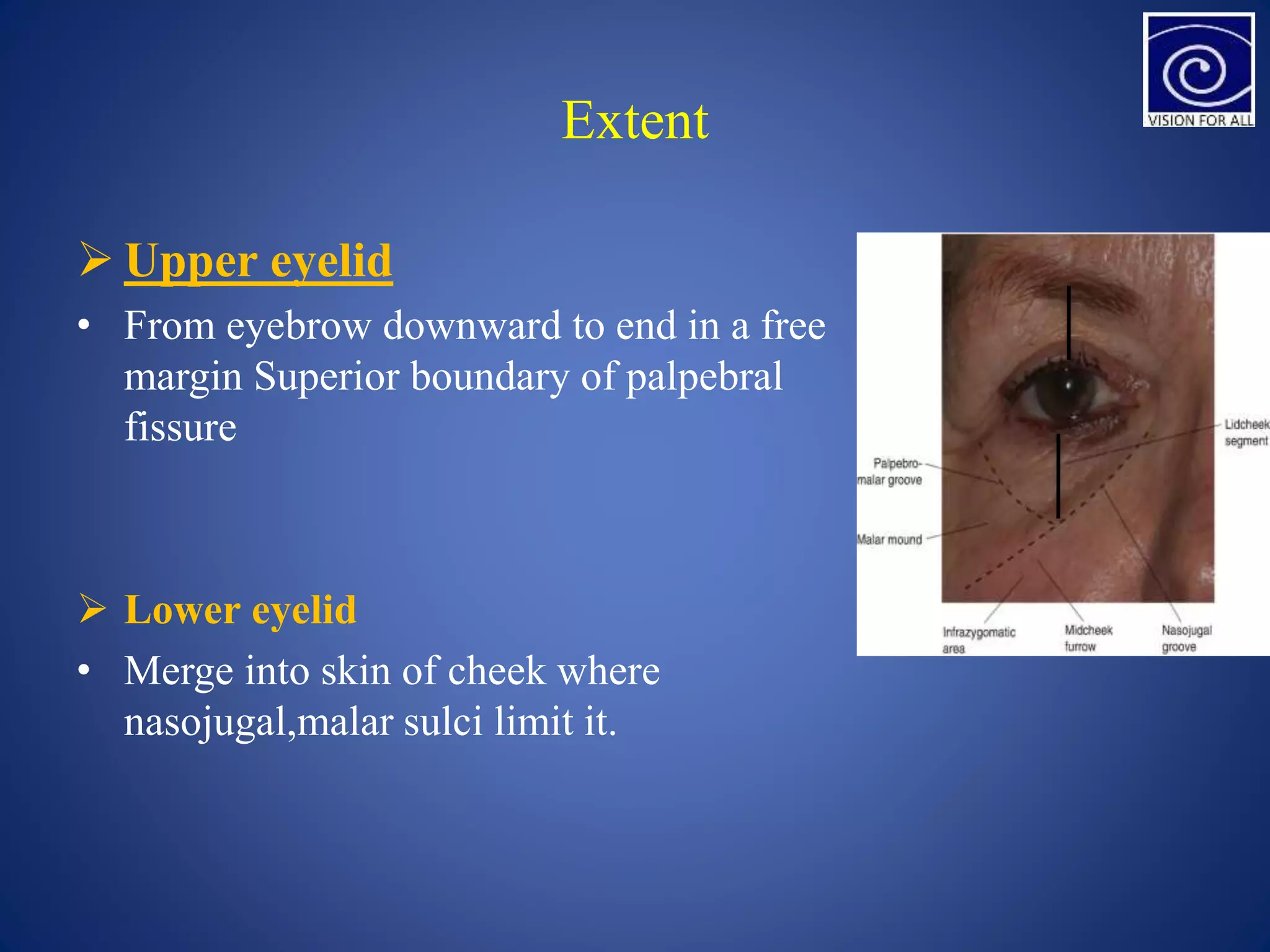
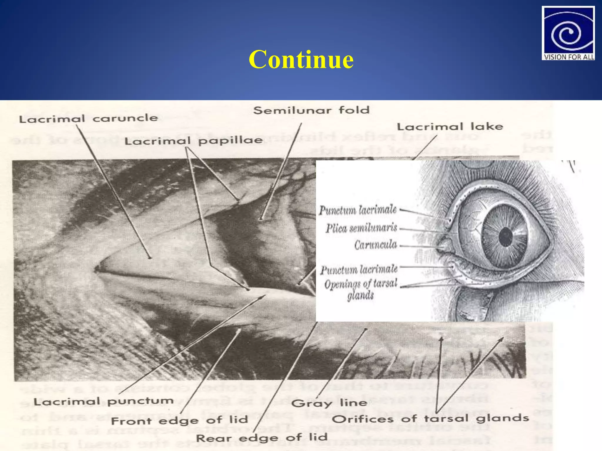
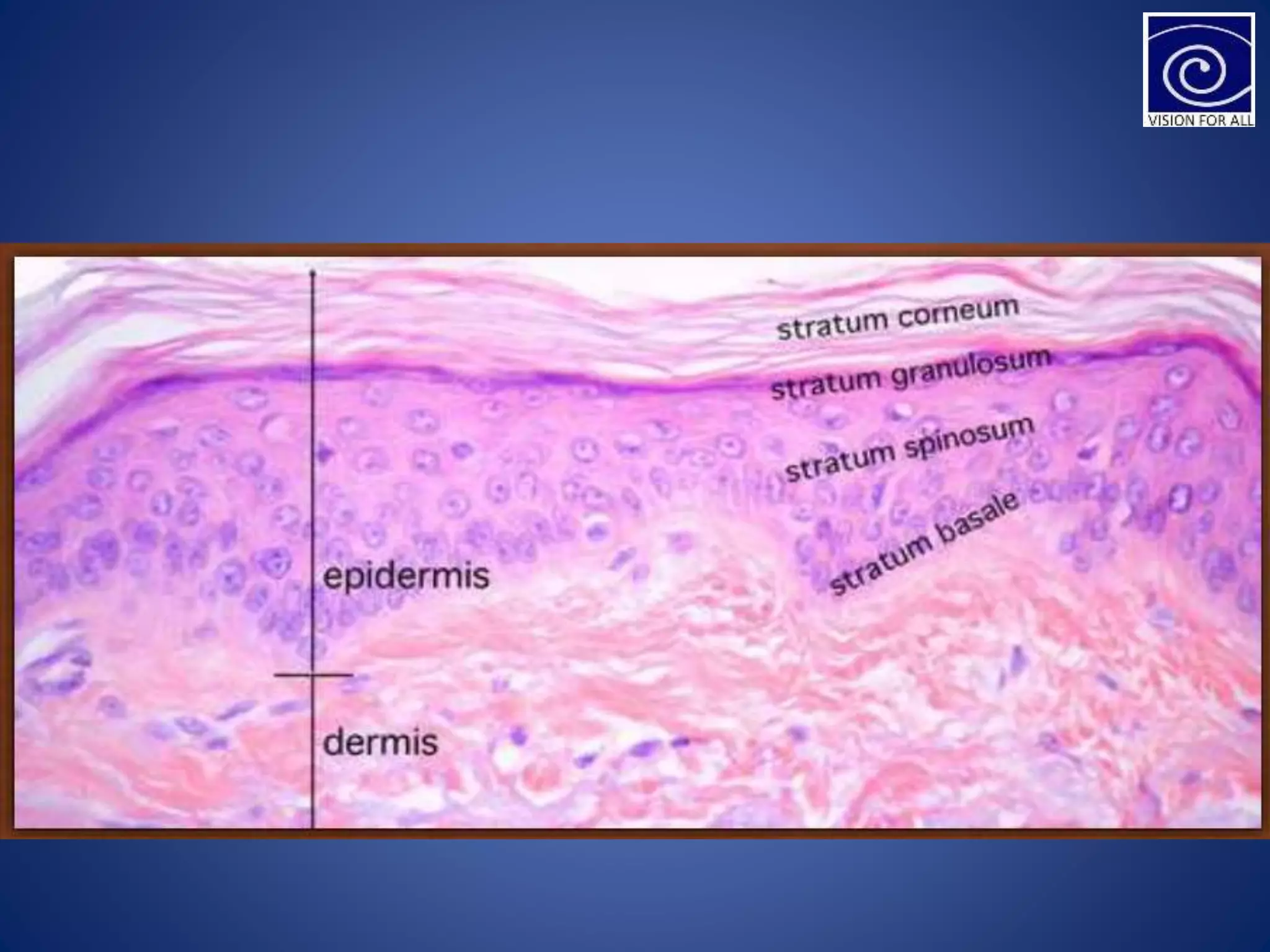
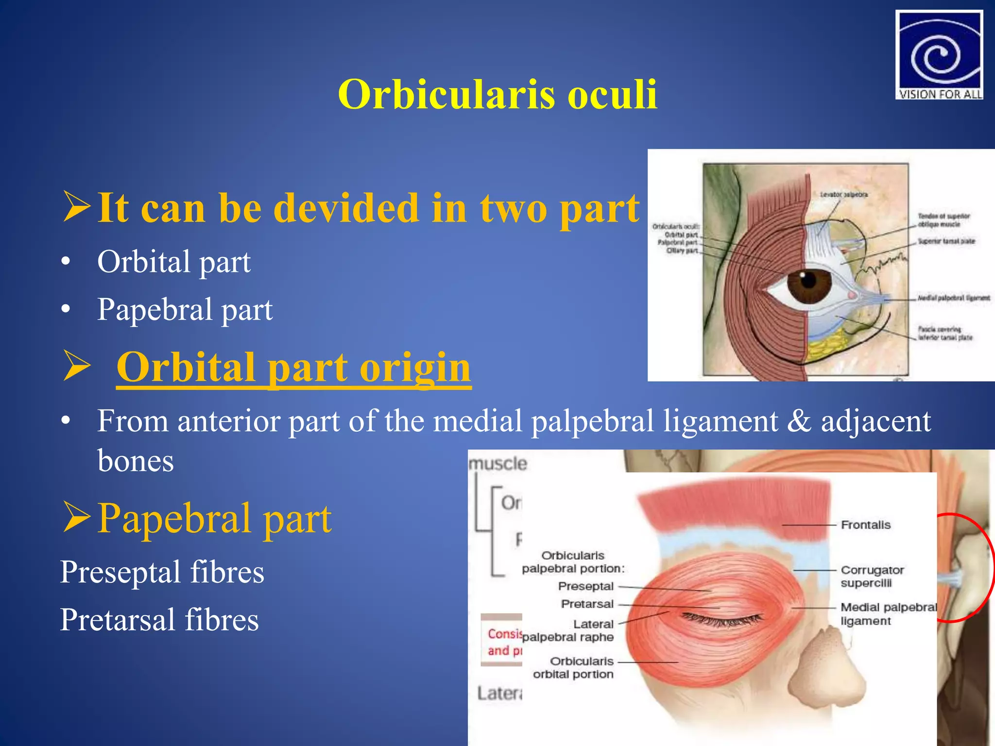
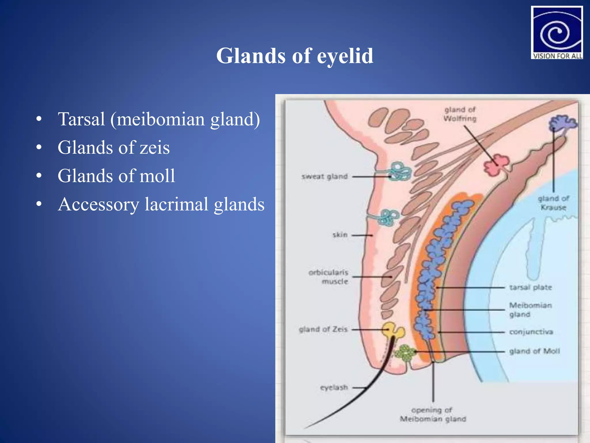
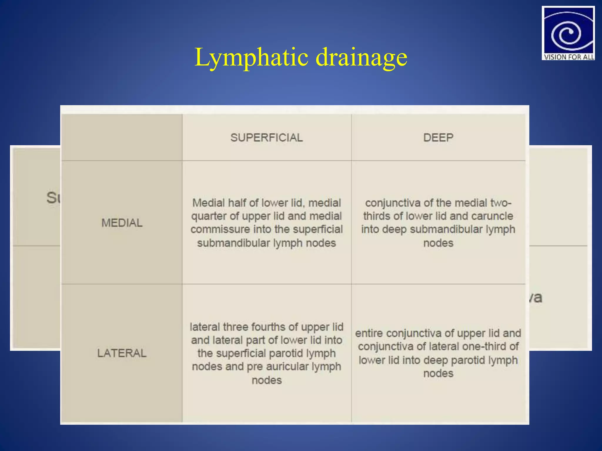
Presenter: MAAZ UL HAQ
C L Gupta Eye Institute
1
Eyelid
Definition of Eyelid
• Eyelids are the mobile tissue curtains
• These act as shutters protection the eyes
• Spread the tear film over the cornea and
conjuctiva
• Important contribution to the facial features
• Relay much useful information regarding –
wakefulness/attention
Gross Anatomy
• Extent
• Lid folds
• Position of eyelids
• Canthi
• Eyelid Margins
• Eyelashes
• Palpebral aperture or fissure
Extent
 Upper eyelid
• From eyebrow downward to end in a free
margin Superior boundary of palpebral
fissure
 Lower eyelid
• Merge into skin of cheek where
nasojugal,malar sulci limit it.
Lid folds
 Superior lid fold
• Between orbital & tarsal
portion
• Formed by fibrous slips,
from tendon of levator
 Inferior lid fold
• On skin of lower eyelids
• Fibrous slips from fascia of
inferior rectus
Position of eyelids
• In primary position of gaze
• Upper eyelid covers 1/6th of cornea
• Lower eyelid just touches the cornea
Canthi
The two eyelids meet each other at
medial and lateral angle
Lateral cantus
 5-7mm from lateral orbital margin
 1cm from frontzgomatic suture
 60 degree with wide open
 30-40 degree with eyes open in
normal way
Medial cantus
 Rounded in structure
Continue
Continue
• the two eyelids are separated by lacus lacrimalis, in
the centre of which is a small pinkish elevation
• the caruncula lacrimalis.it is a small area of tissue
derived from skin,contains large modified sweat
glands and sebaceous glands.
A semilunar fold called plica semilunaris lies on
lateral side of caruncle.
Continue
Eyelid margin
• The opposing margins of the eyelids are
nearly flat some 2 mm in width
• Each lid margin is divided into two parts
• Lacrimal papilla- ls a small elevation
present on the medial side which is
contained the hole – lacrimal puctum in its
center
• Ciliary portion-
• Rounded anterior
• Sharp posterior
•
Continue
• Inter marginal strip- between the two
borders.
• Grey line-(junction of the skin and conjunctiva)
• Inter marginal strip-
• divides the strip into anterior strip which bears
the lashes
• posterior strip which contains openings of
meibomian glands
Eyelashes
• 2-3 rows
• When lids close eyelashes do not interlace
• Upper lid : 100-150
• Lower lid : 50-75
Cilia
• 20 – 120 microns
• They taper throughout their length to end in a fine sharp point
• Lifespan 5 months.
• Replacement is fully grown in 10 wks.
• They are darker then the scalp hair and tend to remain so throughout life
Palpebral aperture
• Elliptical space b/w upper & lower lid
• margins
At Birth
• Horizontally– 18 to 21 mm
• Vertically -- 8mm
In Adults
• 28 to 3o mm (hor)
• 9 to 11 mm (ver)
Structure
• Skin
• Layer of subcutaneous areolar tissue
• Layer of striated muscles(orbicularis oculi)
• Sub muscular areolar tissue
• Fibrous layer and tarsal plate.
• Septum orbitale
• Layer of non-striated muscle fibres
• Conjunctiva
skin
• The skin covering the eyelids is elastic ,having a fine texture
•
• Is thinnest in the body and folds easily thereby contributing to
the case and speed of mobility of the upper eyelids
• Nasal part of the skin in smooth shinning and greasy in
comparison to the temporal part
Epidermis
• Epidermis - 6-7 layers of stratified squamous epithelium
• 1. Keratin layer (stratum corneum or horny layer) -
• 2. Granular cell layer (stratum granulosum) - .
• 3. Prickle cell layer (stratum spinosum) -
• 4. Basal cell layer (stratum basale) –
Dermis
thin layer of dense connective tissue with rich network elastic
fibres, blood vessels, lymphatics and nerves. Melanocytes are
also present which increase their pigment production in
response to chronic edema or inflammation
Subcutaneous areolar tissue
• Loose connective tissue No fat
• Readily distended during oedema or blood.
• Non existent - ciliary margin, lid folds and medial and lateral
angles
Layer of striated muscles
• Consists of orbicularis muscle which forms a
thin oval sheet across the eyelids
• • Levator palpebrae superioris also present
Orbicularis oculi
It can be devided in two part
• Orbital part
• Papebral part
 Orbital part origin
• From anterior part of the medial palpebral ligament & adjacent
bones
Papebral part
Preseptal fibres
Pretarsal fibres
Function of orbicularisoculi
 Orbital part
• Forced closure of eyelids
• Thus pull eyebrows downwards
 Palpebral part
• Helps in gentle closure during blinking, sleep, softvoluntary
closure
Entire muscle supplied by branches of 7th
nerve
Levator palpebrae superioris
 Origin
• At apex of orbit from the Under surface of lesser wing of
sphenoid above Annulus of Zinn by a Short tendon which is
Blended with origin of SR
 Course & attachments
• Muscle has a flat ribbon like belly.
• It passes forwards below the roof of the orbit, above the
superior rectus
Nerve supply & action of LPS
• Branch of superior division of 3rd nerve
• Acts as Elevator of upper lid
Submuscular areolar tissue
• Layer of loose connective tissue between orbicularis muscle
and fibrous layer
• (consisting of tarsal plate and septum orbitale). The nerves and
vessels of the lids lie in
• this layer and so to anaesthetise the lid, injection is made in
this plane
• This layer splits the eyelid into two - anterior lamina and
posterior lamina
Fibrous layer
• Framework of the lids
• Consists of the following –
• Tarsal plate
• Septum orbitale
• Medial palpebral ligamen
• Lateral palpebral ligament
Continue
• Tarsal plates
• Tarsi are firm plates of dense fibrous tissue that form the
skeleton of the eyelids giving them shape and firmness
• Size
• Tarsi about 29mm long
• Tarsi 1 mm thick
• Upper tarsus 10-11mm in height
• Lower tarsus is 4-5mm in height
Continue
• Border of tarsal plates
• Free borders are straight, whereas the opposite attached
borders are convex
• Superior border of upper tarsus –
• Septum orbital and Muller’s muscle
• Inferior border of lower tarsus –
• Orbital septum,Capsulopalpebral fascia and inferior
palpebral
Surface
• Anterior surface of each tarsus is convex Posterior surface is
concave In upper lid
 Extremities
• Lateral ends are attached to Whitnall’s tubercle by lateral
palpebral ligament and medial ends attached by medial
palpebral ligaments to anterior lacrimal crest
 Tarsal ( meibomian)
• Glands are embedded in the subtance of the tarsal plates
Layer of non striated muscle fibres
• The layer consists of smooth muscle fibres of Muller’s muscle
• (superior and inferior palpebral muscles) which lie deep to the
septum orbitale in the upper and lower eyelids, respectively
• • Origin - inferior terminal striated fibres of the LPS muscle in
the and gets inserted into the orbital margin of tarsal plate
• Supplied by sympathetic nerve fibres
Glands of eyelid
• Tarsal (meibomian gland)
• Glands of zeis
• Glands of moll
• Accessory lacrimal glands
Tarsal (meibomian gland)
• Modified sweat glands present in the posterior
part of the stroma of the tarsal plates Arranged
in a single row vertically parallel to each other
20 to 30 each lid
• Structure
• Each tarsal gland consist of a central duct
which ran straight perpendicular to the lid
margin
• Occupies the entire thickness of the tarsal plate
• Into central central open 10-15 acini from the
side
Continue
• Opening of meibomian glands arrange in
single row
• On the lid margin between grey line and
posterior border of the lids
• Secretions are oily in nature (sebum) and
have the following function -
• Prevent outflow
• Prevent evaporation - smooth movements
• Ensure the air tight closure of the eyelids
Glands of zeis
• Modified sebaceous glands attached to the eyelash follicle
directly
• Two glands associated with each cilium
• Structure - single cul-de-sac or two/three lobules are present
 Secretions-
• Zeis gland (sebum) prevent the eyelashes from becoming dry
and brittle
• Its also contributes towards oily layer of tear film
Glands of moll
• Modified sweat glands which lie between the cilia
• • Lower > Upper
• Structure - Each gland is 1.5-2mm long and has unbranched
spiral shape.
• It has a fundus, a body, an ampullary portion and a neck.
• Duct of the gland passes through the dermis, epidermis and
may terminate separately between the two lashes or between
the lash and its epithelium covering or into the ducts of Zeis
gland
Accessory lacrimal gland of wolfring
• These are microscopic accessory lacrimal glands present along
the upper border of superior tarsus and along with the lower
border of the inferior tarsus.
• These are 2 - 5 in upper lid and 2 -3 in lower lid
Arterial supply
• Mainly supplied by the medial and lateral palpebral arteries
which are branches of the dorsal nasal and lacrimal arteries
• The superior and inferior arteries pierce through the above
septum orbitale and below medial palpebral ligament and enter
the upper and lower eyelids respectively
Vessels and nerves of the eyelid
Lymphatic drainage
Nerve supply
References
• ocular anatomy and physiology AK khurana
Eyelid

Eyelid

  • 1.
    Presenter: MAAZ ULHAQ C L Gupta Eye Institute 1 Eyelid
  • 2.
    Definition of Eyelid •Eyelids are the mobile tissue curtains • These act as shutters protection the eyes • Spread the tear film over the cornea and conjuctiva • Important contribution to the facial features • Relay much useful information regarding – wakefulness/attention
  • 3.
    Gross Anatomy • Extent •Lid folds • Position of eyelids • Canthi • Eyelid Margins • Eyelashes • Palpebral aperture or fissure
  • 4.
    Extent  Upper eyelid •From eyebrow downward to end in a free margin Superior boundary of palpebral fissure  Lower eyelid • Merge into skin of cheek where nasojugal,malar sulci limit it.
  • 5.
    Lid folds  Superiorlid fold • Between orbital & tarsal portion • Formed by fibrous slips, from tendon of levator  Inferior lid fold • On skin of lower eyelids • Fibrous slips from fascia of inferior rectus
  • 6.
    Position of eyelids •In primary position of gaze • Upper eyelid covers 1/6th of cornea • Lower eyelid just touches the cornea
  • 7.
    Canthi The two eyelidsmeet each other at medial and lateral angle Lateral cantus  5-7mm from lateral orbital margin  1cm from frontzgomatic suture  60 degree with wide open  30-40 degree with eyes open in normal way Medial cantus  Rounded in structure
  • 8.
  • 9.
    Continue • the twoeyelids are separated by lacus lacrimalis, in the centre of which is a small pinkish elevation • the caruncula lacrimalis.it is a small area of tissue derived from skin,contains large modified sweat glands and sebaceous glands. A semilunar fold called plica semilunaris lies on lateral side of caruncle.
  • 10.
  • 11.
    Eyelid margin • Theopposing margins of the eyelids are nearly flat some 2 mm in width • Each lid margin is divided into two parts • Lacrimal papilla- ls a small elevation present on the medial side which is contained the hole – lacrimal puctum in its center • Ciliary portion- • Rounded anterior • Sharp posterior •
  • 12.
    Continue • Inter marginalstrip- between the two borders. • Grey line-(junction of the skin and conjunctiva) • Inter marginal strip- • divides the strip into anterior strip which bears the lashes • posterior strip which contains openings of meibomian glands
  • 14.
    Eyelashes • 2-3 rows •When lids close eyelashes do not interlace • Upper lid : 100-150 • Lower lid : 50-75 Cilia • 20 – 120 microns • They taper throughout their length to end in a fine sharp point • Lifespan 5 months. • Replacement is fully grown in 10 wks. • They are darker then the scalp hair and tend to remain so throughout life
  • 15.
    Palpebral aperture • Ellipticalspace b/w upper & lower lid • margins At Birth • Horizontally– 18 to 21 mm • Vertically -- 8mm In Adults • 28 to 3o mm (hor) • 9 to 11 mm (ver)
  • 16.
    Structure • Skin • Layerof subcutaneous areolar tissue • Layer of striated muscles(orbicularis oculi) • Sub muscular areolar tissue • Fibrous layer and tarsal plate. • Septum orbitale • Layer of non-striated muscle fibres • Conjunctiva
  • 18.
    skin • The skincovering the eyelids is elastic ,having a fine texture • • Is thinnest in the body and folds easily thereby contributing to the case and speed of mobility of the upper eyelids • Nasal part of the skin in smooth shinning and greasy in comparison to the temporal part
  • 19.
    Epidermis • Epidermis -6-7 layers of stratified squamous epithelium • 1. Keratin layer (stratum corneum or horny layer) - • 2. Granular cell layer (stratum granulosum) - . • 3. Prickle cell layer (stratum spinosum) - • 4. Basal cell layer (stratum basale) – Dermis thin layer of dense connective tissue with rich network elastic fibres, blood vessels, lymphatics and nerves. Melanocytes are also present which increase their pigment production in response to chronic edema or inflammation
  • 20.
    Subcutaneous areolar tissue •Loose connective tissue No fat • Readily distended during oedema or blood. • Non existent - ciliary margin, lid folds and medial and lateral angles
  • 22.
    Layer of striatedmuscles • Consists of orbicularis muscle which forms a thin oval sheet across the eyelids • • Levator palpebrae superioris also present
  • 23.
    Orbicularis oculi It canbe devided in two part • Orbital part • Papebral part  Orbital part origin • From anterior part of the medial palpebral ligament & adjacent bones Papebral part Preseptal fibres Pretarsal fibres
  • 24.
    Function of orbicularisoculi Orbital part • Forced closure of eyelids • Thus pull eyebrows downwards  Palpebral part • Helps in gentle closure during blinking, sleep, softvoluntary closure Entire muscle supplied by branches of 7th nerve
  • 25.
    Levator palpebrae superioris Origin • At apex of orbit from the Under surface of lesser wing of sphenoid above Annulus of Zinn by a Short tendon which is Blended with origin of SR  Course & attachments • Muscle has a flat ribbon like belly. • It passes forwards below the roof of the orbit, above the superior rectus
  • 26.
    Nerve supply &action of LPS • Branch of superior division of 3rd nerve • Acts as Elevator of upper lid
  • 27.
    Submuscular areolar tissue •Layer of loose connective tissue between orbicularis muscle and fibrous layer • (consisting of tarsal plate and septum orbitale). The nerves and vessels of the lids lie in • this layer and so to anaesthetise the lid, injection is made in this plane • This layer splits the eyelid into two - anterior lamina and posterior lamina
  • 28.
    Fibrous layer • Frameworkof the lids • Consists of the following – • Tarsal plate • Septum orbitale • Medial palpebral ligamen • Lateral palpebral ligament
  • 29.
    Continue • Tarsal plates •Tarsi are firm plates of dense fibrous tissue that form the skeleton of the eyelids giving them shape and firmness • Size • Tarsi about 29mm long • Tarsi 1 mm thick • Upper tarsus 10-11mm in height • Lower tarsus is 4-5mm in height
  • 30.
    Continue • Border oftarsal plates • Free borders are straight, whereas the opposite attached borders are convex • Superior border of upper tarsus – • Septum orbital and Muller’s muscle • Inferior border of lower tarsus – • Orbital septum,Capsulopalpebral fascia and inferior palpebral
  • 31.
    Surface • Anterior surfaceof each tarsus is convex Posterior surface is concave In upper lid  Extremities • Lateral ends are attached to Whitnall’s tubercle by lateral palpebral ligament and medial ends attached by medial palpebral ligaments to anterior lacrimal crest  Tarsal ( meibomian) • Glands are embedded in the subtance of the tarsal plates
  • 32.
    Layer of nonstriated muscle fibres • The layer consists of smooth muscle fibres of Muller’s muscle • (superior and inferior palpebral muscles) which lie deep to the septum orbitale in the upper and lower eyelids, respectively • • Origin - inferior terminal striated fibres of the LPS muscle in the and gets inserted into the orbital margin of tarsal plate • Supplied by sympathetic nerve fibres
  • 34.
    Glands of eyelid •Tarsal (meibomian gland) • Glands of zeis • Glands of moll • Accessory lacrimal glands
  • 35.
    Tarsal (meibomian gland) •Modified sweat glands present in the posterior part of the stroma of the tarsal plates Arranged in a single row vertically parallel to each other 20 to 30 each lid • Structure • Each tarsal gland consist of a central duct which ran straight perpendicular to the lid margin • Occupies the entire thickness of the tarsal plate • Into central central open 10-15 acini from the side
  • 36.
    Continue • Opening ofmeibomian glands arrange in single row • On the lid margin between grey line and posterior border of the lids • Secretions are oily in nature (sebum) and have the following function - • Prevent outflow • Prevent evaporation - smooth movements • Ensure the air tight closure of the eyelids
  • 37.
    Glands of zeis •Modified sebaceous glands attached to the eyelash follicle directly • Two glands associated with each cilium • Structure - single cul-de-sac or two/three lobules are present  Secretions- • Zeis gland (sebum) prevent the eyelashes from becoming dry and brittle • Its also contributes towards oily layer of tear film
  • 38.
    Glands of moll •Modified sweat glands which lie between the cilia • • Lower > Upper • Structure - Each gland is 1.5-2mm long and has unbranched spiral shape. • It has a fundus, a body, an ampullary portion and a neck. • Duct of the gland passes through the dermis, epidermis and may terminate separately between the two lashes or between the lash and its epithelium covering or into the ducts of Zeis gland
  • 39.
    Accessory lacrimal glandof wolfring • These are microscopic accessory lacrimal glands present along the upper border of superior tarsus and along with the lower border of the inferior tarsus. • These are 2 - 5 in upper lid and 2 -3 in lower lid
  • 40.
    Arterial supply • Mainlysupplied by the medial and lateral palpebral arteries which are branches of the dorsal nasal and lacrimal arteries • The superior and inferior arteries pierce through the above septum orbitale and below medial palpebral ligament and enter the upper and lower eyelids respectively
  • 41.
    Vessels and nervesof the eyelid
  • 42.
  • 44.
  • 46.
    References • ocular anatomyand physiology AK khurana

Editor's Notes

  • #3 The hypothalamus, a peanut-sized structure deep inside the brain, contains groups of nerve cells that act as control centers affecting sleep and arousal.  Within the hypothalamus is the suprachiasmatic nucleus (SCN) – clusters of thousands of cells that receive information about light exposure directly from the eyes and control your behavioral rhythm.  Some people with damage to the SCN sleep erratically throughout the day because they are not able to match their circadian rhythms with the light-dark cycle.  Most blind people maintain some ability to sense light and are able to modify their sleep/wake cycle The suprachiasmatic nucleus or nuclei (SCN) is a tiny region of the brain in the hypothalamus, situated directly above the optic chiasm. It is responsible for controlling circadian rhythms.
  • #6  flexible but inelastic cord of strong fibrous collagen tissue attaching a muscle to a bone. the hamstring of a quadruped. Two other folds is there – Nasojugal sulcus , Malar sulcus
  • #8 The zygomaticofrontal suture (or frontozygomatic suture) is the cranial suture between the zygomatic bone and the frontal bone. The suture can be palpated just lateral to the eye
  • #10 A sebaceous gland is a microscopic exocrine gland in the skin that opens into a hair follicle to secrete an oily or waxy matter, called sebum, which lubricates the hair and skin of mammals.[1] In humans, sebaceous glands occur in the greatest number on the face and scalp n humans, sebaceous glands occur in the greatest number on the face and scalp, but also on all parts of the skin except the palms of the hands and soles of the feet. In the eyelids, meibomian glands, also called tarsal glands, are a type of sebaceous gland that secrete a special type of sebum into tears. 
  • #12 Sweat glands occur all over the body, but are most numerous on the forehead, the armpits, the palms and the soles of the feet. Sweat is mainly water, but it also contains some salts. Its main function is to control body temperature. As the water in the sweat evaporates, the surface of the skin cool
  • #15 Sudden hair loss that starts with one or more circular bald patches that may overlap. Alopecia areata occurs when the immune system attacks hair follicles and may be brought on by severe stress.The main symptom is hair loss.Treatment may address any underlying conditions and includes topical scalp medication. becoming thinner or narrower towards one end
  • #20 1. Keratin layer (stratum corneum or horny layer) - consists of flat cells devoid of nuclei. 2. Granular cell layer (stratum granulosum) - one or two layers consists of flattened cells with keratohyaline granules. 3. Prickle cell layer (stratum spinosum) - polygonal cells with abundant eosinophilic cytoplasm 4. Basal cell layer (stratum basale) - single layer of columnar shaped proliferatingcells