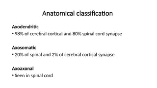
Anatomical classification
Axodendritic
• 98%of cerebral cortical and 80% spinal cord synapse
Axosomatic
• 20% of spinal and 2% of cerebral cortical synapse
Axoaxonal
• Seen in spinal cord
5.
Physiological / Functionalclassification
Chemical
• Synaptic cleft present
• NT from presynaptic neuron excite/ inhibit postsynaptic neuron
Electrical
• Pre and post synaptic cells come closer to form gap junction
• Ions pass through freely
• In lateral vestibular nucleus
Conjoint
• Both electrical and chemical transmission occurs
6.
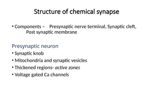
Structure of chemicalsynapse
• Components – Presynaptic nerve terminal, Synaptic cleft,
Post synaptic membrane
Presynaptic neuron
• Synaptic knob
• Mitochondria and synaptic vesicles
• Thickened regions- active zones
• Voltage gated Ca channels
7.
Synaptic cleft
• 20-40nm wide
Postsynaptic neuron
• Post synaptic density
• Receptor proteins
8.
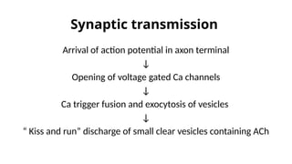
Synaptic transmission
Arrival ofaction potential in axon terminal
↓
Opening of voltage gated Ca channels
↓
Ca trigger fusion and exocytosis of vesicles
↓
“ Kiss and run” discharge of small clear vesicles containing ACh
9.
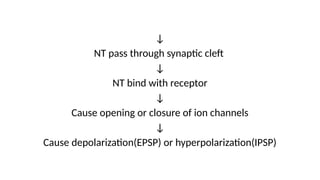
↓
NT pass throughsynaptic cleft
↓
NT bind with receptor
↓
Cause opening or closure of ion channels
↓
Cause depolarization(EPSP) or hyperpolarization(IPSP)
10.
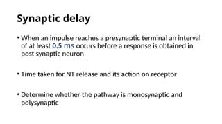
Synaptic delay
• Whenan impulse reaches a presynaptic terminal an interval
of at least 0.5 ms occurs before a response is obtained in
post synaptic neuron
• Time taken for NT release and its action on receptor
• Determine whether the pathway is monosynaptic and
polysynaptic
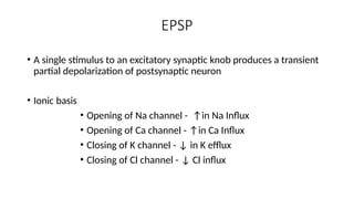
EPSP
• A singlestimulus to an excitatory synaptic knob produces a transient
partial depolarization of postsynaptic neuron
• Ionic basis
• Opening of Na channel - ↑in Na Influx
• Opening of Ca channel - ↑in Ca Influx
• Closing of K channel - ↓ in K efflux
• Closing of Cl channel - ↓ Cl influx
13.
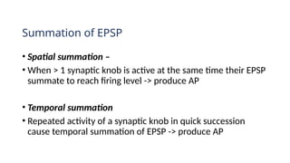
Summation of EPSP
•Spatial summation –
• When > 1 synaptic knob is active at the same time their EPSP
summate to reach firing level -> produce AP
• Temporal summation
• Repeated activity of a synaptic knob in quick succession
cause temporal summation of EPSP -> produce AP
14.
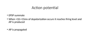
Action potential
• EPSPsummate
• When +10-+15mv of depolarization occurs it reaches firing level and
AP is produced
• AP is propagated
15.
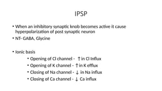
IPSP
• When aninhibitory synaptic knob becomes active it cause
hyperpolarization of post synaptic neuron
• NT- GABA, Glycine
• Ionic basis
• Opening of Cl channel - ↑in Cl Influx
• Opening of K channel - ↑in K efflux
• Closing of Na channel - ↓ in Na influx
• Closing of Ca channel - ↓ Ca influx
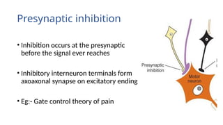
Presynaptic inhibition
• Inhibitionoccurs at the presynaptic terminals
before the signal ever reaches the synapse
• Inhibitory interneuron terminals form
axoaxonal synapse on excitatory ending
• Eg:- Gate control theory of pain
18.
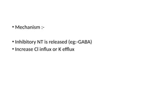
• Mechanism :-
•Inhibitory NT is released (eg:-GABA)
• Increase Cl influx or K efflux
19.
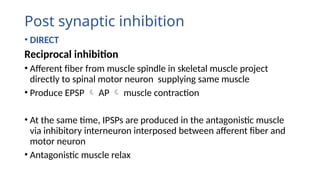
Post synaptic inhibition
•DIRECT
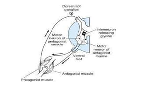
Reciprocal inhibition
• Afferent fiber from muscle spindle in skeletal muscle project
directly to spinal motor neuron supplying same muscle
• Produce EPSP AP muscle contraction
• At the same time, IPSPs are produced in the antagonistic muscle
via inhibitory interneuron interposed between afferent fiber and
motor neuron
• Antagonistic muscle relax
21.
Renshaw cell inhibition
•Spinal motor neuron gives off collateral
that synapse with an inhibitory interneuron
• Interneuron terminate on spinal motor
neurons
• Impulses in motor neuron activate
interneuron to secrete inhibitory NT
• This decreases discharge from spinal motor
neuron
22.
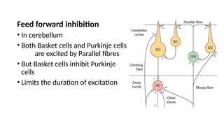
Feed forward inhibition
•In cerebellum
• Both Basket cells and Purkinje cells
are excited by Parallel fibres
• But Basket cells inhibit Purkinje
cells
• Limits the duration of excitation
23.
• INDIRECT
• Inhibitiondue to effect of previous discharge of post synaptic
neuron
• Post synaptic neuron is in its absolute refractory period
• Or during after hyper polarization it is less excitable
24.
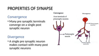
PROPERTIES OF SYNAPSE
Convergence
•Many pre synaptic terminals
converge on a single post
synaptic neuron
Divergence
• A single pre synaptic neuron
makes contact with many post
synaptic neurons
25.
One way conduction
•Neurotransmitter vesicles are present only in presynaptic neuron
Synaptic delay
Synaptic inhibition
Summation
Fatigue
• Repeated stimulation of presynaptic neurons leads to gradual
decrease and final disappearance of postsynaptic response
• d/t exhaustion of chemical transmitter
26.
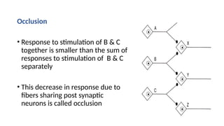
Occlusion
• Response tostimulation of B & C
together is smaller than the sum of
responses to stimulation of B & C
separately
• This decrease in response due to pre synaptic
fibers sharing post synaptic
neurons is called occlusion
27.
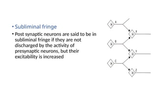
• Subliminal fringe
•Post synaptic neurons are said to be in
subliminal fringe if they are not
discharged by the activity of
presynaptic neurons, but their
excitability is increased
28.
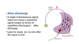
• After discharge
•A single instantaneous signal
input can cause a sustained
signal output (a series of
repetitive discharges) – after
discharge
• Lasts for many ms to min after
the input is over
29.
Synaptic plasticity
• Refersto capability of being easily moulded or changed
• Synaptic transmission can be increased or decreased on the
basis of past experience
• Basis of learning and memory
Editor's Notes
#4 Dendrodentritic between mitral and granule cells of olfactory bulb