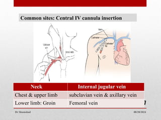
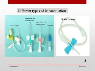
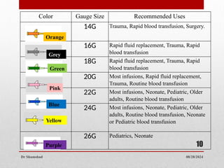
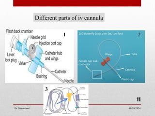
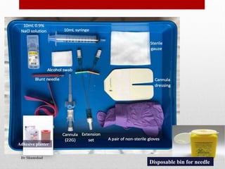
08/28/2024
Dr Shamshad
1
Intravenous Cannulation (IV)
Dr. Shamshad
Majmaah university
KSA
08/28/2024
Dr Shamshad
2
Objectives
1. Identify the common sites of IV cannula insertion
(peripheral & central).
2. Identify the different sizes and shapes of IV cannulae.
3. Insert an IV cannula (peripheral), in a manikin in a
professional way.
• Intravenous (IV) cannulation:
A technique in which a cannula is placed inside a
vein to provide venous access.
 Venous access allows sampling of blood, as well as
administration of fluids, medications, parenteral
nutrition, chemotherapy, and blood products.
08/28/2024
Dr Shamshad
3
Patient and Site selection
Depends on : Duration of IV therapy, patient’s activity level,
condition of the extremities.
Avoid Veins: Not resilient , feel hard or cordlike (often
thrombosed).
Avoid arms : Patients who have undergone radical mastectomy,
same side as the surgery (circulation may be impaired , edema and
complications like thrombosis) .
Pediatric patients: Lower extremities veins used
Neonates patients : Scalp veins sets are commonly.
.
08/28/2024
Dr Shamshad
4
08/28/2024
Dr Shamshad
5
Ease of access:
• Use of the non-dominant extremity
• Avoiding joint areas
• Avoiding use of the lower extremities
Contraindications:
• Pre-existing Vascular Compromise Lymphatic or venous
drainage has been compromised, i.e. lymph node dissection
accompanying mastectomy, A-V fistulas, injured extremities,
thrombosis.
• Regional Infections - overlying cellulitis or infection
08/28/2024
Dr Shamshad
6
Common sites: Peripheral IV cannula insertion
Head Upper limb Lower limb
External
jugular
vein
Cephalic vein Greater saphenous vein
Basilic vein Dorsal digital vein
Median vein
Metacarpal vein
08/28/2024
Dr Shamshad
7
Common sites: Central IV cannula insertion
Neck Internal jugular vein
Chest & upper limb subclavian vein & axillary vein
Lower limb: Groin Femoral vein
08/28/2024
Dr Shamshad
8
Different types of iv cannulation
Scalp vein set
08/28/2024
Dr Shamshad
10
Color Gauge Size Recommended Uses
14G Trauma, Rapid blood transfusion, Surgery.
16G Rapid fluid replacement, Trauma, Rapid
blood transfusion
18G Rapid fluid replacement, Trauma, Rapid
blood transfusion
20G Most infusions, Rapid fluid replacement,
Trauma, Routine blood transfusion
22G Most infusions, Neonate, Pediatric, Older
adults, Routine blood transfusion
24G Most infusions, Neonate, Pediatric, Older
adults, Routine blood transfusion, Neonate
or Pediatric blood transfusion
26G Pediatrics, Neonate
Orange
Grey
Green
Pink
Blue
Yellow
Purple
08/28/2024
Dr Shamshad
11
Different parts of iv cannula
1 2
3
08/28/2024
Dr Shamshad
12
Adhesive plaster
Disposable bin for needle
08/28/2024
Dr Shamshad
13
1. Introduce yourself to the patient and
clarify the patient’s identity.
2. Explain the procedure to the patient
and gain informed consent to continue.
3. Explain that cannulation may cause
some discomfort but for short period
of time.
Procedure
08/28/2024
Dr Shamshad
14
4. Sanitize our hands using alcohol cleanser.
08/28/2024
Dr Shamshad
15
5. Put on your gloves, clean the patient’s skin with the alcohol wipe and let it
dry
6. Position the arm so that it is comfortable for the patient and identify a vein.
7. Apply the tourniquet and re-check the vein.
08/28/2024
Dr Shamshad
16
8. Remove the cannula from its packaging and remove
the needle cover ensuring not to touch the needle.
08/28/2024
Dr Shamshad
17
9. Stretch the skin distally and tell the patient to expect a sharp
scratch.
10. Insert the needle, bevel upwards at about 30 degrees.
11. Advance the needle until a flashback of blood is seen in the
hub at the back of the cannula
08/28/2024
Dr Shamshad
18
12. Once this is seen, progress the entire cannula a further 2mm,
then fix the needle, advancing the rest of the cannula into the
vein.
08/28/2024
Dr Shamshad
19
13. Release the tourniquet, apply pressure to the vein at the tip of
the cannula and remove the needle fully. Remove the cap
from the needle and put this on the end of the cannula.
08/28/2024
Dr Shamshad
20
14. Carefully dispose of the needle into the sharps box.
15. Apply the dressing to the cannula to fix it in place and ensure
that the date sticker has been completed and applied.
08/28/2024
Dr Shamshad
21
16. Check that the use-by date on the saline has not passed.
If the date is ok, fill the syringe with saline and flush it
through the cannula to check for patency.
17. If there is any resistance, if it causes any pain, or you
notice any localized tissue swelling; immediately stop
flushing, remove the cannula and start again.
08/28/2024
Dr Shamshad
22
18. Dispose of your gloves and
equipment in the clinical waste
bin, ensure the patient is
comfortable and thank them
08/28/2024
Dr Shamshad
23
Complications :Peripheral IV cannulation
 Infection
 Phlebitis
 Infiltration
 Artery / nerve damage
 Dislocation
 Stenosis
08/28/2024
Dr Shamshad
24
Complications :Central IV cannulation
 Pneumothorax
 Hemothorax
 Air embolism
 Venous thrombosis
 “Pinch off” syndrome
 Infection
 Artery / nerve damage
08/28/2024
Dr Shamshad
25
Ultrasound guided venipuncture
• An established technique for both peripherally
inserted central catheters & central venous cannulation.
• With the increasing availability of portable ultrasound facilities,
this may become an option in the future for difficult peripheral
venous cannulations.
• A hand held Doppler probe has been used to identify accurately
forearm veins of more than 2 mm diameter in patients with invisible
and impalpable veins.
08/28/2024
Dr Shamshad
26
Ultrasound guided venipuncture
08/28/2024
Dr Shamshad
27
Vein Finders: Optical devices use the behavior of Infrared light to
create an image & superimpose project it onto the viewing area.
Infrared light is projected onto the viewing area and is absorbed at
the locations where blood (veins) reside. Where light is absorbed
there is less light reflected.

Procedure of IV cannulation for reference

  • 1.
    08/28/2024 Dr Shamshad 1 Intravenous Cannulation(IV) Dr. Shamshad Majmaah university KSA
  • 2.
    08/28/2024 Dr Shamshad 2 Objectives 1. Identifythe common sites of IV cannula insertion (peripheral & central). 2. Identify the different sizes and shapes of IV cannulae. 3. Insert an IV cannula (peripheral), in a manikin in a professional way.
  • 3.
    • Intravenous (IV)cannulation: A technique in which a cannula is placed inside a vein to provide venous access.  Venous access allows sampling of blood, as well as administration of fluids, medications, parenteral nutrition, chemotherapy, and blood products. 08/28/2024 Dr Shamshad 3
  • 4.
    Patient and Siteselection Depends on : Duration of IV therapy, patient’s activity level, condition of the extremities. Avoid Veins: Not resilient , feel hard or cordlike (often thrombosed). Avoid arms : Patients who have undergone radical mastectomy, same side as the surgery (circulation may be impaired , edema and complications like thrombosis) . Pediatric patients: Lower extremities veins used Neonates patients : Scalp veins sets are commonly. . 08/28/2024 Dr Shamshad 4
  • 5.
    08/28/2024 Dr Shamshad 5 Ease ofaccess: • Use of the non-dominant extremity • Avoiding joint areas • Avoiding use of the lower extremities Contraindications: • Pre-existing Vascular Compromise Lymphatic or venous drainage has been compromised, i.e. lymph node dissection accompanying mastectomy, A-V fistulas, injured extremities, thrombosis. • Regional Infections - overlying cellulitis or infection
  • 6.
    08/28/2024 Dr Shamshad 6 Common sites:Peripheral IV cannula insertion Head Upper limb Lower limb External jugular vein Cephalic vein Greater saphenous vein Basilic vein Dorsal digital vein Median vein Metacarpal vein
  • 7.
    08/28/2024 Dr Shamshad 7 Common sites:Central IV cannula insertion Neck Internal jugular vein Chest & upper limb subclavian vein & axillary vein Lower limb: Groin Femoral vein
  • 8.
    08/28/2024 Dr Shamshad 8 Different typesof iv cannulation Scalp vein set
  • 9.
    08/28/2024 Dr Shamshad 10 Color GaugeSize Recommended Uses 14G Trauma, Rapid blood transfusion, Surgery. 16G Rapid fluid replacement, Trauma, Rapid blood transfusion 18G Rapid fluid replacement, Trauma, Rapid blood transfusion 20G Most infusions, Rapid fluid replacement, Trauma, Routine blood transfusion 22G Most infusions, Neonate, Pediatric, Older adults, Routine blood transfusion 24G Most infusions, Neonate, Pediatric, Older adults, Routine blood transfusion, Neonate or Pediatric blood transfusion 26G Pediatrics, Neonate Orange Grey Green Pink Blue Yellow Purple
  • 10.
  • 11.
  • 12.
    08/28/2024 Dr Shamshad 13 1. Introduceyourself to the patient and clarify the patient’s identity. 2. Explain the procedure to the patient and gain informed consent to continue. 3. Explain that cannulation may cause some discomfort but for short period of time. Procedure
  • 13.
    08/28/2024 Dr Shamshad 14 4. Sanitizeour hands using alcohol cleanser.
  • 14.
    08/28/2024 Dr Shamshad 15 5. Puton your gloves, clean the patient’s skin with the alcohol wipe and let it dry 6. Position the arm so that it is comfortable for the patient and identify a vein. 7. Apply the tourniquet and re-check the vein.
  • 15.
    08/28/2024 Dr Shamshad 16 8. Removethe cannula from its packaging and remove the needle cover ensuring not to touch the needle.
  • 16.
    08/28/2024 Dr Shamshad 17 9. Stretchthe skin distally and tell the patient to expect a sharp scratch. 10. Insert the needle, bevel upwards at about 30 degrees. 11. Advance the needle until a flashback of blood is seen in the hub at the back of the cannula
  • 17.
    08/28/2024 Dr Shamshad 18 12. Oncethis is seen, progress the entire cannula a further 2mm, then fix the needle, advancing the rest of the cannula into the vein.
  • 18.
    08/28/2024 Dr Shamshad 19 13. Releasethe tourniquet, apply pressure to the vein at the tip of the cannula and remove the needle fully. Remove the cap from the needle and put this on the end of the cannula.
  • 19.
    08/28/2024 Dr Shamshad 20 14. Carefullydispose of the needle into the sharps box. 15. Apply the dressing to the cannula to fix it in place and ensure that the date sticker has been completed and applied.
  • 20.
    08/28/2024 Dr Shamshad 21 16. Checkthat the use-by date on the saline has not passed. If the date is ok, fill the syringe with saline and flush it through the cannula to check for patency. 17. If there is any resistance, if it causes any pain, or you notice any localized tissue swelling; immediately stop flushing, remove the cannula and start again.
  • 21.
    08/28/2024 Dr Shamshad 22 18. Disposeof your gloves and equipment in the clinical waste bin, ensure the patient is comfortable and thank them
  • 22.
    08/28/2024 Dr Shamshad 23 Complications :PeripheralIV cannulation  Infection  Phlebitis  Infiltration  Artery / nerve damage  Dislocation  Stenosis
  • 23.
    08/28/2024 Dr Shamshad 24 Complications :CentralIV cannulation  Pneumothorax  Hemothorax  Air embolism  Venous thrombosis  “Pinch off” syndrome  Infection  Artery / nerve damage
  • 24.
    08/28/2024 Dr Shamshad 25 Ultrasound guidedvenipuncture • An established technique for both peripherally inserted central catheters & central venous cannulation. • With the increasing availability of portable ultrasound facilities, this may become an option in the future for difficult peripheral venous cannulations. • A hand held Doppler probe has been used to identify accurately forearm veins of more than 2 mm diameter in patients with invisible and impalpable veins.
  • 25.
  • 26.
    08/28/2024 Dr Shamshad 27 Vein Finders:Optical devices use the behavior of Infrared light to create an image & superimpose project it onto the viewing area. Infrared light is projected onto the viewing area and is absorbed at the locations where blood (veins) reside. Where light is absorbed there is less light reflected.