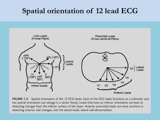
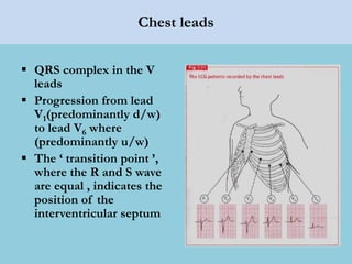
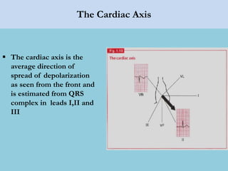
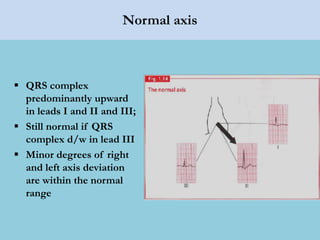
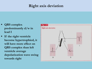
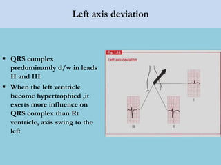
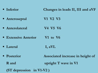
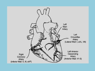
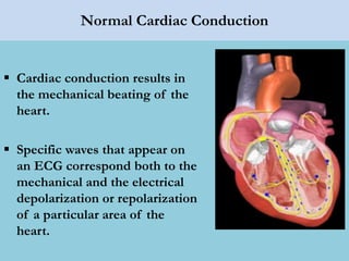
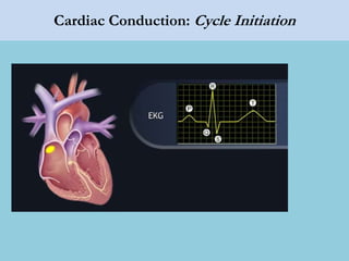
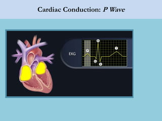
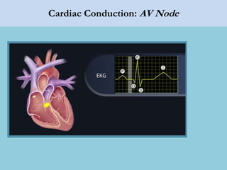
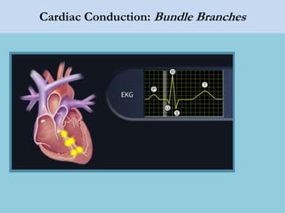
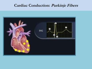
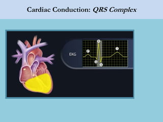
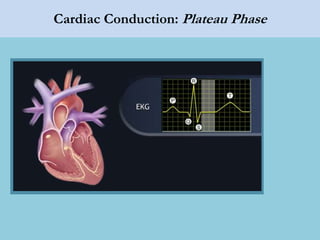
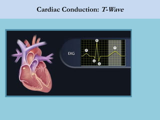
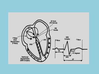
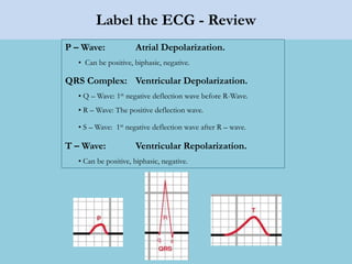
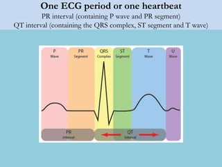
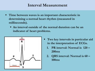
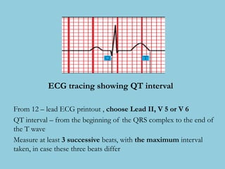
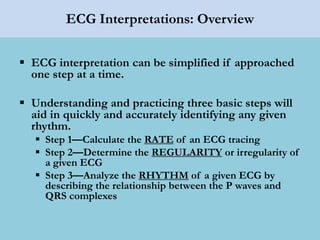
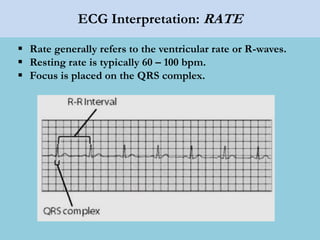
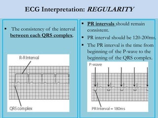
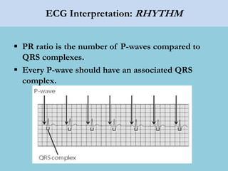
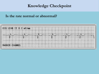
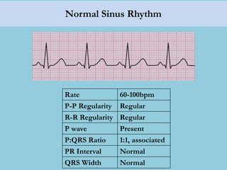
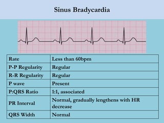
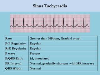
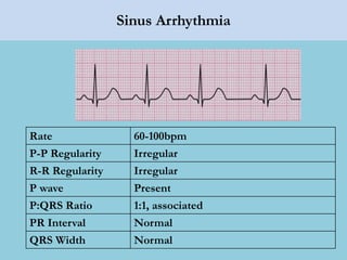
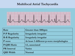
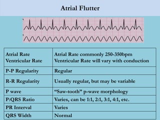
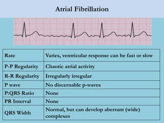
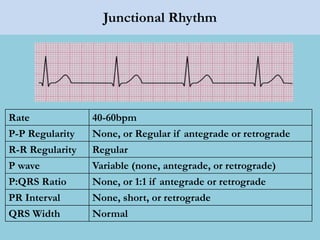
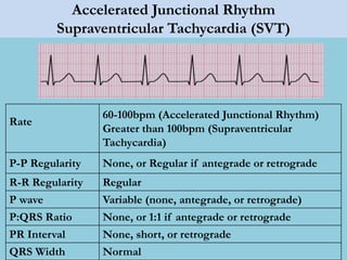
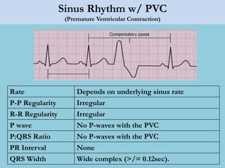
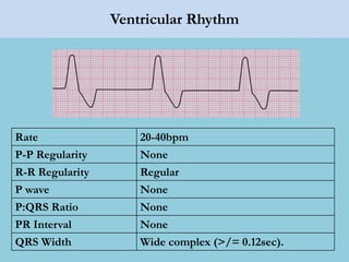
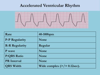
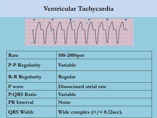
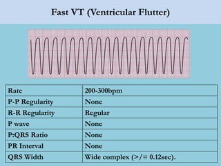
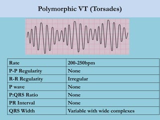
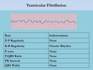
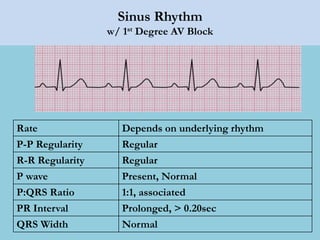
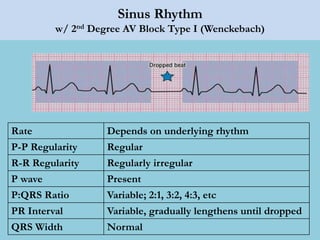
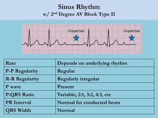
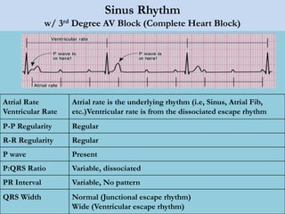
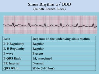
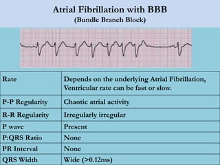
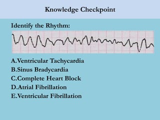
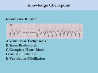
BASIC ECG INTERPRETATION
Department of Cardiology
Yangon General Hospital
Spatial orientation of 12 lead ECG
Chest leads
 QRS complex in the V
leads
 Progression from lead
V1(predominantly d/w)
to lead V6 where
(predominantly u/w)
 The ‘ transition point ’,
where the R and S wave
are equal , indicates the
position of the
interventricular septum
The Cardiac Axis
 The cardiac axis is the
average direction of
spread of depolarization
as seen from the front and
is estimated from QRS
complex in leads I,II and
III
Normal axis
 QRS complex
predominantly upward
in leads I and II and III;
 Still normal if QRS
complex d/w in lead III
 Minor degrees of right
and left axis deviation
are within the normal
range
Right axis deviation
 QRS complex
predominantly d/w in
lead I
 If the right ventricle
become hypertrophied, it
will have more effect on
QRS complex than left
ventricle average
depolarization wave swing
towards right
Left axis deviation
 QRS complex
predominantly d/w in leads
II and III
 When the left ventricle
become hypertrophied ,it
exerts more influence on
QRS complex than Rt
ventricle, axis swing to the
left
 Inferior Changes in leads II, III and aVF
 Anteroseptal V1 V2 V3
 Anterolateral V4 V5 V6
 Extensive Anterior V1 to V6
 Lateral I, aVL
 Posterior Associated increase in height of
R and upright T wave in V1
(ST depression in V1-V2 )
Normal Cardiac Conduction
 Cardiac conduction results in
the mechanical beating of the
heart.
 Specific waves that appear on
an ECG correspond both to the
mechanical and the electrical
depolarization or repolarization
of a particular area of the
heart.
Cardiac Conduction: Cycle Initiation
Cardiac Conduction: P Wave
Cardiac Conduction: AV Node
Cardiac Conduction: Bundle Branches
Cardiac Conduction: Purkinje Fibers
Cardiac Conduction: QRS Complex
Cardiac Conduction: Plateau Phase
Cardiac Conduction: T-Wave
Label the ECG - Review
P – Wave: Atrial Depolarization.
• Can be positive, biphasic, negative.
QRS Complex: Ventricular Depolarization.
• Q – Wave: 1st negative deflection wave before R-Wave.
• R – Wave: The positive deflection wave.
• S – Wave: 1st negative deflection wave after R – wave.
T – Wave: Ventricular Repolarization.
• Can be positive, biphasic, negative.
One ECG period or one heartbeat
PR interval (containing P wave and PR segment)
QT interval (containing the QRS complex, ST segment and T wave)
Interval Measurement
 Time between waves is an important characteristic in
determining a normal heart rhythm (measured in
milliseconds).
 An interval outside of the normal duration can be an
indicator of heart problems.
 Two key intervals in particular aid
in the interpretation of ECGs.
1. PR interval: Normal is 120 –
200ms
2. QRS interval: Normal is 60 –
100ms
ECG tracing showing QT interval
From 12 – lead ECG printout , choose Lead II, V 5 or V 6
QT interval – from the beginning of the QRS complex to the end of
the T wave
Measure at least 3 successive beats, with the maximum interval
taken, in case these three beats differ
Reading ECG Squares
Intervals and Timing
Horizontal axis – time:
 Each small square = 40
ms
 Each block = 200 ms (5
ea. 40 ms squares)
Converting this to a rate in
bpm:
 1 min = 60,000 ms, so:
 60,000/ms = bpm
– 60,000/600ms = 100
bpm
Calculating Heart Rate
1) Measure Cycle Length (CL).
1) (# small boxes from R – R) (40ms) = CL .
2) Calculate HR
• 60,000/CL = “x” BPM
(20)(40ms) = 800ms
60,000/800 = 75 bpm
(25)(40ms) = 1000ms
60,000/1000 = 60 bpm
(12)(40ms) = 480ms
60,000/480 = 125 bpm
Rate – 6 second Rule
1) Count the # of complexes on a 6 second strip.
2) Multiply by 10 to get approx “bpm”
0 1 2 3 4 5 6 7
Approximately 70bpm
ECG Interpretations: Overview
 ECG interpretation can be simplified if approached
one step at a time.
 Understanding and practicing three basic steps will
aid in quickly and accurately identifying any given
rhythm.
 Step 1—Calculate the RATE of an ECG tracing
 Step 2—Determine the REGULARITY or irregularity of
a given ECG
 Step 3—Analyze the RHYTHM of a given ECG by
describing the relationship between the P waves and
QRS complexes
ECG Interpretation: RATE
 Rate generally refers to the ventricular rate or R-waves.
 Resting rate is typically 60 – 100 bpm.
 Focus is placed on the QRS complex.
ECG Interpretation: REGULARITY
 The consistency of the interval
between each QRS complex.
 PR intervals should remain
consistent.
 PR interval should be 120-200ms.
 The PR interval is the time from
beginning of the P-wave to the
beginning of the QRS complex.
ECG Interpretation: RHYTHM
 PR ratio is the number of P-waves compared to
QRS complexes.
 Every P-wave should have an associated QRS
complex.
Knowledge Checkpoint
Is the rate normal or abnormal?
Normal Sinus Rhythm
Rate 60-100bpm
P-P Regularity Regular
R-R Regularity Regular
P wave Present
P:QRS Ratio 1:1, associated
PR Interval Normal
QRS Width Normal
Sinus Bradycardia
Rate Less than 60bpm
P-P Regularity Regular
R-R Regularity Regular
P wave Present
P:QRS Ratio 1:1, associated
PR Interval
Normal, gradually lengthens with HR
decrease
QRS Width Normal
Sinus Tachycardia
Rate Greater than 100bpm, Gradual onset
P-P Regularity Regular
R-R Regularity Regular
P wave Present
P:QRS Ratio 1:1, associated
PR Interval Normal, gradually shortens with HR increase
QRS Width Normal
Sinus Arrhythmia
Rate 60-100bpm
P-P Regularity Irregular
R-R Regularity Irregular
P wave Present
P:QRS Ratio 1:1, associated
PR Interval Normal
QRS Width Normal
Sinus Pause/Arrest
Rate Varies
P-P Regularity Irregular
R-R Regularity Irregular
P wave Present, except during pause
P:QRS Ratio 1:1, associated
PR Interval Normal
QRS Width Normal
Sinus Node Exit Block
Rate Varies
P-P Regularity Irregular
R-R Regularity Irregular
P wave Present, except during dropped beats
P:QRS Ratio 1:1, associated
PR Interval Normal
QRS Width Normal
Sinus Rhythm w/ PAC
(Premature Atrial Contraction)
Rate Depends on underlying sinus rate
P-P Regularity Irregular
R-R Regularity Irregular
P wave Present, may be different morphology during PAC
P:QRS Ratio 1:1, associated
PR Interval Normal, varies during PAC
QRS Width Normal
Atrial Tachycardia
Rate 100-180bpm, Sudden onset
P-P Regularity Regular
R-R Regularity Regular
P wave Morphology will differ from sinus p-wave
P:QRS Ratio 1:1, associated
PR Interval Interval of ectopic focus will differ from sinus PR
QRS Width
Normal, but can develop aberrant (wide)
complexes
Multifocal Atrial Tachycardia
Rate Greater than 100bpm
P-P Regularity Irregularly irregular
R-R Regularity Irregularly irregular
P wave At least 3 different p-wave morphologies
P:QRS Ratio 1:1, associated
PR Interval Varies
QRS Width Normal
Atrial Flutter
Atrial Rate
Ventricular Rate
Atrial Rate commonly 250-350bpm
Ventricular Rate will vary with conduction
P-P Regularity Regular
R-R Regularity Usually regular, but may be variable
P wave “Saw-tooth” p-wave morphology
P:QRS Ratio Varies, can be 1:1, 2:1, 3:1, 4:1, etc.
PR Interval Varies
QRS Width Normal
Atrial Fibrillation
Rate Varies, ventricular response can be fast or slow
P-P Regularity Chaotic atrial activity
R-R Regularity Irregularly irregular
P wave No discernable p-waves
P:QRS Ratio None
PR Interval None
QRS Width
Normal, but can develop aberrant (wide)
complexes
Junctional Rhythm
Rate 40-60bpm
P-P Regularity None, or Regular if antegrade or retrograde
R-R Regularity Regular
P wave Variable (none, antegrade, or retrograde)
P:QRS Ratio None, or 1:1 if antegrade or retrograde
PR Interval None, short, or retrograde
QRS Width Normal
Accelerated Junctional Rhythm
Supraventricular Tachycardia (SVT)
Rate
60-100bpm (Accelerated Junctional Rhythm)
Greater than 100bpm (Supraventricular
Tachycardia)
P-P Regularity None, or Regular if antegrade or retrograde
R-R Regularity Regular
P wave Variable (none, antegrade, or retrograde)
P:QRS Ratio None, or 1:1 if antegrade or retrograde
PR Interval None, short, or retrograde
QRS Width Normal
Sinus Rhythm w/ PVC
(Premature Ventricular Contraction)
Rate Depends on underlying sinus rate
P-P Regularity Irregular
R-R Regularity Irregular
P wave No P-waves with the PVC
P:QRS Ratio No P-waves with the PVC
PR Interval None
QRS Width Wide complex (>/= 0.12sec).
Ventricular Rhythm
Rate 20-40bpm
P-P Regularity None
R-R Regularity Regular
P wave None
P:QRS Ratio None
PR Interval None
QRS Width Wide complex (>/= 0.12sec).
Accelerated Ventricular Rhythm
Rate 40-100bpm
P-P Regularity None
R-R Regularity Regular
P wave None
P:QRS Ratio None
PR Interval None
QRS Width Wide complex (>/= 0.12sec).
Ventricular Tachycardia
Rate 100-200bpm
P-P Regularity Variable
R-R Regularity Regular
P wave Dissociated atrial rate
P:QRS Ratio Variable
PR Interval None
QRS Width Wide complex (>/= 0.12sec).
Fast VT (Ventricular Flutter)
Rate 200-300bpm
P-P Regularity None
R-R Regularity Regular
P wave None
P:QRS Ratio None
PR Interval None
QRS Width Wide complex (>/= 0.12sec).
Polymorphic VT (Torsades)
Rate 200-250bpm
P-P Regularity None
R-R Regularity Irregular
P wave None
P:QRS Ratio None
PR Interval None
QRS Width Variable with wide complexes
Ventricular Fibrillation
Rate Indeterminate
P-P Regularity None
R-R Regularity Chaotic Rhythm
P wave None
P:QRS Ratio None
PR Interval None
QRS Width None
Sinus Rhythm
w/ 1st Degree AV Block
Rate Depends on underlying rhythm
P-P Regularity Regular
R-R Regularity Regular
P wave Present, Normal
P:QRS Ratio 1:1, associated
PR Interval Prolonged, > 0.20sec
QRS Width Normal
Sinus Rhythm
w/ 2nd Degree AV Block Type I (Wenckebach)
Rate Depends on underlying rhythm
P-P Regularity Regular
R-R Regularity Regularly irregular
P wave Present
P:QRS Ratio Variable; 2:1, 3:2, 4:3, etc
PR Interval Variable, gradually lengthens until dropped
QRS Width Normal
Sinus Rhythm
w/ 2nd Degree AV Block Type II
Rate Depends on underlying rhythm
P-P Regularity Regular
R-R Regularity Regularly irregular
P wave Present
P:QRS Ratio Variable; 2:1, 3:2, 4:3, etc
PR Interval Normal for conducted beats
QRS Width Normal
Sinus Rhythm
w/ 3rd Degree AV Block (Complete Heart Block)
Atrial Rate
Ventricular Rate
Atrial rate is the underlying rhythm (i.e, Sinus, Atrial Fib,
etc.)Ventricular rate is from the dissociated escape rhythm
P-P Regularity Regular
R-R Regularity Regular
P wave Present
P:QRS Ratio Variable, dissociated
PR Interval Variable, No pattern
QRS Width Normal (Junctional escape rhythm)
Wide (Ventricular escape rhythm)
Sinus Rhythm w/ BBB
(Bundle Branch Block)
Rate Depends on the underlying sinus rhythm
P-P Regularity Regular
R-R Regularity Regular
P wave Present
P:QRS Ratio 1:1, associated
PR Interval Normal
QRS Width Wide (>0.12ms)
Atrial Fibrillation with BBB
(Bundle Branch Block)
Rate Depends on the underlying Atrial Fibrillation,
Ventricular rate can be fast or slow.
P-P Regularity Chaotic atrial activity
R-R Regularity Irregularly irregular
P wave Present
P:QRS Ratio None
PR Interval None
QRS Width Wide (>0.12ms)
Knowledge Checkpoint
Identify the Rhythm:
A.Ventricular Tachycardia
B.Sinus Bradycardia
C.Complete Heart Block
D.Atrial Fibrillation
E.Ventricular Fibrillation
Knowledge Checkpoint
Identify the Rhythm:
A.Ventricular Tachycardia
B.Sinus Bradycardia
C.Complete Heart Block
D.Atrial Fibrillation
E.Ventricular Fibrillation
Knowledge Checkpoint
Identify the Rhythm:
A.Ventricular Tachycardia
B.Sinus Bradycardia
C.Complete Heart Block
D.Atrial Fibrillation
E.Ventricular Fibrillation
Knowledge Checkpoint
Identify the Rhythm:
A.Ventricular Tachycardia
B.Sinus Bradycardia
C.Complete Heart Block
D.Atrial Fibrillation
E.Ventricular Fibrillation
The End

Lec 14 basic ecg interpretation for mohs

  • 1.
    BASIC ECG INTERPRETATION Departmentof Cardiology Yangon General Hospital
  • 2.
  • 3.
    Chest leads  QRScomplex in the V leads  Progression from lead V1(predominantly d/w) to lead V6 where (predominantly u/w)  The ‘ transition point ’, where the R and S wave are equal , indicates the position of the interventricular septum
  • 4.
    The Cardiac Axis The cardiac axis is the average direction of spread of depolarization as seen from the front and is estimated from QRS complex in leads I,II and III
  • 5.
    Normal axis  QRScomplex predominantly upward in leads I and II and III;  Still normal if QRS complex d/w in lead III  Minor degrees of right and left axis deviation are within the normal range
  • 6.
    Right axis deviation QRS complex predominantly d/w in lead I  If the right ventricle become hypertrophied, it will have more effect on QRS complex than left ventricle average depolarization wave swing towards right
  • 7.
    Left axis deviation QRS complex predominantly d/w in leads II and III  When the left ventricle become hypertrophied ,it exerts more influence on QRS complex than Rt ventricle, axis swing to the left
  • 8.
     Inferior Changesin leads II, III and aVF  Anteroseptal V1 V2 V3  Anterolateral V4 V5 V6  Extensive Anterior V1 to V6  Lateral I, aVL  Posterior Associated increase in height of R and upright T wave in V1 (ST depression in V1-V2 )
  • 10.
    Normal Cardiac Conduction Cardiac conduction results in the mechanical beating of the heart.  Specific waves that appear on an ECG correspond both to the mechanical and the electrical depolarization or repolarization of a particular area of the heart.
  • 11.
  • 12.
  • 13.
  • 14.
  • 15.
  • 16.
  • 17.
  • 18.
  • 20.
    Label the ECG- Review P – Wave: Atrial Depolarization. • Can be positive, biphasic, negative. QRS Complex: Ventricular Depolarization. • Q – Wave: 1st negative deflection wave before R-Wave. • R – Wave: The positive deflection wave. • S – Wave: 1st negative deflection wave after R – wave. T – Wave: Ventricular Repolarization. • Can be positive, biphasic, negative.
  • 21.
    One ECG periodor one heartbeat PR interval (containing P wave and PR segment) QT interval (containing the QRS complex, ST segment and T wave)
  • 22.
    Interval Measurement  Timebetween waves is an important characteristic in determining a normal heart rhythm (measured in milliseconds).  An interval outside of the normal duration can be an indicator of heart problems.  Two key intervals in particular aid in the interpretation of ECGs. 1. PR interval: Normal is 120 – 200ms 2. QRS interval: Normal is 60 – 100ms
  • 23.
    ECG tracing showingQT interval From 12 – lead ECG printout , choose Lead II, V 5 or V 6 QT interval – from the beginning of the QRS complex to the end of the T wave Measure at least 3 successive beats, with the maximum interval taken, in case these three beats differ
  • 24.
    Reading ECG Squares Intervalsand Timing Horizontal axis – time:  Each small square = 40 ms  Each block = 200 ms (5 ea. 40 ms squares) Converting this to a rate in bpm:  1 min = 60,000 ms, so:  60,000/ms = bpm – 60,000/600ms = 100 bpm
  • 25.
    Calculating Heart Rate 1)Measure Cycle Length (CL). 1) (# small boxes from R – R) (40ms) = CL . 2) Calculate HR • 60,000/CL = “x” BPM (20)(40ms) = 800ms 60,000/800 = 75 bpm (25)(40ms) = 1000ms 60,000/1000 = 60 bpm (12)(40ms) = 480ms 60,000/480 = 125 bpm
  • 26.
    Rate – 6second Rule 1) Count the # of complexes on a 6 second strip. 2) Multiply by 10 to get approx “bpm” 0 1 2 3 4 5 6 7 Approximately 70bpm
  • 27.
    ECG Interpretations: Overview ECG interpretation can be simplified if approached one step at a time.  Understanding and practicing three basic steps will aid in quickly and accurately identifying any given rhythm.  Step 1—Calculate the RATE of an ECG tracing  Step 2—Determine the REGULARITY or irregularity of a given ECG  Step 3—Analyze the RHYTHM of a given ECG by describing the relationship between the P waves and QRS complexes
  • 28.
    ECG Interpretation: RATE Rate generally refers to the ventricular rate or R-waves.  Resting rate is typically 60 – 100 bpm.  Focus is placed on the QRS complex.
  • 29.
    ECG Interpretation: REGULARITY The consistency of the interval between each QRS complex.  PR intervals should remain consistent.  PR interval should be 120-200ms.  The PR interval is the time from beginning of the P-wave to the beginning of the QRS complex.
  • 30.
    ECG Interpretation: RHYTHM PR ratio is the number of P-waves compared to QRS complexes.  Every P-wave should have an associated QRS complex.
  • 31.
    Knowledge Checkpoint Is therate normal or abnormal?
  • 32.
    Normal Sinus Rhythm Rate60-100bpm P-P Regularity Regular R-R Regularity Regular P wave Present P:QRS Ratio 1:1, associated PR Interval Normal QRS Width Normal
  • 33.
    Sinus Bradycardia Rate Lessthan 60bpm P-P Regularity Regular R-R Regularity Regular P wave Present P:QRS Ratio 1:1, associated PR Interval Normal, gradually lengthens with HR decrease QRS Width Normal
  • 34.
    Sinus Tachycardia Rate Greaterthan 100bpm, Gradual onset P-P Regularity Regular R-R Regularity Regular P wave Present P:QRS Ratio 1:1, associated PR Interval Normal, gradually shortens with HR increase QRS Width Normal
  • 35.
    Sinus Arrhythmia Rate 60-100bpm P-PRegularity Irregular R-R Regularity Irregular P wave Present P:QRS Ratio 1:1, associated PR Interval Normal QRS Width Normal
  • 36.
    Sinus Pause/Arrest Rate Varies P-PRegularity Irregular R-R Regularity Irregular P wave Present, except during pause P:QRS Ratio 1:1, associated PR Interval Normal QRS Width Normal
  • 37.
    Sinus Node ExitBlock Rate Varies P-P Regularity Irregular R-R Regularity Irregular P wave Present, except during dropped beats P:QRS Ratio 1:1, associated PR Interval Normal QRS Width Normal
  • 38.
    Sinus Rhythm w/PAC (Premature Atrial Contraction) Rate Depends on underlying sinus rate P-P Regularity Irregular R-R Regularity Irregular P wave Present, may be different morphology during PAC P:QRS Ratio 1:1, associated PR Interval Normal, varies during PAC QRS Width Normal
  • 39.
    Atrial Tachycardia Rate 100-180bpm,Sudden onset P-P Regularity Regular R-R Regularity Regular P wave Morphology will differ from sinus p-wave P:QRS Ratio 1:1, associated PR Interval Interval of ectopic focus will differ from sinus PR QRS Width Normal, but can develop aberrant (wide) complexes
  • 40.
    Multifocal Atrial Tachycardia RateGreater than 100bpm P-P Regularity Irregularly irregular R-R Regularity Irregularly irregular P wave At least 3 different p-wave morphologies P:QRS Ratio 1:1, associated PR Interval Varies QRS Width Normal
  • 41.
    Atrial Flutter Atrial Rate VentricularRate Atrial Rate commonly 250-350bpm Ventricular Rate will vary with conduction P-P Regularity Regular R-R Regularity Usually regular, but may be variable P wave “Saw-tooth” p-wave morphology P:QRS Ratio Varies, can be 1:1, 2:1, 3:1, 4:1, etc. PR Interval Varies QRS Width Normal
  • 42.
    Atrial Fibrillation Rate Varies,ventricular response can be fast or slow P-P Regularity Chaotic atrial activity R-R Regularity Irregularly irregular P wave No discernable p-waves P:QRS Ratio None PR Interval None QRS Width Normal, but can develop aberrant (wide) complexes
  • 43.
    Junctional Rhythm Rate 40-60bpm P-PRegularity None, or Regular if antegrade or retrograde R-R Regularity Regular P wave Variable (none, antegrade, or retrograde) P:QRS Ratio None, or 1:1 if antegrade or retrograde PR Interval None, short, or retrograde QRS Width Normal
  • 44.
    Accelerated Junctional Rhythm SupraventricularTachycardia (SVT) Rate 60-100bpm (Accelerated Junctional Rhythm) Greater than 100bpm (Supraventricular Tachycardia) P-P Regularity None, or Regular if antegrade or retrograde R-R Regularity Regular P wave Variable (none, antegrade, or retrograde) P:QRS Ratio None, or 1:1 if antegrade or retrograde PR Interval None, short, or retrograde QRS Width Normal
  • 45.
    Sinus Rhythm w/PVC (Premature Ventricular Contraction) Rate Depends on underlying sinus rate P-P Regularity Irregular R-R Regularity Irregular P wave No P-waves with the PVC P:QRS Ratio No P-waves with the PVC PR Interval None QRS Width Wide complex (>/= 0.12sec).
  • 46.
    Ventricular Rhythm Rate 20-40bpm P-PRegularity None R-R Regularity Regular P wave None P:QRS Ratio None PR Interval None QRS Width Wide complex (>/= 0.12sec).
  • 47.
    Accelerated Ventricular Rhythm Rate40-100bpm P-P Regularity None R-R Regularity Regular P wave None P:QRS Ratio None PR Interval None QRS Width Wide complex (>/= 0.12sec).
  • 48.
    Ventricular Tachycardia Rate 100-200bpm P-PRegularity Variable R-R Regularity Regular P wave Dissociated atrial rate P:QRS Ratio Variable PR Interval None QRS Width Wide complex (>/= 0.12sec).
  • 49.
    Fast VT (VentricularFlutter) Rate 200-300bpm P-P Regularity None R-R Regularity Regular P wave None P:QRS Ratio None PR Interval None QRS Width Wide complex (>/= 0.12sec).
  • 50.
    Polymorphic VT (Torsades) Rate200-250bpm P-P Regularity None R-R Regularity Irregular P wave None P:QRS Ratio None PR Interval None QRS Width Variable with wide complexes
  • 51.
    Ventricular Fibrillation Rate Indeterminate P-PRegularity None R-R Regularity Chaotic Rhythm P wave None P:QRS Ratio None PR Interval None QRS Width None
  • 52.
    Sinus Rhythm w/ 1stDegree AV Block Rate Depends on underlying rhythm P-P Regularity Regular R-R Regularity Regular P wave Present, Normal P:QRS Ratio 1:1, associated PR Interval Prolonged, > 0.20sec QRS Width Normal
  • 53.
    Sinus Rhythm w/ 2ndDegree AV Block Type I (Wenckebach) Rate Depends on underlying rhythm P-P Regularity Regular R-R Regularity Regularly irregular P wave Present P:QRS Ratio Variable; 2:1, 3:2, 4:3, etc PR Interval Variable, gradually lengthens until dropped QRS Width Normal
  • 54.
    Sinus Rhythm w/ 2ndDegree AV Block Type II Rate Depends on underlying rhythm P-P Regularity Regular R-R Regularity Regularly irregular P wave Present P:QRS Ratio Variable; 2:1, 3:2, 4:3, etc PR Interval Normal for conducted beats QRS Width Normal
  • 55.
    Sinus Rhythm w/ 3rdDegree AV Block (Complete Heart Block) Atrial Rate Ventricular Rate Atrial rate is the underlying rhythm (i.e, Sinus, Atrial Fib, etc.)Ventricular rate is from the dissociated escape rhythm P-P Regularity Regular R-R Regularity Regular P wave Present P:QRS Ratio Variable, dissociated PR Interval Variable, No pattern QRS Width Normal (Junctional escape rhythm) Wide (Ventricular escape rhythm)
  • 56.
    Sinus Rhythm w/BBB (Bundle Branch Block) Rate Depends on the underlying sinus rhythm P-P Regularity Regular R-R Regularity Regular P wave Present P:QRS Ratio 1:1, associated PR Interval Normal QRS Width Wide (>0.12ms)
  • 57.
    Atrial Fibrillation withBBB (Bundle Branch Block) Rate Depends on the underlying Atrial Fibrillation, Ventricular rate can be fast or slow. P-P Regularity Chaotic atrial activity R-R Regularity Irregularly irregular P wave Present P:QRS Ratio None PR Interval None QRS Width Wide (>0.12ms)
  • 58.
    Knowledge Checkpoint Identify theRhythm: A.Ventricular Tachycardia B.Sinus Bradycardia C.Complete Heart Block D.Atrial Fibrillation E.Ventricular Fibrillation
  • 59.
    Knowledge Checkpoint Identify theRhythm: A.Ventricular Tachycardia B.Sinus Bradycardia C.Complete Heart Block D.Atrial Fibrillation E.Ventricular Fibrillation
  • 60.
    Knowledge Checkpoint Identify theRhythm: A.Ventricular Tachycardia B.Sinus Bradycardia C.Complete Heart Block D.Atrial Fibrillation E.Ventricular Fibrillation
  • 61.
    Knowledge Checkpoint Identify theRhythm: A.Ventricular Tachycardia B.Sinus Bradycardia C.Complete Heart Block D.Atrial Fibrillation E.Ventricular Fibrillation
  • 62.