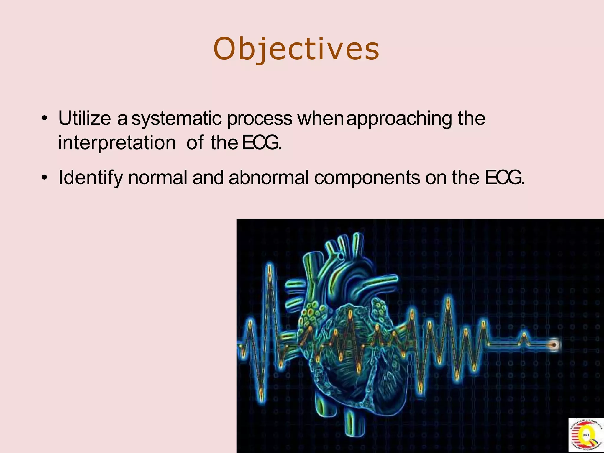
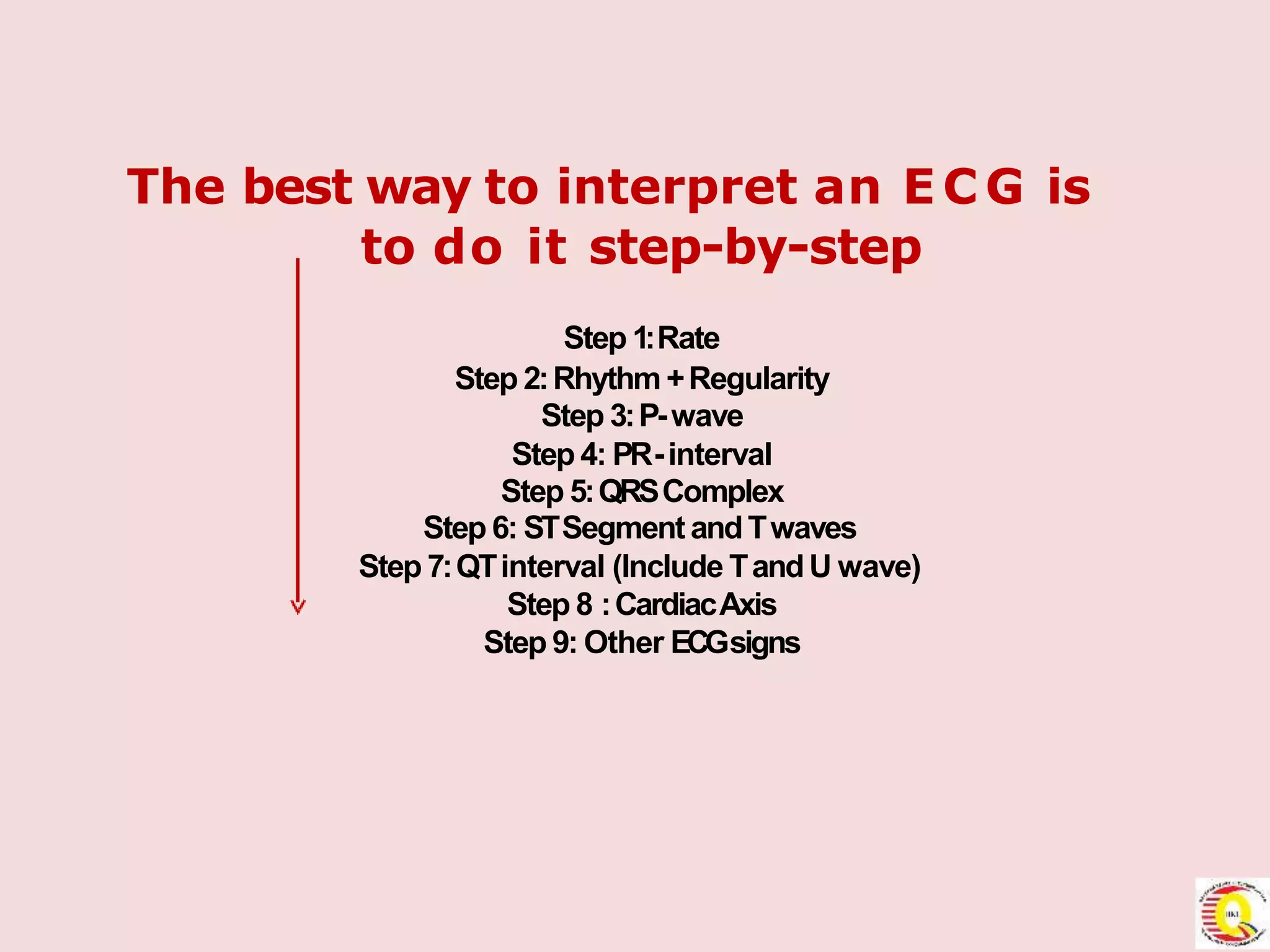
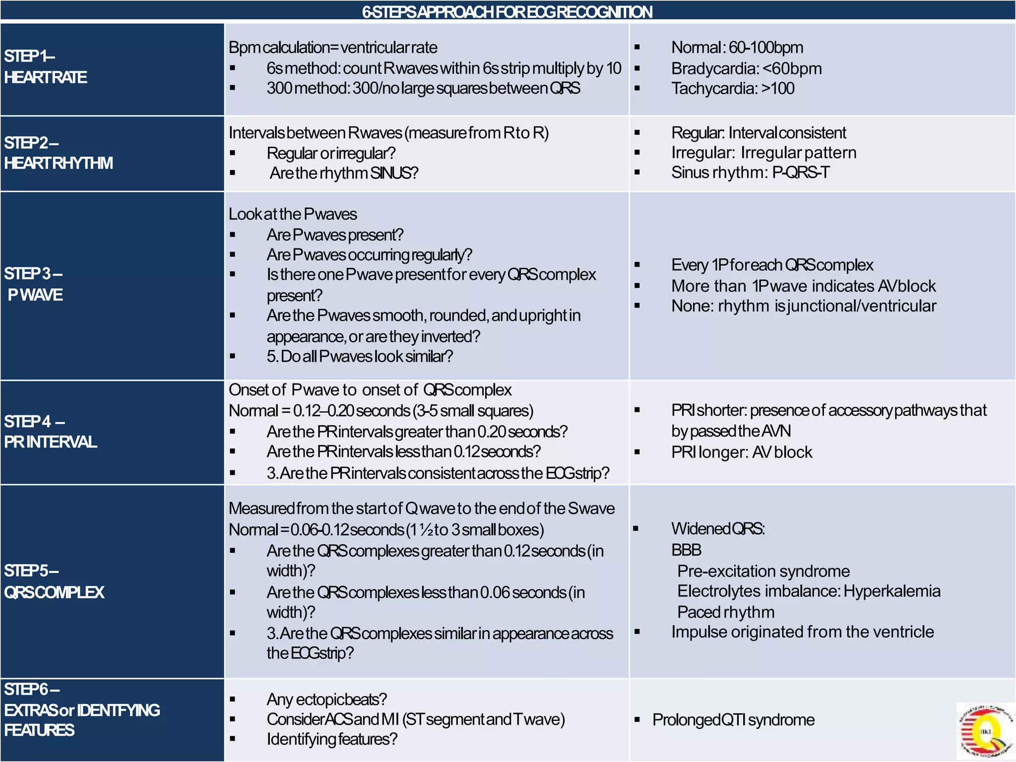
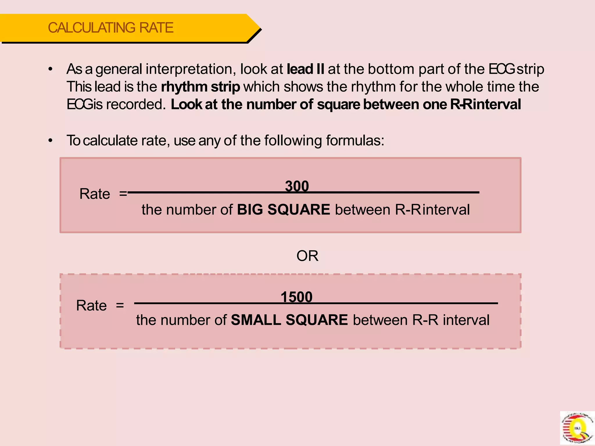
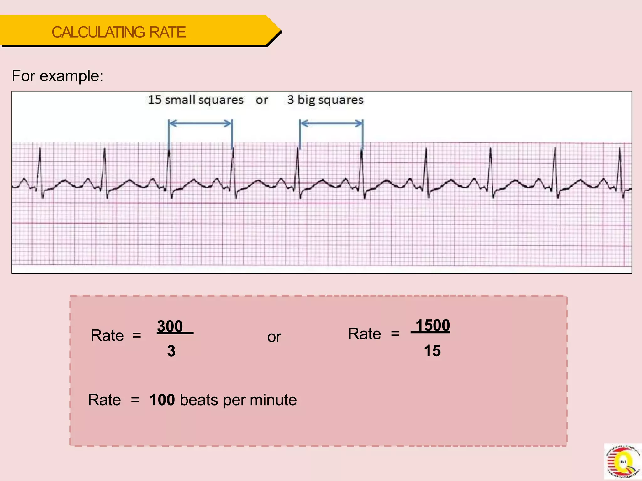
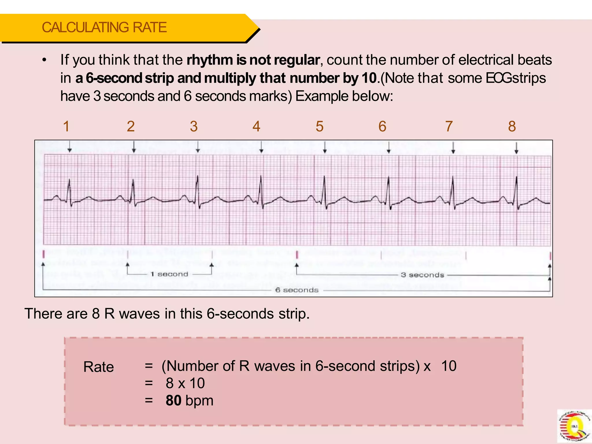
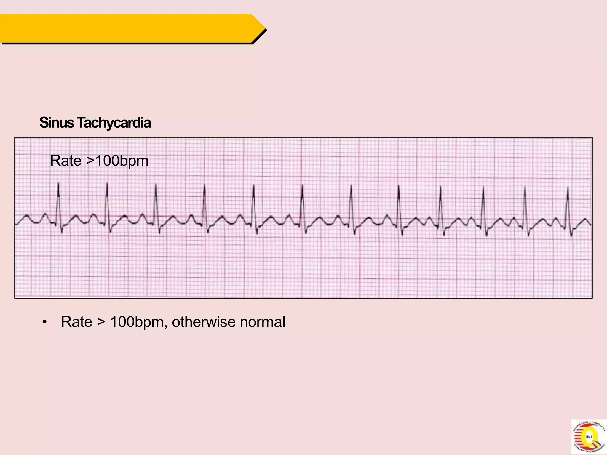
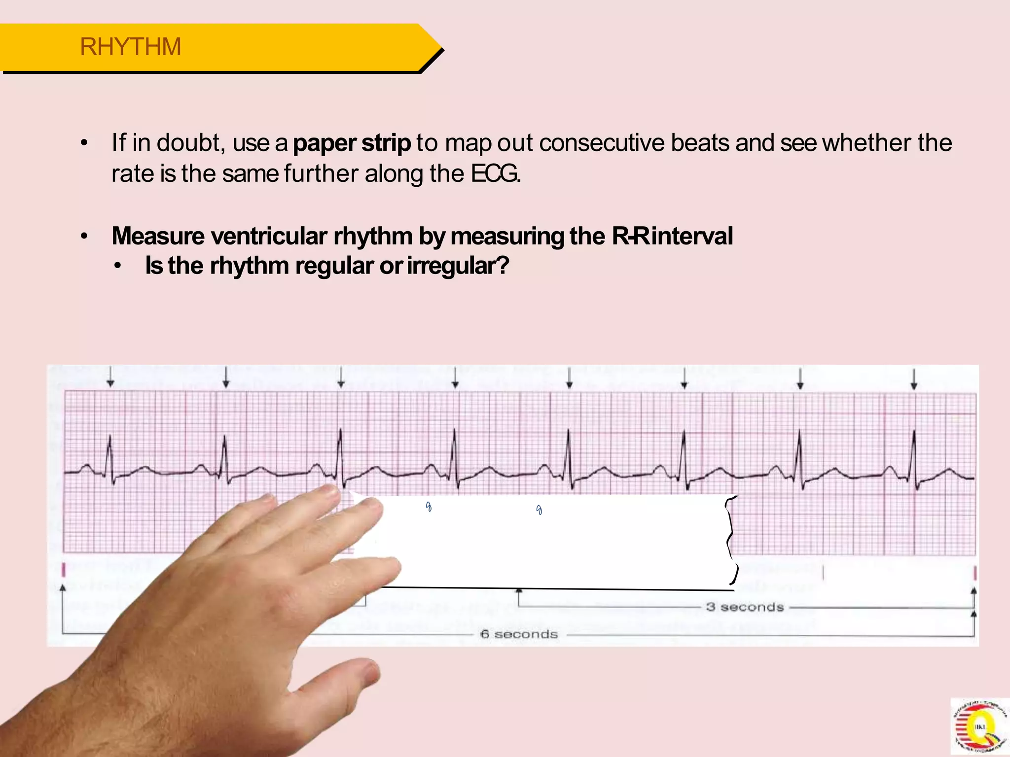
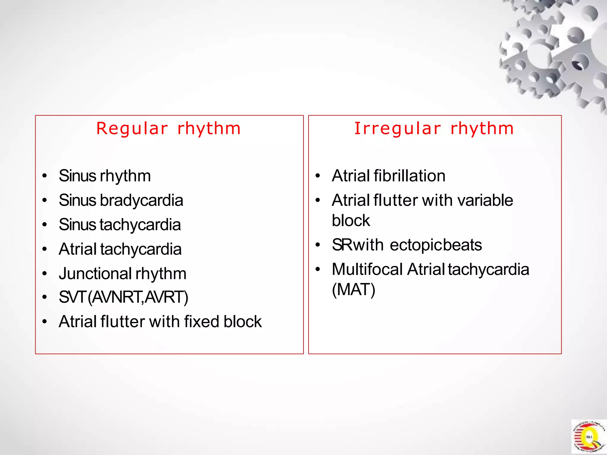
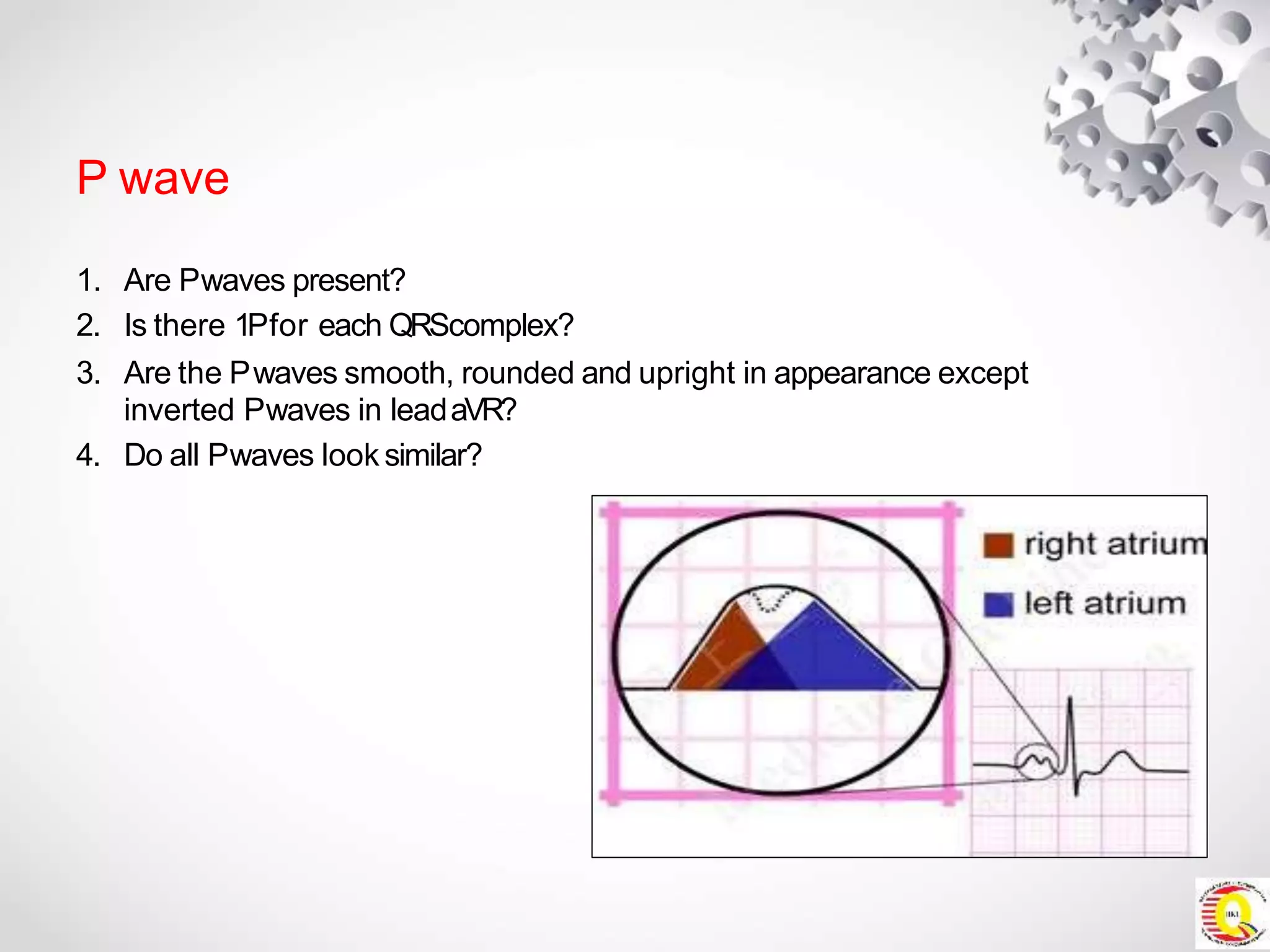
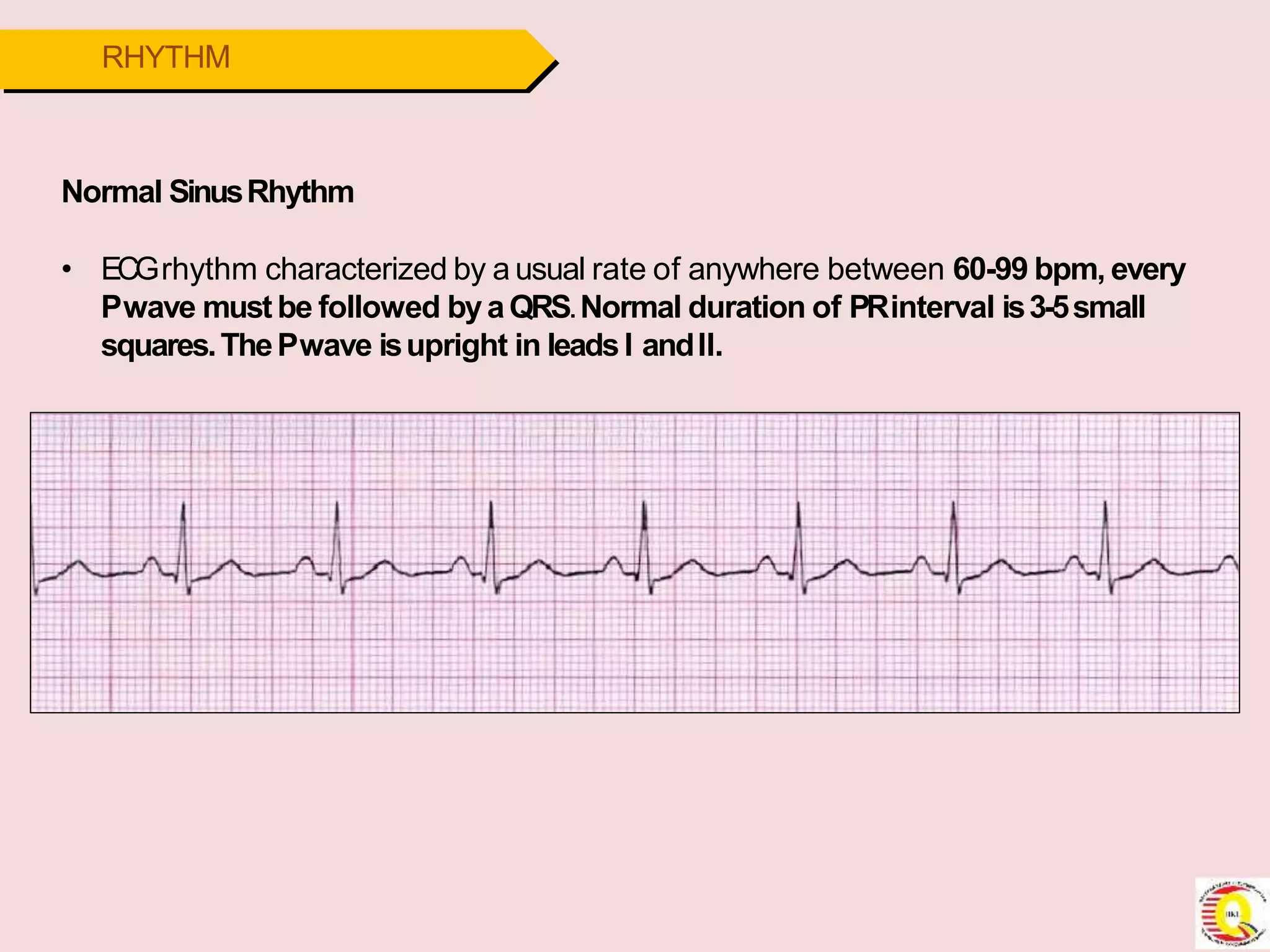
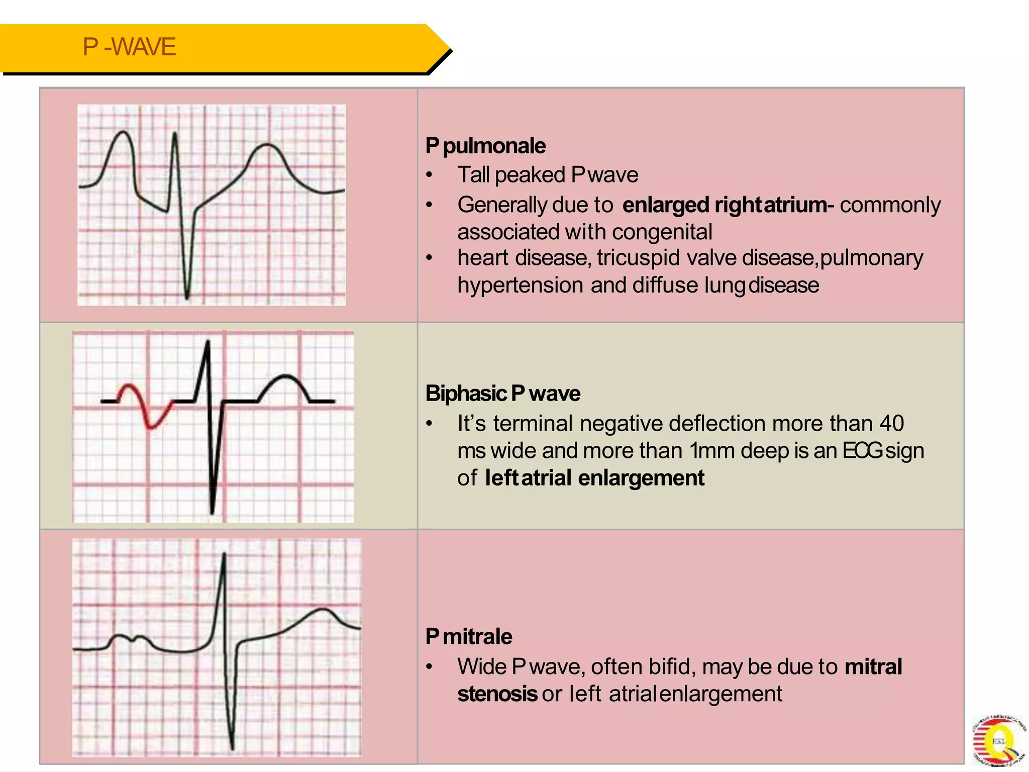
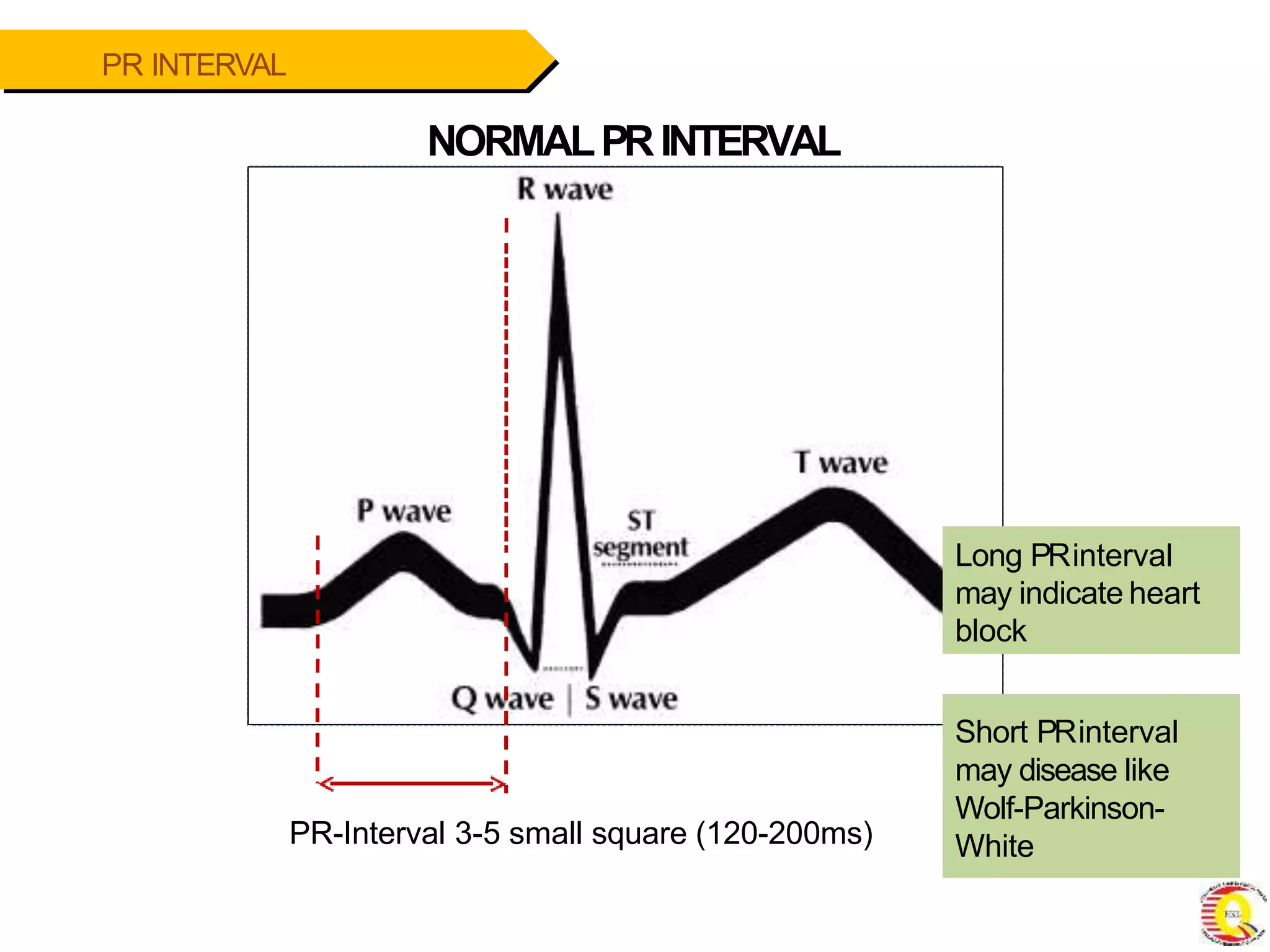
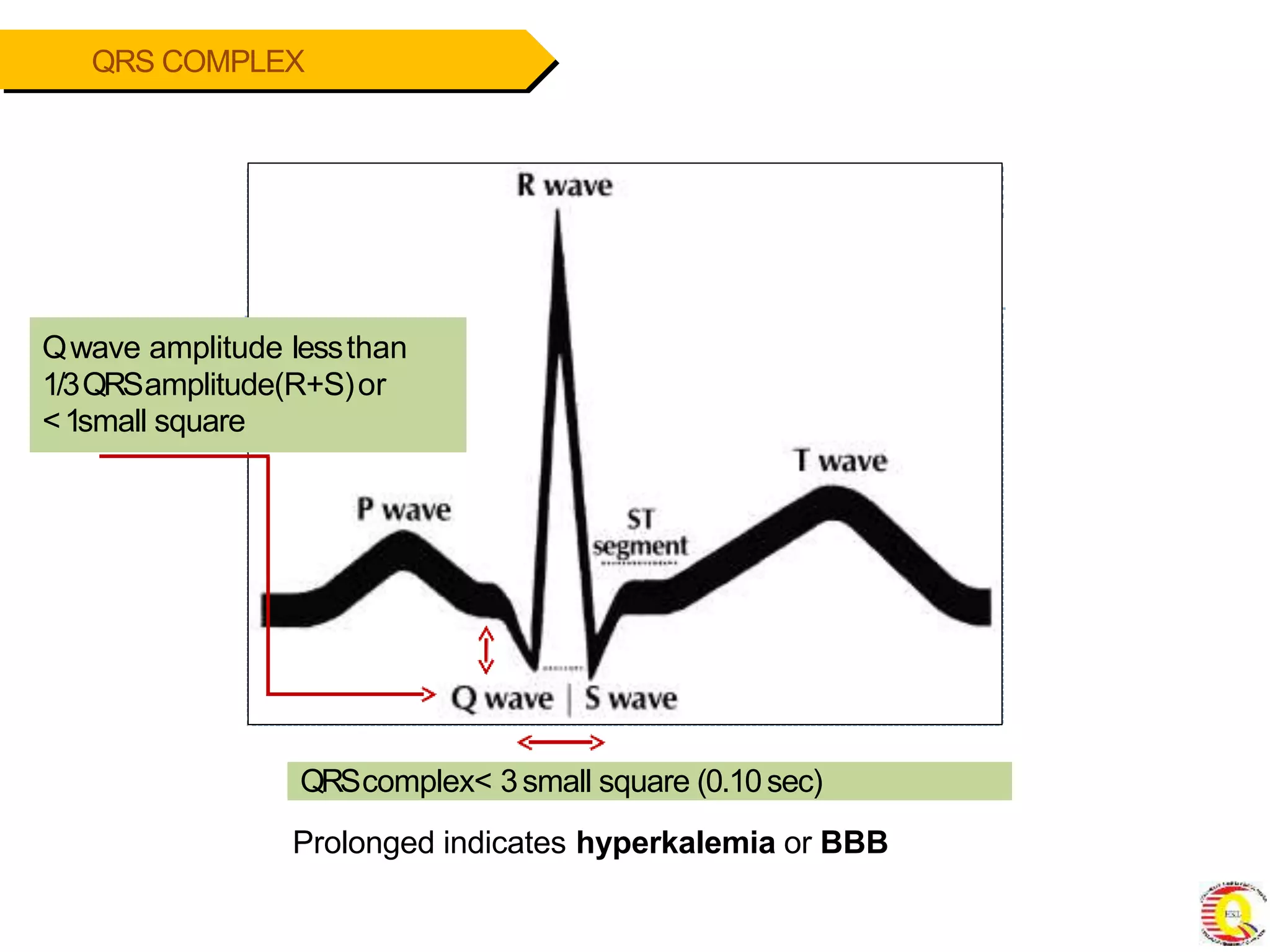
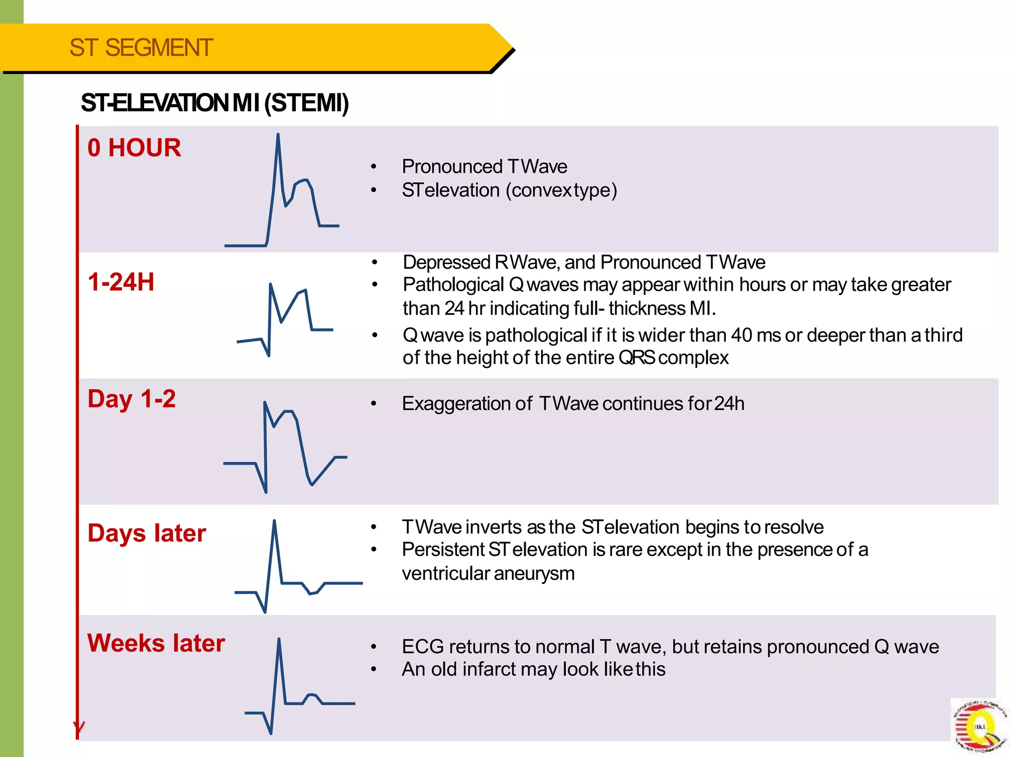
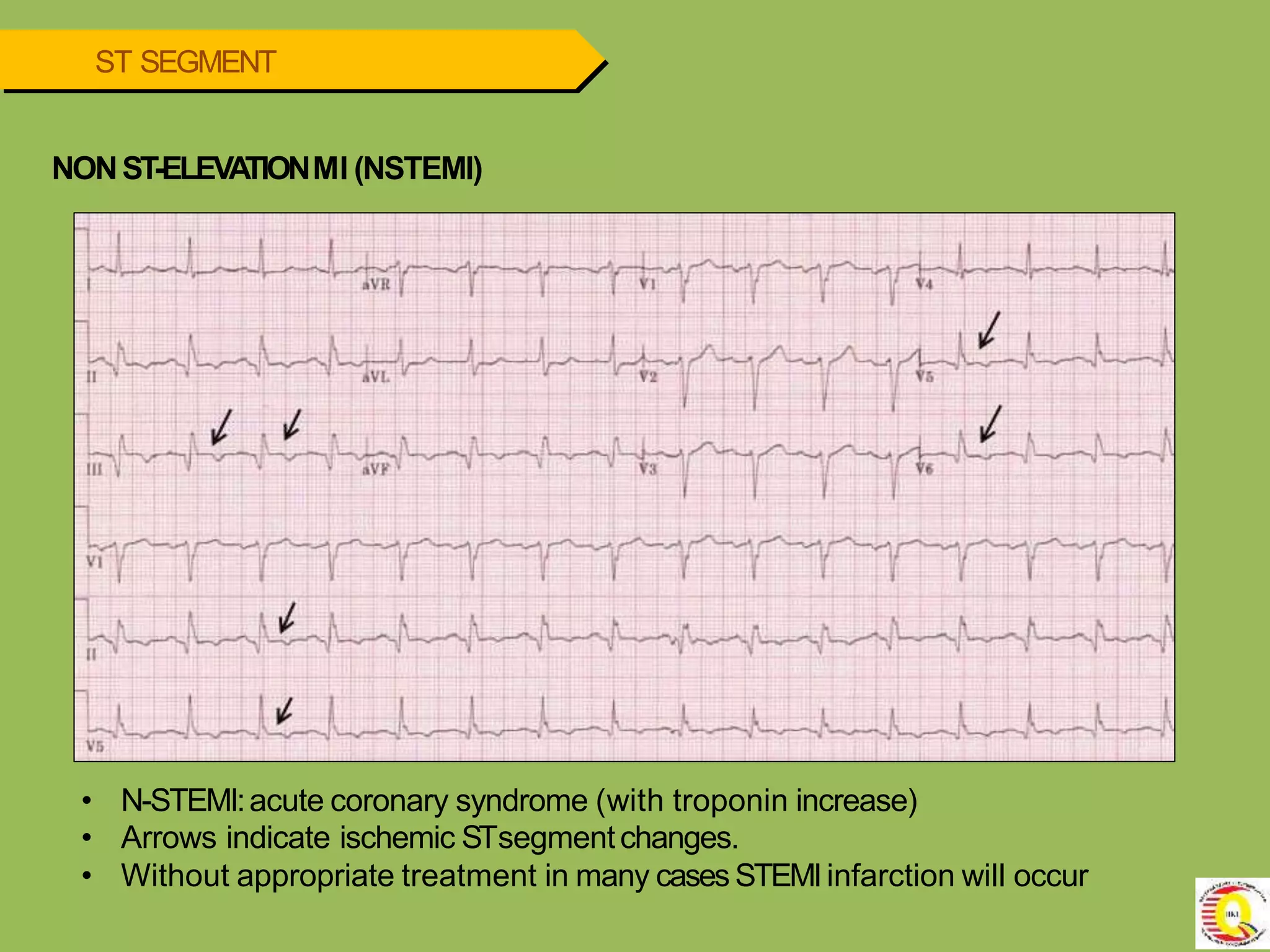
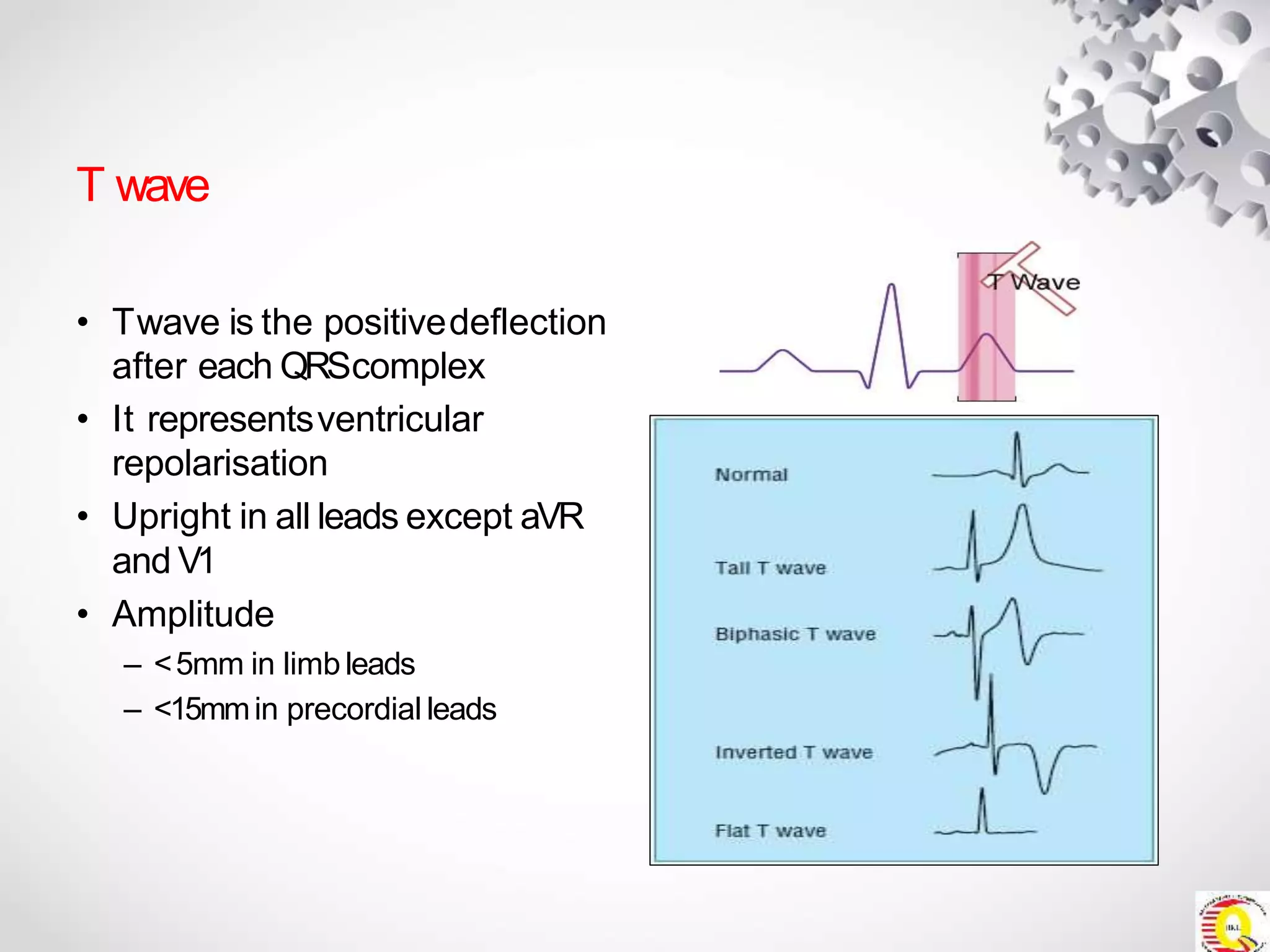
SlideShare a Scribd company logo
MODULE2
Interpreting E C G rhythm STRIPS
Objectives
• Utilize asystematic process whenapproaching the
interpretation of theECG.
• Identify normal and abnormal components on the ECG.
ECG
interpretation
Cardiac Anatomy
▪ 2upper chambers
▪ Right and left atria
▪ 2lower chambers
▪ Right and left ventricle
▪ 2Atrioventricular valves
(Mitral &Tricuspid)
▪ Open with ventriculardiastole
▪ Closewith ventricularsystole
▪ 2Semilunar Valves(Aortic &Pulmonic)
▪ Open with ventricularsystole
▪ Open with ventriculardiastole
Anatomy
Coronary Arteries
▪ 2major vessels of thecoronary circulation
▪ Left main coronaryartery
▪ Left anterior descending and circumflexbranches
▪ Right main coronaryartery
▪ Left and right coronary arteries originate at the base of the aorta
from openings called the coronary ostia behind the aortic valve
leaflets.
Anatomy Coronary Arteries
Normal Conduction System
Sinoatrial node
Internodalpathway
A
Vnode
Bundle of His
BundleBranches
Purkinjefibers
Pacemakers of the Heart
• SA Node -Dominant pacemakerwith an intrinsic rate of
60 -100beats/minute.
• AVNode -Back-up pacemaker with an intrinsic rate of
40 -60 beats/minute.
• Ventricular cells-Back-up pacemaker with an intrinsic rate
of 20 -45bpm.
ECGs?
Basic E C G Interpretation
STANDARD CALLIBRATION
Speed = 25mm/s
Amplitude = 0.1mV/mm
• 1large square: 0.2s(200ms)
1small square: 0.04s(40ms)
• 1mV:10mm high
• 0.1mV amplitude
OBTAINA NECG,ACT CONFIDENT,READ THE PT DETAILS
• Some ECGmachines come with interpretation software.
• Thisone says the patient is fine. DONOTtotally trust this software.
ECGLIMB LEADREVERSAL
• LeadI or aVLis completely inverted (P wave, QRScomplex andTwave).
• Lead aVRoften becomespositive.
• The P-wave is unexpectedly larger in lead I than lead II (it is usually the other way around).
The best way to interpret an EC G is
to do it step-by-step
Step 1:Rate
Step 2:Rhythm +Regularity
Step 3:P-wave
Step 4: PR-interval
Step 5:QRSComplex
Step 6: STSegment andTwaves
Step 7:QTinterval (Include TandU wave)
Step 8 :CardiacAxis
Step 9: Other ECGsigns
THE NORMAL SIZE
<3small square
<3-5 small square
<2large square
<2small square
PQRSTConfiguration
One completecardiac cycle =P
,Q, R, S, (QRS
complex), and Twave
P wave First deflectionRounded
and upright in lead II
Amplitude: 0.5-2.5mn
Duration: 0.10s
Atrial depolarization
PR Interval Beginning of Pwaveto
beginning of QRS
complex
Duration: 0.12s-0.2s
Atrial depolarization +conductiondelay
through AVjunction (AVN+ Bundle ofHis)
QRS Complex Beginning of Qwaveto
the end of Swave
Duration: 0.06-0.12s
Ventricular depolarization
ST Segment Between Swave tothe
beginning of Twave
Normal STsegmentis
flat (isoelectric)
Isoelectric, ventricle stilldepolarized
QT Interval Beginning of QRS
complex to the end of T
wave
Duration: ½of the
distance of RRinterval
Ventricular depolarization +repolarization
Mechanical contraction of ventricle
T Wave End of STsegmentto
the end ofTwave
Rounded and
asymmetrical
Upright in lead II
Amplitude: <5mm
Ventricular repolarization
6-STEPSAPPROACHFORECGRECOGNITION
STEP1–
HEARTRA
TE
Bpmcalculation=ventricularrate
 6smethod:countRwaveswithin6sstripmultiplyby10
 300method:300/nolargesquaresbetweenQRS
 Normal:60-100bpm
 Bradycardia:<60bpm
 Tachycardia:>100
STEP2–
HEARTRHYTHM
IntervalsbetweenRwaves(measurefromRtoR)
 Regularorirregular?
 AretherhythmSINUS?
 Regular: Intervalconsistent
 Irregular: Irregularpattern
 Sinus rhythm: P-QRS-T
STEP3–
PWAVE
LookatthePwaves
 ArePwavespresent?
 ArePwavesoccurringregularly?
 IsthereonePwavepresentforeveryQRScomplex
present?
 ArethePwavessmooth,rounded,anduprightin
appearance,oraretheyinverted?
 5.DoallPwaveslooksimilar?
 Every1PforeachQRScomplex
 More than 1Pwave indicates AVblock
 None: rhythm isjunctional/ventricular
STEP4 –
PRINTERVAL
Onset of Pwave to onset of QRScomplex
Normal=0.12–0.20seconds(3-5smallsquares)
 ArethePRintervalsgreaterthan0.20seconds?
 ArethePRintervalslessthan0.12seconds?
 3.ArethePRintervalsconsistentacrosstheECGstrip?
 PRIshorter:presenceof accessorypathwaysthat
bypassedtheAVN
 PRIlonger: AVblock
STEP5–
QRSCOMPLEX
Measuredfromthestartof Qwaveto theendof theSwave
Normal=0.06-0.12seconds(1½to 3smallboxes)
 AretheQRScomplexesgreaterthan0.12seconds(in
width)?
 AretheQRScomplexeslessthan0.06seconds(in
width)?
 3.AretheQRScomplexessimilarinappearanceacross
theECGstrip?
 WidenedQRS:
BBB
Pre-excitation syndrome
Electrolytes imbalance:Hyperkalemia
Pacedrhythm
 Impulse originated from the ventricle
STEP6–
EXTRASorIDENTFYING
FEA
TURES
 Anyectopicbeats?
 ConsiderACSandMI(STsegmentandTwave)
 Identifyingfeatures?
 ProlongedQTIsyndrome
STEP 1: RATE
CALCULATING RATE
• Asa general interpretation, look at lead II at the bottom part of the ECGstrip
Thislead is the rhythm strip which shows the rhythm for the whole time the
ECGis recorded. Lookat the number of squarebetween one R-Rinterval
• Tocalculate rate, use any of the following formulas:
OR
Rate =
300
the number of BIG SQUARE between R-Rinterval
1500
the number of SMALL SQUARE between R-R interval
Rate =
CALCULATING RATE
For example:
Rate =
300
3
Rate =
1500
15
or
Rate = 100 beats per minute
CALCULATING RATE
• If you think that the rhythm isnot regular, count the number of electrical beats
in a6-secondstrip andmultiply that number by10.(Note that some ECGstrips
have 3seconds and 6 seconds marks) Example below:
1 2 3 4 5 6 7 8
= (Number of R waves in 6-second strips) x 10
= 8 x 10
= 80 bpm
Rate
There are 8 R waves in this 6-seconds strip.
INTERPRETATION
Interpretation Bpm Causes
Normal 60-99
Bradycardia <60 Hypothermia, increased vagal tone (due to vagal
stimulation or e.g. drugs), athletes (fit people)
hypothyroidism, beta blockade, markedintracranial
hypertension, obstructive jaundice, and even in uremia,
structural SAnode disease, or ischaemia.
Tachycardia >100 Any cause ofadrenergic stimulation (including
pain); thyrotoxicosis; hypovolaemia; vagolyticdrugs
(e.g. atropine) anaemia, pregnancy; vasodilatordrugs,
including many hypotensive agents;FEVER,
myocarditis
SinusBradycardia
• Rate < 60bpm, otherwise normal
SinusTachycardia
Rate >100bpm
• Rate > 100bpm, otherwise normal
STEP 2: RHYTHM
RHYTHM
• If in doubt, use apaper strip to map out consecutive beats and see whether the
rate is the same further along the ECG.
• Measure ventricular rhythm bymeasuring the R-Rinterval
• Isthe rhythm regular orirregular?
Regular rhythm
• Sinus rhythm
• Sinus bradycardia
• Sinustachycardia
• Atrial tachycardia
• Junctional rhythm
• SVT(AVNRT,AVRT)
• Atrial flutter with fixed block
Irregular rhythm
• Atrial fibrillation
• Atrial flutter with variable
block
• SRwith ectopicbeats
• Multifocal Atrialtachycardia
(MAT)
STEP 3: P- WAVE
P -WAVE
Normal P-wave
• 3small square wide, and 2.5small square high
• Always positive in lead I and II in NSR
• Always negative in lead aVRin NSR
• Commonly biphasic in leadV1
P wave
1. Are Pwaves present?
2. Is there 1Pfor each QRScomplex?
3. Are the Pwaves smooth, rounded and upright in appearance except
inverted Pwaves in leadaVR?
4. Do all Pwaves look similar?
INTERPRETATION
• Sinus rhythm: Every 1Pfor each QRScomplex
• More than 1Pwave indicates AVblock
• None: Rhythm is junctional or ventricular in origin
RHYTHM
Normal SinusRhythm
• ECGrhythm characterized by a usual rate of anywhere between 60-99 bpm, every
Pwave must be followed byaQRS.Normal duration of PRinterval is3-5small
squares.ThePwave isupright in leadsI andII.
P -WAVE
Ppulmonale
• Tall peaked Pwave
• Generally due to enlarged rightatrium- commonly
associated with congenital
• heart disease, tricuspid valve disease,pulmonary
hypertension and diffuse lungdisease
BiphasicPwave
• It’s terminal negative deflection more than 40
ms wide and more than 1mm deep is an ECGsign
of leftatrial enlargement
Pmitrale
• Wide Pwave, often bifid, may be due to mitral
stenosisor left atrialenlargement
STEP 4: PR- INTERVAL
PR INTERVAL
NORMALPRINTERVAL
PR-Interval 3-5 small square (120-200ms)
Long PRinterval
may indicate heart
block
Short PRinterval
may disease like
Wolf-Parkinson-
White
PR-INTERVAL
Wolff–Parkinson–White Syndrome
Wolf ParkinsonWhiteSyndrome
• One beat from arhythm strip in V2
demonstrating characteristic findingsin
WPWsyndrome
• Note the characteristic:
• Delta wave (above the blue bar),
the short PRinterval (red bar) of 0.08
seconds, and the longQRScomplex
(green) at 0.12seconds
• Accessory pathway (Bundle of
Kent) allows early activation of the
ventricle (delta wave and short PRI)
STEP 5: QRS-COMPLEX
QRScomplex< 3small square (0.10sec)
QRS COMPLEX
Prolonged indicates hyperkalemia or BBB
Qwave amplitude lessthan
1/3QRSamplitude(R+S)or
<1small square
INTERPRETATON
• Narrow complexes (QRS<100ms) are supraventricular inorigin.
• Broad complexes (QRS>100ms) may beeither:
– Ventricular in origin
– Due to aberrant conduction e.g. due to bundle branch block
– Electrolytes imbalance e.g.Hyperkalaemia
– Ventricular pacedrhythm
QRS COMPLEX
Left Bundle BranchBlock(LBBB)
indirect activation causes left ventricle contracts
later than the right ventricle.
QSor rScomplex in V1-W-shaped RsR'wave in
V6-M-shaped
Right bundle branchblock (RBBB)
Indirect activation causesrightventricle
contracts later than the left ventricle
Terminal Rwave (rSR’) in V1-M-shaped
Slurred Swave in V6 -W-shaped
Mnemonic: WILLIAM Mnemonic: MARROW
STEP 6: ST-SEGMENT
and T WAVE
ST SEGMENT
NORMALSTSEGMENT
STsegment <2-3small square (80 to 120ms)
STsegment is isoelectric and
at thesame level as
subsequent TPsegment
ST SEGMENT
WE DECIDE THIS BY LOOKING AT THE ST SEGMENT IN ALL LEADS
Flat (isoelectric) ± Same level with subsequent TP segment
Elevation or depression of STsegment by 1mm or
more, measured at Jpoint is abnormal.
Jpoint is the point between QRSandSTsegment
NORMAL
STSEGMENT
OK,WEGONNATALKABOUTMYOCARDIAL INFARCTION
(MI)
THEREARE2TYPESOFMI
ST-ELEVATION MI (STEMI) NON ST-ELEVATION MI (NSTEMI)
AND
ST-SEGMENT
Localizing MI
Look at ST changes, Q wave in all leads.
Grouping the leads into anatomical location,we
Ischemic change can be attributed to
differentcoronary arteries supplying the
area.
Location of
MI
Lead with
ST changes
Affected
coronary
artery
Anterior V1, V2, V3,
V4
LAD
Septum V1, V2 LAD
left lateral I, aVL, V5,
V6
Left
circumflex
inferior II, III, aVF RCA
Right atrium aVR, V1 RCA
*Posterior Posterior
chest leads
RCA
*Right
ventricle
Right sided
leads
RCA
* T
ohelp identify MI,
right sided andposterior
leads can beapplied
haveI.this:
II.
III.
aVR V1
aVL V2
aVF V3
V4
V5
V6
(LAD)
ST SEGMENT
ST-ELEVA
TIONMI(STEMI)
0 HOUR
• Pronounced TWave
• STelevation (convextype)
1-24H
• Depressed RWave, and Pronounced TWave
• Pathological Qwaves may appear within hours or may take greater
than 24hr indicating full- thickness MI.
• Qwave is pathological if it is wider than 40 ms or deeper than athird
of the height of the entire QRScomplex
Day 1-2 • Exaggeration of TWave continues for24h
Days later • TWave inverts asthe STelevation begins toresolve
• Persistent STelevation is rare except in the presenceof a
ventricular aneurysm
Weeks later • ECG returns to normal T wave, but retains pronounced Q wave
• An old infarct may look likethis
I
II
III
aVR
aVL
aVF
V1
V2
V3
V4
V5
V6
Inferior MI
ST SEGMENT
How about thisone?
ST SEGMENT
NONST-ELEVA
TIONMI (NSTEMI)
• NSTEMIis also known assubendocardial or non Q-wave MI
• In apt with Acute Coronary Syndrome (ACS)in which the ECGdoes not show ST
elevation, NSTEMI(subendocardial MI)is suspected if
• ST Depression (A)
• T wave inversion with or without ST depression (B)
• Q wave and ST elevation will never happen
To confirm a NSTEMI, do Troponin test:
• If positive: NSTEMI
• If negative: Unstable angina
ST SEGMENT
NONST-ELEVA
TIONMI (NSTEMI)
• N-STEMI:acute coronary syndrome (with troponin increase)
• Arrows indicate ischemic STsegmentchanges.
• Without appropriate treatment in many cases STEMIinfarction will occur
MYOCARDIALISCHEMIA
• 1mmST-segmentdepression
• Symmetrical
• Tall Twave
• Long QT-interval
• STdepression ismore suggestive of myocardialischemia
than infarction
ST SEGMENT
T wave
• Twave is the positivedeflection
after each QRScomplex
• It representsventricular
repolarisation
• Upright in all leads except aVR
and V1
• Amplitude
– <5mm in limb leads
– <15mmin precordial leads
Step 7: QT- INTERVAL
QT- INTERVAL
• QTinterval decreases when heart rate increases
• Asageneral guide theQTinterval shouldbe 0.35- 0.45 s,(<2 large square) and
shouldnot be more than half of the interval between adjacent Rwaves(R-R
interval)
< 2 large square
T-WAVE
Normal T wave
• Asymmetrical, the first half having more gradual slope
than the secondhalf
• >1/8and <2/3 of the amplitude of corresponding Rwave
Amplitude rarely exceeds10mm
• Abnormal Twaves are symmetrical, tall, peaked, biphasic,
or inverted
LONGQT SYNDROME
• LQTis a rare inborn heart condition in
which repolarization of the heart is
delayed following aheartbeat
• Example: Jervell and Lange-Nielsen
Syndrome or Romano-WardSyndrome
STEP 8: CARDIAC AXIS
CARDIAC AXIS
• Todetermine cardiac axis look at QRScomplexes of lead I , II, III.
Axis Lead I Lead II Lead III
Normal Positive Positive Positive/Negative
Right axis.
deviation
Negative Positive Positive
Left axis
deviation
Positive Negative Negative
• Remember, positive(upgoing) QRScomplex means theimpulse
travels towards thelead.
• Negative means movingaway
Positive
Positive
Positive
Normal Axis Deviation
CARDIACAXIS
Negative
Positive
Positive
RightAxis Deviation
CARDIACAXIS
Positive
Negative
Negative
Left AxisDeviation
CARDIACAXIS
I (0º)
I
I
aVF
aVF (+90º)
(180º)
NORMAL AXIS
LEFT AXIS
DEVIATION
RIGHT AXIS
DEVIATION
EXTREME
RIGHT AXIS
DEVIATION
I
aVF
I
aVF
Cardiac Axis Causes
Left axis deviation Normal variation in pregnancy, obesity; Ascites,
abdominal distention, tumour; left anterior
hemiblock, leftventricular hypertrophy, QWolff-
Parkinson-White syndrome, Inferior MI
Right axis deviation Normal finding in children and tall thin adults,
chronic lung disease(COPD), left posterior
hemiblock, Wolff-Parkinson-White syndrome,
anterolateral MI
North West Emphysema, hyperkalaemia. leadtransposition,
artificial cardiac pacing, ventriculartachycardia
CARDIACAXIS
STEP 9: OTHER ECG
SIGNS
LEFT VENTRICULAR
HYPERTROPHY
RIGHT VENTRICULAR
HYPERTROPHY
AND
So, we have to start looking at the S waves and R waves
Left ventricular hypertrophy (LVH)
Criteria:
Example:
• Todetermine LVH,use one of the following:
Sokolow & Lyon Criteria: S (V1) + R(V5 or V6) > 35mm
Cornell Criteria: S (V3) + R (aVL) > 28 mm (men) or > 20 mm (women)
Others: R (aVL) > 13mm
S (V1) + R(V5) = 15 + 25 = 40mm
S(V3) + R (aVL)= 15 + 14 =29mm
R(aVL) =14 mm
LVH!
• Tall Rwaves in V4and V5with down sloping STsegment depression and Twave
inversion are suggestive of left ventricular hypertrophy (LVH) with strain pattern
• LVHwith strain pattern usually occurs in pressure overload of the left ventricle as
in systemic hypertension or aortic stenosis
Let’s see another example of LVH
LVH strain pattern
Right ventricular hypertrophy (RVH)
Example:
Right axis deviation (QRS axis >100o) V1(R>S), V6 (S>R)
Right ventricular strain T waveinversion
So, it’s RVH!
CommonCausesof LVHand RVH
LVH RVH
• Hypertension (most common
cause) Aortic stenosis
• Aortic regurgitation Mitral
regurgitation
• Coarctation of theaorta
• Hypertrophic cardiomyopathy
• Pulmonary hypertension
• Tetralogy of Fallot
• Pulmonary valve stenosis
• Ventricular septal defect(VSD)
• High altitude
• Cardiac fibrosis COPD
• Athletic heart syndrome
• Narrow and tall peaked Twave (A) is an early sign PRinterval becomes longer
• Pwave loses its amplitude and may disappear QRScomplex widens(B)
• When hyperkalemia is very severe, the widened QRScomplexes merge with their
• corresponding Twaves and the resultant ECGlooks like a series of sine waves (C)
• If untreated, the heart arrests in asystole
HYPOKALAEMIA
• Twave becomes flattened together with appearance of a prominent U wave
• The STsegment may become depressed and the Twaveinverted
• These additional changes are not related to the degree of hypokalemia
HYPERKALAEMIA
E C G RULES
• ProfessorChamberlains10 rules of a normal ECG,afoundation to ECG
interpretation usedall over the world to this date
10RULESOFA NORMALECG
Rule1
PRIshould be 120to 200ms
(3 to 5smallsquares)
Rule2
QRScomplex width should not
exceed 120ms(<3 smallsquares)
Rule3
QRScomplex should be
Rule4
QRSand Twaves tend to have
dominantly uprightin lead 1and the samedirection in the limb
II leads
Rule5
All waves are negative in lead
Rule6
Rwave must grow from V1tov4
AVR and
Swave must grow from V1toV3
and disappear inV6
Rule7
STsegment shouldstart
Rule8
Pwaves should be upright inI,II
isoelectric except V1andV2 and V2to V6
(maybe elevated)
Rule9
No Qwave or only asmall q wave
(<0.04 in width) in I,II,V2 to V6
Rule10
Twave must be upright in 1,II,V2-
V6
THANK YOU

More Related Content

What's hot

All About ECG
All About ECGAll About ECG
All About ECG
Najib Bahurdin
 
Bundle branch blocks
Bundle branch blocksBundle branch blocks
Bundle branch blocks
Sonukurian
 
Wide complex Tachycardia by Dr. Vaibhav Yawalkar
Wide complex Tachycardia by Dr. Vaibhav YawalkarWide complex Tachycardia by Dr. Vaibhav Yawalkar
Wide complex Tachycardia by Dr. Vaibhav Yawalkar
vaibhavyawalkar
 
The secret of ECG
The secret of ECG The secret of ECG
The secret of ECG
Imtinan Barnawi
 
ECG, step by step approach (Updated)
ECG, step by step approach (Updated)ECG, step by step approach (Updated)
ECG, step by step approach (Updated)
Kerolus Shehata
 
Wide complex tacycardia
Wide complex tacycardiaWide complex tacycardia
Wide complex tacycardia
Kush Bhagat
 
Doppler echocardiography
Doppler echocardiographyDoppler echocardiography
Doppler echocardiography
sruthiMeenaxshiSR
 
16 arrhythmias2009
16 arrhythmias200916 arrhythmias2009
16 arrhythmias2009internalmed
 
ATRIAL ARRHYTHMIAS
ATRIAL ARRHYTHMIASATRIAL ARRHYTHMIAS
ATRIAL ARRHYTHMIAS
BabieChong Haokip
 
How to differentiate VT from SVT
How to differentiate VT from SVTHow to differentiate VT from SVT
How to differentiate VT from SVT
Haneen Hassan
 
ECG BY Dr.MOHAMED RAMADAN
ECG BY Dr.MOHAMED RAMADANECG BY Dr.MOHAMED RAMADAN
ECG BY Dr.MOHAMED RAMADAN
Mohamed Ramadan
 
Electrophysiologic study - DR SIVANAND PATEL DM CARDIOLOGY (MAMC & GB PANT ,...
Electrophysiologic study - DR SIVANAND PATEL DM CARDIOLOGY  (MAMC & GB PANT ,...Electrophysiologic study - DR SIVANAND PATEL DM CARDIOLOGY  (MAMC & GB PANT ,...
Electrophysiologic study - DR SIVANAND PATEL DM CARDIOLOGY (MAMC & GB PANT ,...
GB PANT INSTITUTE OF POSTGRADUATE MEDICAL EDUCATION AND RESEARCH , NEW DELHI
 
ECG abnormalities in drugs, temperature, electrolyte , metabolic and temperat...
ECG abnormalities in drugs, temperature, electrolyte , metabolic and temperat...ECG abnormalities in drugs, temperature, electrolyte , metabolic and temperat...
ECG abnormalities in drugs, temperature, electrolyte , metabolic and temperat...
Sumedh Ramteke
 
Junctional arrhythmias
Junctional arrhythmiasJunctional arrhythmias
Junctional arrhythmias
MEEQAT HOSPITAL
 
Genesis of cardiac arrhythmias
Genesis of cardiac arrhythmiasGenesis of cardiac arrhythmias
Genesis of cardiac arrhythmias
Dr Virbhan Balai
 
ELectrophysiology basics part4
ELectrophysiology basics part4ELectrophysiology basics part4
ELectrophysiology basics part4
salah_atta
 

What's hot (20)

All About ECG
All About ECGAll About ECG
All About ECG
 
Bundle branch blocks
Bundle branch blocksBundle branch blocks
Bundle branch blocks
 
Wide complex Tachycardia by Dr. Vaibhav Yawalkar
Wide complex Tachycardia by Dr. Vaibhav YawalkarWide complex Tachycardia by Dr. Vaibhav Yawalkar
Wide complex Tachycardia by Dr. Vaibhav Yawalkar
 
The secret of ECG
The secret of ECG The secret of ECG
The secret of ECG
 
ECG, step by step approach (Updated)
ECG, step by step approach (Updated)ECG, step by step approach (Updated)
ECG, step by step approach (Updated)
 
Wide complex tacycardia
Wide complex tacycardiaWide complex tacycardia
Wide complex tacycardia
 
Doppler echocardiography
Doppler echocardiographyDoppler echocardiography
Doppler echocardiography
 
16 arrhythmias2009
16 arrhythmias200916 arrhythmias2009
16 arrhythmias2009
 
ATRIAL ARRHYTHMIAS
ATRIAL ARRHYTHMIASATRIAL ARRHYTHMIAS
ATRIAL ARRHYTHMIAS
 
How to differentiate VT from SVT
How to differentiate VT from SVTHow to differentiate VT from SVT
How to differentiate VT from SVT
 
ECG BY Dr.MOHAMED RAMADAN
ECG BY Dr.MOHAMED RAMADANECG BY Dr.MOHAMED RAMADAN
ECG BY Dr.MOHAMED RAMADAN
 
Electrophysiologic study - DR SIVANAND PATEL DM CARDIOLOGY (MAMC & GB PANT ,...
Electrophysiologic study - DR SIVANAND PATEL DM CARDIOLOGY  (MAMC & GB PANT ,...Electrophysiologic study - DR SIVANAND PATEL DM CARDIOLOGY  (MAMC & GB PANT ,...
Electrophysiologic study - DR SIVANAND PATEL DM CARDIOLOGY (MAMC & GB PANT ,...
 
ECG abnormalities in drugs, temperature, electrolyte , metabolic and temperat...
ECG abnormalities in drugs, temperature, electrolyte , metabolic and temperat...ECG abnormalities in drugs, temperature, electrolyte , metabolic and temperat...
ECG abnormalities in drugs, temperature, electrolyte , metabolic and temperat...
 
Electrophysiologic Study
Electrophysiologic StudyElectrophysiologic Study
Electrophysiologic Study
 
Junctional arrhythmias
Junctional arrhythmiasJunctional arrhythmias
Junctional arrhythmias
 
Ecg !
Ecg !Ecg !
Ecg !
 
Genesis of cardiac arrhythmias
Genesis of cardiac arrhythmiasGenesis of cardiac arrhythmias
Genesis of cardiac arrhythmias
 
ELectrophysiology basics part4
ELectrophysiology basics part4ELectrophysiology basics part4
ELectrophysiology basics part4
 
ECG
ECGECG
ECG
 
ECG: Wide Complex Tachycardia
ECG: Wide Complex TachycardiaECG: Wide Complex Tachycardia
ECG: Wide Complex Tachycardia
 

Similar to Module 2 interpreting ecg rythm strips-1-converted

1 CARDIAC DYSARHYTHMIAS - INTERPRETATION.pptx
1 CARDIAC DYSARHYTHMIAS - INTERPRETATION.pptx1 CARDIAC DYSARHYTHMIAS - INTERPRETATION.pptx
1 CARDIAC DYSARHYTHMIAS - INTERPRETATION.pptx
PeruguMuniPrathiba
 
Edited_-_ECG_Interpretation_and_Arrhythmia_Recognition_-_Azeren.pptx
Edited_-_ECG_Interpretation_and_Arrhythmia_Recognition_-_Azeren.pptxEdited_-_ECG_Interpretation_and_Arrhythmia_Recognition_-_Azeren.pptx
Edited_-_ECG_Interpretation_and_Arrhythmia_Recognition_-_Azeren.pptx
Muhammad Habib
 
Its all about ECG..from A to Z.its so easy to understand
Its all about ECG..from A to Z.its so easy to understandIts all about ECG..from A to Z.its so easy to understand
Its all about ECG..from A to Z.its so easy to understand
haravenkatdoddi
 
CARDIAC ARRHYTHMIAS
CARDIAC ARRHYTHMIASCARDIAC ARRHYTHMIAS
CARDIAC ARRHYTHMIAS
PRATYUSH KANTI MISRA
 
Ecg 5th year 2016
Ecg 5th year 2016Ecg 5th year 2016
Ecg 5th year 2016
Yousef Biuk
 
PERIOPERATIVE ARRYTHMIAS
PERIOPERATIVE ARRYTHMIASPERIOPERATIVE ARRYTHMIAS
PERIOPERATIVE ARRYTHMIAS
ashishnair22
 
Approach to Bradycardia.pptx
Approach to Bradycardia.pptxApproach to Bradycardia.pptx
Approach to Bradycardia.pptx
Hermonhaile2
 
Electro Cardio Gram - Basics of ECG
Electro Cardio Gram - Basics of ECG Electro Cardio Gram - Basics of ECG
Electro Cardio Gram - Basics of ECG
Dr. Dinesh C. Sharma
 
ECG CME NURUL.pptx
ECG CME NURUL.pptxECG CME NURUL.pptx
ECG CME NURUL.pptx
shinichikudo300206
 
ECG BASICS.pptx
ECG BASICS.pptxECG BASICS.pptx
ECG BASICS.pptx
Nirmal Shanmugam
 
Tachyarrhythmia l.pptx
Tachyarrhythmia l.pptxTachyarrhythmia l.pptx
Tachyarrhythmia l.pptx
Lara Masri
 
Cardiac Arrhythmias
Cardiac ArrhythmiasCardiac Arrhythmias
Cardiac Arrhythmias
Kathiri Venkat
 
Ecg made easy
Ecg made easyEcg made easy
Ecg made easy
RupeshSangroula1
 
ECG Basics
ECG BasicsECG Basics
Arrhythmias 2
Arrhythmias 2Arrhythmias 2
Arrhythmias 2
mona Al-sayed
 
Basic ecg
Basic ecgBasic ecg
Ecg easy way
Ecg easy wayEcg easy way
Basics of EKG Interpretation.ppt
Basics of EKG Interpretation.pptBasics of EKG Interpretation.ppt
Basics of EKG Interpretation.ppt
ssuser20002c
 

Similar to Module 2 interpreting ecg rythm strips-1-converted (20)

1 CARDIAC DYSARHYTHMIAS - INTERPRETATION.pptx
1 CARDIAC DYSARHYTHMIAS - INTERPRETATION.pptx1 CARDIAC DYSARHYTHMIAS - INTERPRETATION.pptx
1 CARDIAC DYSARHYTHMIAS - INTERPRETATION.pptx
 
Edited_-_ECG_Interpretation_and_Arrhythmia_Recognition_-_Azeren.pptx
Edited_-_ECG_Interpretation_and_Arrhythmia_Recognition_-_Azeren.pptxEdited_-_ECG_Interpretation_and_Arrhythmia_Recognition_-_Azeren.pptx
Edited_-_ECG_Interpretation_and_Arrhythmia_Recognition_-_Azeren.pptx
 
Its all about ECG..from A to Z.its so easy to understand
Its all about ECG..from A to Z.its so easy to understandIts all about ECG..from A to Z.its so easy to understand
Its all about ECG..from A to Z.its so easy to understand
 
CARDIAC ARRHYTHMIAS
CARDIAC ARRHYTHMIASCARDIAC ARRHYTHMIAS
CARDIAC ARRHYTHMIAS
 
Ecg 5th year 2016
Ecg 5th year 2016Ecg 5th year 2016
Ecg 5th year 2016
 
PERIOPERATIVE ARRYTHMIAS
PERIOPERATIVE ARRYTHMIASPERIOPERATIVE ARRYTHMIAS
PERIOPERATIVE ARRYTHMIAS
 
Approach to Bradycardia.pptx
Approach to Bradycardia.pptxApproach to Bradycardia.pptx
Approach to Bradycardia.pptx
 
Electro Cardio Gram - Basics of ECG
Electro Cardio Gram - Basics of ECG Electro Cardio Gram - Basics of ECG
Electro Cardio Gram - Basics of ECG
 
ECG CME NURUL.pptx
ECG CME NURUL.pptxECG CME NURUL.pptx
ECG CME NURUL.pptx
 
ECG BASICS.pptx
ECG BASICS.pptxECG BASICS.pptx
ECG BASICS.pptx
 
Ecg basics
Ecg basicsEcg basics
Ecg basics
 
Tachyarrhythmia l.pptx
Tachyarrhythmia l.pptxTachyarrhythmia l.pptx
Tachyarrhythmia l.pptx
 
Cardiac Arrhythmias
Cardiac ArrhythmiasCardiac Arrhythmias
Cardiac Arrhythmias
 
Ecg made easy
Ecg made easyEcg made easy
Ecg made easy
 
ECG Basics
ECG BasicsECG Basics
ECG Basics
 
Arrhythmias 2
Arrhythmias 2Arrhythmias 2
Arrhythmias 2
 
Basic ecg
Basic ecgBasic ecg
Basic ecg
 
Ecg easy way
Ecg easy wayEcg easy way
Ecg easy way
 
Basics of EKG Interpretation.ppt
Basics of EKG Interpretation.pptBasics of EKG Interpretation.ppt
Basics of EKG Interpretation.ppt
 
ECG easy way
ECG easy way ECG easy way
ECG easy way
 

More from Ewei Voon

post-covid 19 management protocol 2021_final25062021.pdf
post-covid 19 management protocol 2021_final25062021.pdfpost-covid 19 management protocol 2021_final25062021.pdf
post-covid 19 management protocol 2021_final25062021.pdf
Ewei Voon
 
DISEASE OUTBREAK 2011.ppt
DISEASE OUTBREAK 2011.pptDISEASE OUTBREAK 2011.ppt
DISEASE OUTBREAK 2011.ppt
Ewei Voon
 
WEEK_8_-_LARAS_BAHASA.pptx
WEEK_8_-_LARAS_BAHASA.pptxWEEK_8_-_LARAS_BAHASA.pptx
WEEK_8_-_LARAS_BAHASA.pptx
Ewei Voon
 
WEEK_7_-_RAGAM_BAHASA.pptx
WEEK_7_-_RAGAM_BAHASA.pptxWEEK_7_-_RAGAM_BAHASA.pptx
WEEK_7_-_RAGAM_BAHASA.pptx
Ewei Voon
 
WEEK_5_-_SINTAKSIS.pptx
WEEK_5_-_SINTAKSIS.pptxWEEK_5_-_SINTAKSIS.pptx
WEEK_5_-_SINTAKSIS.pptx
Ewei Voon
 
WEEK_14_-_PENULISAN.pptx
WEEK_14_-_PENULISAN.pptxWEEK_14_-_PENULISAN.pptx
WEEK_14_-_PENULISAN.pptx
Ewei Voon
 
WEEK_10_-_KESANTUNAN_BAHASA.pptx
WEEK_10_-_KESANTUNAN_BAHASA.pptxWEEK_10_-_KESANTUNAN_BAHASA.pptx
WEEK_10_-_KESANTUNAN_BAHASA.pptx
Ewei Voon
 
WEEK_11_-_KOMUNIKASI_DI_KHALAYAK_2.pptx
WEEK_11_-_KOMUNIKASI_DI_KHALAYAK_2.pptxWEEK_11_-_KOMUNIKASI_DI_KHALAYAK_2.pptx
WEEK_11_-_KOMUNIKASI_DI_KHALAYAK_2.pptx
Ewei Voon
 
WEEK_6_-_SINTAKSIS_2.pptx
WEEK_6_-_SINTAKSIS_2.pptxWEEK_6_-_SINTAKSIS_2.pptx
WEEK_6_-_SINTAKSIS_2.pptx
Ewei Voon
 
WEEK_2_-_DASAR_DAN_FUNGSI_BAHASA_MELAYU.ppt
WEEK_2_-_DASAR_DAN_FUNGSI_BAHASA_MELAYU.pptWEEK_2_-_DASAR_DAN_FUNGSI_BAHASA_MELAYU.ppt
WEEK_2_-_DASAR_DAN_FUNGSI_BAHASA_MELAYU.ppt
Ewei Voon
 
WEEK_4_-_TATABAHASA.pptx
WEEK_4_-_TATABAHASA.pptxWEEK_4_-_TATABAHASA.pptx
WEEK_4_-_TATABAHASA.pptx
Ewei Voon
 
WEEK_12_-_KOMUNIKASI_DI_KHALAYAK_3.pptx
WEEK_12_-_KOMUNIKASI_DI_KHALAYAK_3.pptxWEEK_12_-_KOMUNIKASI_DI_KHALAYAK_3.pptx
WEEK_12_-_KOMUNIKASI_DI_KHALAYAK_3.pptx
Ewei Voon
 
WEEK_1_-_PENGENALAN.pptx
WEEK_1_-_PENGENALAN.pptxWEEK_1_-_PENGENALAN.pptx
WEEK_1_-_PENGENALAN.pptx
Ewei Voon
 
WEEK_13_-_KOMUNIKASI_DI_KHALAYAK.pptx
WEEK_13_-_KOMUNIKASI_DI_KHALAYAK.pptxWEEK_13_-_KOMUNIKASI_DI_KHALAYAK.pptx
WEEK_13_-_KOMUNIKASI_DI_KHALAYAK.pptx
Ewei Voon
 
WEEK_9_-_RETORIK.pptx
WEEK_9_-_RETORIK.pptxWEEK_9_-_RETORIK.pptx
WEEK_9_-_RETORIK.pptx
Ewei Voon
 
WEEK_3_-_SISTEM_EJAAN_DAN_SEBUTAN_BAKU.pptx
WEEK_3_-_SISTEM_EJAAN_DAN_SEBUTAN_BAKU.pptxWEEK_3_-_SISTEM_EJAAN_DAN_SEBUTAN_BAKU.pptx
WEEK_3_-_SISTEM_EJAAN_DAN_SEBUTAN_BAKU.pptx
Ewei Voon
 
Common surgical procedure
Common surgical procedureCommon surgical procedure
Common surgical procedure
Ewei Voon
 
Approach to chest pain
Approach to chest pain Approach to chest pain
Approach to chest pain
Ewei Voon
 
Module 5 tips and tricks- stemi ecg recognition &amp; patterns finale
Module 5  tips and tricks- stemi ecg  recognition &amp; patterns finaleModule 5  tips and tricks- stemi ecg  recognition &amp; patterns finale
Module 5 tips and tricks- stemi ecg recognition &amp; patterns finale
Ewei Voon
 
Module 4 av block and bradyarrythmias-1
Module 4   av block and bradyarrythmias-1Module 4   av block and bradyarrythmias-1
Module 4 av block and bradyarrythmias-1
Ewei Voon
 

More from Ewei Voon (20)

post-covid 19 management protocol 2021_final25062021.pdf
post-covid 19 management protocol 2021_final25062021.pdfpost-covid 19 management protocol 2021_final25062021.pdf
post-covid 19 management protocol 2021_final25062021.pdf
 
DISEASE OUTBREAK 2011.ppt
DISEASE OUTBREAK 2011.pptDISEASE OUTBREAK 2011.ppt
DISEASE OUTBREAK 2011.ppt
 
WEEK_8_-_LARAS_BAHASA.pptx
WEEK_8_-_LARAS_BAHASA.pptxWEEK_8_-_LARAS_BAHASA.pptx
WEEK_8_-_LARAS_BAHASA.pptx
 
WEEK_7_-_RAGAM_BAHASA.pptx
WEEK_7_-_RAGAM_BAHASA.pptxWEEK_7_-_RAGAM_BAHASA.pptx
WEEK_7_-_RAGAM_BAHASA.pptx
 
WEEK_5_-_SINTAKSIS.pptx
WEEK_5_-_SINTAKSIS.pptxWEEK_5_-_SINTAKSIS.pptx
WEEK_5_-_SINTAKSIS.pptx
 
WEEK_14_-_PENULISAN.pptx
WEEK_14_-_PENULISAN.pptxWEEK_14_-_PENULISAN.pptx
WEEK_14_-_PENULISAN.pptx
 
WEEK_10_-_KESANTUNAN_BAHASA.pptx
WEEK_10_-_KESANTUNAN_BAHASA.pptxWEEK_10_-_KESANTUNAN_BAHASA.pptx
WEEK_10_-_KESANTUNAN_BAHASA.pptx
 
WEEK_11_-_KOMUNIKASI_DI_KHALAYAK_2.pptx
WEEK_11_-_KOMUNIKASI_DI_KHALAYAK_2.pptxWEEK_11_-_KOMUNIKASI_DI_KHALAYAK_2.pptx
WEEK_11_-_KOMUNIKASI_DI_KHALAYAK_2.pptx
 
WEEK_6_-_SINTAKSIS_2.pptx
WEEK_6_-_SINTAKSIS_2.pptxWEEK_6_-_SINTAKSIS_2.pptx
WEEK_6_-_SINTAKSIS_2.pptx
 
WEEK_2_-_DASAR_DAN_FUNGSI_BAHASA_MELAYU.ppt
WEEK_2_-_DASAR_DAN_FUNGSI_BAHASA_MELAYU.pptWEEK_2_-_DASAR_DAN_FUNGSI_BAHASA_MELAYU.ppt
WEEK_2_-_DASAR_DAN_FUNGSI_BAHASA_MELAYU.ppt
 
WEEK_4_-_TATABAHASA.pptx
WEEK_4_-_TATABAHASA.pptxWEEK_4_-_TATABAHASA.pptx
WEEK_4_-_TATABAHASA.pptx
 
WEEK_12_-_KOMUNIKASI_DI_KHALAYAK_3.pptx
WEEK_12_-_KOMUNIKASI_DI_KHALAYAK_3.pptxWEEK_12_-_KOMUNIKASI_DI_KHALAYAK_3.pptx
WEEK_12_-_KOMUNIKASI_DI_KHALAYAK_3.pptx
 
WEEK_1_-_PENGENALAN.pptx
WEEK_1_-_PENGENALAN.pptxWEEK_1_-_PENGENALAN.pptx
WEEK_1_-_PENGENALAN.pptx
 
WEEK_13_-_KOMUNIKASI_DI_KHALAYAK.pptx
WEEK_13_-_KOMUNIKASI_DI_KHALAYAK.pptxWEEK_13_-_KOMUNIKASI_DI_KHALAYAK.pptx
WEEK_13_-_KOMUNIKASI_DI_KHALAYAK.pptx
 
WEEK_9_-_RETORIK.pptx
WEEK_9_-_RETORIK.pptxWEEK_9_-_RETORIK.pptx
WEEK_9_-_RETORIK.pptx
 
WEEK_3_-_SISTEM_EJAAN_DAN_SEBUTAN_BAKU.pptx
WEEK_3_-_SISTEM_EJAAN_DAN_SEBUTAN_BAKU.pptxWEEK_3_-_SISTEM_EJAAN_DAN_SEBUTAN_BAKU.pptx
WEEK_3_-_SISTEM_EJAAN_DAN_SEBUTAN_BAKU.pptx
 
Common surgical procedure
Common surgical procedureCommon surgical procedure
Common surgical procedure
 
Approach to chest pain
Approach to chest pain Approach to chest pain
Approach to chest pain
 
Module 5 tips and tricks- stemi ecg recognition &amp; patterns finale
Module 5  tips and tricks- stemi ecg  recognition &amp; patterns finaleModule 5  tips and tricks- stemi ecg  recognition &amp; patterns finale
Module 5 tips and tricks- stemi ecg recognition &amp; patterns finale
 
Module 4 av block and bradyarrythmias-1
Module 4   av block and bradyarrythmias-1Module 4   av block and bradyarrythmias-1
Module 4 av block and bradyarrythmias-1
 

Recently uploaded

Pharynx and Clinical Correlations BY Dr.Rabia Inam Gandapore.pptx
Pharynx and Clinical Correlations BY Dr.Rabia Inam Gandapore.pptxPharynx and Clinical Correlations BY Dr.Rabia Inam Gandapore.pptx
Pharynx and Clinical Correlations BY Dr.Rabia Inam Gandapore.pptx
Dr. Rabia Inam Gandapore
 
BENIGN PROSTATIC HYPERPLASIA.BPH. BPHpdf
BENIGN PROSTATIC HYPERPLASIA.BPH. BPHpdfBENIGN PROSTATIC HYPERPLASIA.BPH. BPHpdf
BENIGN PROSTATIC HYPERPLASIA.BPH. BPHpdf
DR SETH JOTHAM
 
micro teaching on communication m.sc nursing.pdf
micro teaching on communication m.sc nursing.pdfmicro teaching on communication m.sc nursing.pdf
micro teaching on communication m.sc nursing.pdf
Anurag Sharma
 
ARTHROLOGY PPT NCISM SYLLABUS AYURVEDA STUDENTS
ARTHROLOGY PPT NCISM SYLLABUS AYURVEDA STUDENTSARTHROLOGY PPT NCISM SYLLABUS AYURVEDA STUDENTS
ARTHROLOGY PPT NCISM SYLLABUS AYURVEDA STUDENTS
Dr. Vinay Pareek
 
NVBDCP.pptx Nation vector borne disease control program
NVBDCP.pptx Nation vector borne disease control programNVBDCP.pptx Nation vector borne disease control program
NVBDCP.pptx Nation vector borne disease control program
Sapna Thakur
 
Charaka Samhita Sutra sthana Chapter 15 Upakalpaniyaadhyaya
Charaka Samhita Sutra sthana Chapter 15 UpakalpaniyaadhyayaCharaka Samhita Sutra sthana Chapter 15 Upakalpaniyaadhyaya
Charaka Samhita Sutra sthana Chapter 15 Upakalpaniyaadhyaya
Dr KHALID B.M
 
Tom Selleck Health: A Comprehensive Look at the Iconic Actor’s Wellness Journey
Tom Selleck Health: A Comprehensive Look at the Iconic Actor’s Wellness JourneyTom Selleck Health: A Comprehensive Look at the Iconic Actor’s Wellness Journey
Tom Selleck Health: A Comprehensive Look at the Iconic Actor’s Wellness Journey
greendigital
 
For Better Surat #ℂall #Girl Service ❤85270-49040❤ Surat #ℂall #Girls
For Better Surat #ℂall #Girl Service ❤85270-49040❤ Surat #ℂall #GirlsFor Better Surat #ℂall #Girl Service ❤85270-49040❤ Surat #ℂall #Girls
For Better Surat #ℂall #Girl Service ❤85270-49040❤ Surat #ℂall #Girls
Savita Shen $i11
 
Ocular injury ppt Upendra pal optometrist upums saifai etawah
Ocular injury  ppt  Upendra pal  optometrist upums saifai etawahOcular injury  ppt  Upendra pal  optometrist upums saifai etawah
Ocular injury ppt Upendra pal optometrist upums saifai etawah
pal078100
 
Knee anatomy and clinical tests 2024.pdf
Knee anatomy and clinical tests 2024.pdfKnee anatomy and clinical tests 2024.pdf
Knee anatomy and clinical tests 2024.pdf
vimalpl1234
 
Ophthalmology Clinical Tests for OSCE exam
Ophthalmology Clinical Tests for OSCE examOphthalmology Clinical Tests for OSCE exam
Ophthalmology Clinical Tests for OSCE exam
KafrELShiekh University
 
Pulmonary Thromboembolism - etilogy, types, medical- Surgical and nursing man...
Pulmonary Thromboembolism - etilogy, types, medical- Surgical and nursing man...Pulmonary Thromboembolism - etilogy, types, medical- Surgical and nursing man...
Pulmonary Thromboembolism - etilogy, types, medical- Surgical and nursing man...
VarunMahajani
 
Superficial & Deep Fascia of the NECK.pptx
Superficial & Deep Fascia of the NECK.pptxSuperficial & Deep Fascia of the NECK.pptx
Superficial & Deep Fascia of the NECK.pptx
Dr. Rabia Inam Gandapore
 
How to Give Better Lectures: Some Tips for Doctors
How to Give Better Lectures: Some Tips for DoctorsHow to Give Better Lectures: Some Tips for Doctors
How to Give Better Lectures: Some Tips for Doctors
LanceCatedral
 
New Drug Discovery and Development .....
New Drug Discovery and Development .....New Drug Discovery and Development .....
New Drug Discovery and Development .....
NEHA GUPTA
 
Triangles of Neck and Clinical Correlation by Dr. RIG.pptx
Triangles of Neck and Clinical Correlation by Dr. RIG.pptxTriangles of Neck and Clinical Correlation by Dr. RIG.pptx
Triangles of Neck and Clinical Correlation by Dr. RIG.pptx
Dr. Rabia Inam Gandapore
 
HOT NEW PRODUCT! BIG SALES FAST SHIPPING NOW FROM CHINA!! EU KU DB BK substit...
HOT NEW PRODUCT! BIG SALES FAST SHIPPING NOW FROM CHINA!! EU KU DB BK substit...HOT NEW PRODUCT! BIG SALES FAST SHIPPING NOW FROM CHINA!! EU KU DB BK substit...
HOT NEW PRODUCT! BIG SALES FAST SHIPPING NOW FROM CHINA!! EU KU DB BK substit...
GL Anaacs
 
Ozempic: Preoperative Management of Patients on GLP-1 Receptor Agonists
Ozempic: Preoperative Management of Patients on GLP-1 Receptor Agonists  Ozempic: Preoperative Management of Patients on GLP-1 Receptor Agonists
Ozempic: Preoperative Management of Patients on GLP-1 Receptor Agonists
Saeid Safari
 
Phone Us ❤85270-49040❤ #ℂall #gIRLS In Surat By Surat @ℂall @Girls Hotel With...
Phone Us ❤85270-49040❤ #ℂall #gIRLS In Surat By Surat @ℂall @Girls Hotel With...Phone Us ❤85270-49040❤ #ℂall #gIRLS In Surat By Surat @ℂall @Girls Hotel With...
Phone Us ❤85270-49040❤ #ℂall #gIRLS In Surat By Surat @ℂall @Girls Hotel With...
Savita Shen $i11
 
Maxilla, Mandible & Hyoid Bone & Clinical Correlations by Dr. RIG.pptx
Maxilla, Mandible & Hyoid Bone & Clinical Correlations by Dr. RIG.pptxMaxilla, Mandible & Hyoid Bone & Clinical Correlations by Dr. RIG.pptx
Maxilla, Mandible & Hyoid Bone & Clinical Correlations by Dr. RIG.pptx
Dr. Rabia Inam Gandapore
 

Recently uploaded (20)

Pharynx and Clinical Correlations BY Dr.Rabia Inam Gandapore.pptx
Pharynx and Clinical Correlations BY Dr.Rabia Inam Gandapore.pptxPharynx and Clinical Correlations BY Dr.Rabia Inam Gandapore.pptx
Pharynx and Clinical Correlations BY Dr.Rabia Inam Gandapore.pptx
 
BENIGN PROSTATIC HYPERPLASIA.BPH. BPHpdf
BENIGN PROSTATIC HYPERPLASIA.BPH. BPHpdfBENIGN PROSTATIC HYPERPLASIA.BPH. BPHpdf
BENIGN PROSTATIC HYPERPLASIA.BPH. BPHpdf
 
micro teaching on communication m.sc nursing.pdf
micro teaching on communication m.sc nursing.pdfmicro teaching on communication m.sc nursing.pdf
micro teaching on communication m.sc nursing.pdf
 
ARTHROLOGY PPT NCISM SYLLABUS AYURVEDA STUDENTS
ARTHROLOGY PPT NCISM SYLLABUS AYURVEDA STUDENTSARTHROLOGY PPT NCISM SYLLABUS AYURVEDA STUDENTS
ARTHROLOGY PPT NCISM SYLLABUS AYURVEDA STUDENTS
 
NVBDCP.pptx Nation vector borne disease control program
NVBDCP.pptx Nation vector borne disease control programNVBDCP.pptx Nation vector borne disease control program
NVBDCP.pptx Nation vector borne disease control program
 
Charaka Samhita Sutra sthana Chapter 15 Upakalpaniyaadhyaya
Charaka Samhita Sutra sthana Chapter 15 UpakalpaniyaadhyayaCharaka Samhita Sutra sthana Chapter 15 Upakalpaniyaadhyaya
Charaka Samhita Sutra sthana Chapter 15 Upakalpaniyaadhyaya
 
Tom Selleck Health: A Comprehensive Look at the Iconic Actor’s Wellness Journey
Tom Selleck Health: A Comprehensive Look at the Iconic Actor’s Wellness JourneyTom Selleck Health: A Comprehensive Look at the Iconic Actor’s Wellness Journey
Tom Selleck Health: A Comprehensive Look at the Iconic Actor’s Wellness Journey
 
For Better Surat #ℂall #Girl Service ❤85270-49040❤ Surat #ℂall #Girls
For Better Surat #ℂall #Girl Service ❤85270-49040❤ Surat #ℂall #GirlsFor Better Surat #ℂall #Girl Service ❤85270-49040❤ Surat #ℂall #Girls
For Better Surat #ℂall #Girl Service ❤85270-49040❤ Surat #ℂall #Girls
 
Ocular injury ppt Upendra pal optometrist upums saifai etawah
Ocular injury  ppt  Upendra pal  optometrist upums saifai etawahOcular injury  ppt  Upendra pal  optometrist upums saifai etawah
Ocular injury ppt Upendra pal optometrist upums saifai etawah
 
Knee anatomy and clinical tests 2024.pdf
Knee anatomy and clinical tests 2024.pdfKnee anatomy and clinical tests 2024.pdf
Knee anatomy and clinical tests 2024.pdf
 
Ophthalmology Clinical Tests for OSCE exam
Ophthalmology Clinical Tests for OSCE examOphthalmology Clinical Tests for OSCE exam
Ophthalmology Clinical Tests for OSCE exam
 
Pulmonary Thromboembolism - etilogy, types, medical- Surgical and nursing man...
Pulmonary Thromboembolism - etilogy, types, medical- Surgical and nursing man...Pulmonary Thromboembolism - etilogy, types, medical- Surgical and nursing man...
Pulmonary Thromboembolism - etilogy, types, medical- Surgical and nursing man...
 
Superficial & Deep Fascia of the NECK.pptx
Superficial & Deep Fascia of the NECK.pptxSuperficial & Deep Fascia of the NECK.pptx
Superficial & Deep Fascia of the NECK.pptx
 
How to Give Better Lectures: Some Tips for Doctors
How to Give Better Lectures: Some Tips for DoctorsHow to Give Better Lectures: Some Tips for Doctors
How to Give Better Lectures: Some Tips for Doctors
 
New Drug Discovery and Development .....
New Drug Discovery and Development .....New Drug Discovery and Development .....
New Drug Discovery and Development .....
 
Triangles of Neck and Clinical Correlation by Dr. RIG.pptx
Triangles of Neck and Clinical Correlation by Dr. RIG.pptxTriangles of Neck and Clinical Correlation by Dr. RIG.pptx
Triangles of Neck and Clinical Correlation by Dr. RIG.pptx
 
HOT NEW PRODUCT! BIG SALES FAST SHIPPING NOW FROM CHINA!! EU KU DB BK substit...
HOT NEW PRODUCT! BIG SALES FAST SHIPPING NOW FROM CHINA!! EU KU DB BK substit...HOT NEW PRODUCT! BIG SALES FAST SHIPPING NOW FROM CHINA!! EU KU DB BK substit...
HOT NEW PRODUCT! BIG SALES FAST SHIPPING NOW FROM CHINA!! EU KU DB BK substit...
 
Ozempic: Preoperative Management of Patients on GLP-1 Receptor Agonists
Ozempic: Preoperative Management of Patients on GLP-1 Receptor Agonists  Ozempic: Preoperative Management of Patients on GLP-1 Receptor Agonists
Ozempic: Preoperative Management of Patients on GLP-1 Receptor Agonists
 
Phone Us ❤85270-49040❤ #ℂall #gIRLS In Surat By Surat @ℂall @Girls Hotel With...
Phone Us ❤85270-49040❤ #ℂall #gIRLS In Surat By Surat @ℂall @Girls Hotel With...Phone Us ❤85270-49040❤ #ℂall #gIRLS In Surat By Surat @ℂall @Girls Hotel With...
Phone Us ❤85270-49040❤ #ℂall #gIRLS In Surat By Surat @ℂall @Girls Hotel With...
 
Maxilla, Mandible & Hyoid Bone & Clinical Correlations by Dr. RIG.pptx
Maxilla, Mandible & Hyoid Bone & Clinical Correlations by Dr. RIG.pptxMaxilla, Mandible & Hyoid Bone & Clinical Correlations by Dr. RIG.pptx
Maxilla, Mandible & Hyoid Bone & Clinical Correlations by Dr. RIG.pptx
 

Module 2 interpreting ecg rythm strips-1-converted

  • 1. MODULE2 Interpreting E C G rhythm STRIPS
  • 2. Objectives • Utilize asystematic process whenapproaching the interpretation of theECG. • Identify normal and abnormal components on the ECG.
  • 4. Cardiac Anatomy ▪ 2upper chambers ▪ Right and left atria ▪ 2lower chambers ▪ Right and left ventricle ▪ 2Atrioventricular valves (Mitral &Tricuspid) ▪ Open with ventriculardiastole ▪ Closewith ventricularsystole ▪ 2Semilunar Valves(Aortic &Pulmonic) ▪ Open with ventricularsystole ▪ Open with ventriculardiastole
  • 6. ▪ 2major vessels of thecoronary circulation ▪ Left main coronaryartery ▪ Left anterior descending and circumflexbranches ▪ Right main coronaryartery ▪ Left and right coronary arteries originate at the base of the aorta from openings called the coronary ostia behind the aortic valve leaflets. Anatomy Coronary Arteries
  • 7. Normal Conduction System Sinoatrial node Internodalpathway A Vnode Bundle of His BundleBranches Purkinjefibers
  • 8. Pacemakers of the Heart • SA Node -Dominant pacemakerwith an intrinsic rate of 60 -100beats/minute. • AVNode -Back-up pacemaker with an intrinsic rate of 40 -60 beats/minute. • Ventricular cells-Back-up pacemaker with an intrinsic rate of 20 -45bpm.
  • 10. Basic E C G Interpretation STANDARD CALLIBRATION Speed = 25mm/s Amplitude = 0.1mV/mm • 1large square: 0.2s(200ms) 1small square: 0.04s(40ms) • 1mV:10mm high • 0.1mV amplitude
  • 12. OBTAINA NECG,ACT CONFIDENT,READ THE PT DETAILS • Some ECGmachines come with interpretation software. • Thisone says the patient is fine. DONOTtotally trust this software.
  • 13. ECGLIMB LEADREVERSAL • LeadI or aVLis completely inverted (P wave, QRScomplex andTwave). • Lead aVRoften becomespositive. • The P-wave is unexpectedly larger in lead I than lead II (it is usually the other way around).
  • 14. The best way to interpret an EC G is to do it step-by-step Step 1:Rate Step 2:Rhythm +Regularity Step 3:P-wave Step 4: PR-interval Step 5:QRSComplex Step 6: STSegment andTwaves Step 7:QTinterval (Include TandU wave) Step 8 :CardiacAxis Step 9: Other ECGsigns
  • 15. THE NORMAL SIZE <3small square <3-5 small square <2large square <2small square
  • 16. PQRSTConfiguration One completecardiac cycle =P ,Q, R, S, (QRS complex), and Twave P wave First deflectionRounded and upright in lead II Amplitude: 0.5-2.5mn Duration: 0.10s Atrial depolarization PR Interval Beginning of Pwaveto beginning of QRS complex Duration: 0.12s-0.2s Atrial depolarization +conductiondelay through AVjunction (AVN+ Bundle ofHis) QRS Complex Beginning of Qwaveto the end of Swave Duration: 0.06-0.12s Ventricular depolarization ST Segment Between Swave tothe beginning of Twave Normal STsegmentis flat (isoelectric) Isoelectric, ventricle stilldepolarized QT Interval Beginning of QRS complex to the end of T wave Duration: ½of the distance of RRinterval Ventricular depolarization +repolarization Mechanical contraction of ventricle T Wave End of STsegmentto the end ofTwave Rounded and asymmetrical Upright in lead II Amplitude: <5mm Ventricular repolarization
  • 17. 6-STEPSAPPROACHFORECGRECOGNITION STEP1– HEARTRA TE Bpmcalculation=ventricularrate  6smethod:countRwaveswithin6sstripmultiplyby10  300method:300/nolargesquaresbetweenQRS  Normal:60-100bpm  Bradycardia:<60bpm  Tachycardia:>100 STEP2– HEARTRHYTHM IntervalsbetweenRwaves(measurefromRtoR)  Regularorirregular?  AretherhythmSINUS?  Regular: Intervalconsistent  Irregular: Irregularpattern  Sinus rhythm: P-QRS-T STEP3– PWAVE LookatthePwaves  ArePwavespresent?  ArePwavesoccurringregularly?  IsthereonePwavepresentforeveryQRScomplex present?  ArethePwavessmooth,rounded,anduprightin appearance,oraretheyinverted?  5.DoallPwaveslooksimilar?  Every1PforeachQRScomplex  More than 1Pwave indicates AVblock  None: rhythm isjunctional/ventricular STEP4 – PRINTERVAL Onset of Pwave to onset of QRScomplex Normal=0.12–0.20seconds(3-5smallsquares)  ArethePRintervalsgreaterthan0.20seconds?  ArethePRintervalslessthan0.12seconds?  3.ArethePRintervalsconsistentacrosstheECGstrip?  PRIshorter:presenceof accessorypathwaysthat bypassedtheAVN  PRIlonger: AVblock STEP5– QRSCOMPLEX Measuredfromthestartof Qwaveto theendof theSwave Normal=0.06-0.12seconds(1½to 3smallboxes)  AretheQRScomplexesgreaterthan0.12seconds(in width)?  AretheQRScomplexeslessthan0.06seconds(in width)?  3.AretheQRScomplexessimilarinappearanceacross theECGstrip?  WidenedQRS: BBB Pre-excitation syndrome Electrolytes imbalance:Hyperkalemia Pacedrhythm  Impulse originated from the ventricle STEP6– EXTRASorIDENTFYING FEA TURES  Anyectopicbeats?  ConsiderACSandMI(STsegmentandTwave)  Identifyingfeatures?  ProlongedQTIsyndrome
  • 19. CALCULATING RATE • Asa general interpretation, look at lead II at the bottom part of the ECGstrip Thislead is the rhythm strip which shows the rhythm for the whole time the ECGis recorded. Lookat the number of squarebetween one R-Rinterval • Tocalculate rate, use any of the following formulas: OR Rate = 300 the number of BIG SQUARE between R-Rinterval 1500 the number of SMALL SQUARE between R-R interval Rate =
  • 20. CALCULATING RATE For example: Rate = 300 3 Rate = 1500 15 or Rate = 100 beats per minute
  • 21. CALCULATING RATE • If you think that the rhythm isnot regular, count the number of electrical beats in a6-secondstrip andmultiply that number by10.(Note that some ECGstrips have 3seconds and 6 seconds marks) Example below: 1 2 3 4 5 6 7 8 = (Number of R waves in 6-second strips) x 10 = 8 x 10 = 80 bpm Rate There are 8 R waves in this 6-seconds strip.
  • 22. INTERPRETATION Interpretation Bpm Causes Normal 60-99 Bradycardia <60 Hypothermia, increased vagal tone (due to vagal stimulation or e.g. drugs), athletes (fit people) hypothyroidism, beta blockade, markedintracranial hypertension, obstructive jaundice, and even in uremia, structural SAnode disease, or ischaemia. Tachycardia >100 Any cause ofadrenergic stimulation (including pain); thyrotoxicosis; hypovolaemia; vagolyticdrugs (e.g. atropine) anaemia, pregnancy; vasodilatordrugs, including many hypotensive agents;FEVER, myocarditis
  • 23. SinusBradycardia • Rate < 60bpm, otherwise normal
  • 24. SinusTachycardia Rate >100bpm • Rate > 100bpm, otherwise normal
  • 26. RHYTHM • If in doubt, use apaper strip to map out consecutive beats and see whether the rate is the same further along the ECG. • Measure ventricular rhythm bymeasuring the R-Rinterval • Isthe rhythm regular orirregular?
  • 27. Regular rhythm • Sinus rhythm • Sinus bradycardia • Sinustachycardia • Atrial tachycardia • Junctional rhythm • SVT(AVNRT,AVRT) • Atrial flutter with fixed block Irregular rhythm • Atrial fibrillation • Atrial flutter with variable block • SRwith ectopicbeats • Multifocal Atrialtachycardia (MAT)
  • 28. STEP 3: P- WAVE
  • 29. P -WAVE Normal P-wave • 3small square wide, and 2.5small square high • Always positive in lead I and II in NSR • Always negative in lead aVRin NSR • Commonly biphasic in leadV1
  • 30. P wave 1. Are Pwaves present? 2. Is there 1Pfor each QRScomplex? 3. Are the Pwaves smooth, rounded and upright in appearance except inverted Pwaves in leadaVR? 4. Do all Pwaves look similar?
  • 31. INTERPRETATION • Sinus rhythm: Every 1Pfor each QRScomplex • More than 1Pwave indicates AVblock • None: Rhythm is junctional or ventricular in origin
  • 32. RHYTHM Normal SinusRhythm • ECGrhythm characterized by a usual rate of anywhere between 60-99 bpm, every Pwave must be followed byaQRS.Normal duration of PRinterval is3-5small squares.ThePwave isupright in leadsI andII.
  • 33. P -WAVE Ppulmonale • Tall peaked Pwave • Generally due to enlarged rightatrium- commonly associated with congenital • heart disease, tricuspid valve disease,pulmonary hypertension and diffuse lungdisease BiphasicPwave • It’s terminal negative deflection more than 40 ms wide and more than 1mm deep is an ECGsign of leftatrial enlargement Pmitrale • Wide Pwave, often bifid, may be due to mitral stenosisor left atrialenlargement
  • 34. STEP 4: PR- INTERVAL
  • 35. PR INTERVAL NORMALPRINTERVAL PR-Interval 3-5 small square (120-200ms) Long PRinterval may indicate heart block Short PRinterval may disease like Wolf-Parkinson- White
  • 36. PR-INTERVAL Wolff–Parkinson–White Syndrome Wolf ParkinsonWhiteSyndrome • One beat from arhythm strip in V2 demonstrating characteristic findingsin WPWsyndrome • Note the characteristic: • Delta wave (above the blue bar), the short PRinterval (red bar) of 0.08 seconds, and the longQRScomplex (green) at 0.12seconds • Accessory pathway (Bundle of Kent) allows early activation of the ventricle (delta wave and short PRI)
  • 38. QRScomplex< 3small square (0.10sec) QRS COMPLEX Prolonged indicates hyperkalemia or BBB Qwave amplitude lessthan 1/3QRSamplitude(R+S)or <1small square
  • 39. INTERPRETATON • Narrow complexes (QRS<100ms) are supraventricular inorigin. • Broad complexes (QRS>100ms) may beeither: – Ventricular in origin – Due to aberrant conduction e.g. due to bundle branch block – Electrolytes imbalance e.g.Hyperkalaemia – Ventricular pacedrhythm
  • 40. QRS COMPLEX Left Bundle BranchBlock(LBBB) indirect activation causes left ventricle contracts later than the right ventricle. QSor rScomplex in V1-W-shaped RsR'wave in V6-M-shaped Right bundle branchblock (RBBB) Indirect activation causesrightventricle contracts later than the left ventricle Terminal Rwave (rSR’) in V1-M-shaped Slurred Swave in V6 -W-shaped Mnemonic: WILLIAM Mnemonic: MARROW
  • 42. ST SEGMENT NORMALSTSEGMENT STsegment <2-3small square (80 to 120ms) STsegment is isoelectric and at thesame level as subsequent TPsegment
  • 43. ST SEGMENT WE DECIDE THIS BY LOOKING AT THE ST SEGMENT IN ALL LEADS Flat (isoelectric) ± Same level with subsequent TP segment Elevation or depression of STsegment by 1mm or more, measured at Jpoint is abnormal. Jpoint is the point between QRSandSTsegment NORMAL STSEGMENT OK,WEGONNATALKABOUTMYOCARDIAL INFARCTION (MI) THEREARE2TYPESOFMI ST-ELEVATION MI (STEMI) NON ST-ELEVATION MI (NSTEMI) AND
  • 44.
  • 45. ST-SEGMENT Localizing MI Look at ST changes, Q wave in all leads. Grouping the leads into anatomical location,we Ischemic change can be attributed to differentcoronary arteries supplying the area. Location of MI Lead with ST changes Affected coronary artery Anterior V1, V2, V3, V4 LAD Septum V1, V2 LAD left lateral I, aVL, V5, V6 Left circumflex inferior II, III, aVF RCA Right atrium aVR, V1 RCA *Posterior Posterior chest leads RCA *Right ventricle Right sided leads RCA * T ohelp identify MI, right sided andposterior leads can beapplied haveI.this: II. III. aVR V1 aVL V2 aVF V3 V4 V5 V6 (LAD)
  • 46. ST SEGMENT ST-ELEVA TIONMI(STEMI) 0 HOUR • Pronounced TWave • STelevation (convextype) 1-24H • Depressed RWave, and Pronounced TWave • Pathological Qwaves may appear within hours or may take greater than 24hr indicating full- thickness MI. • Qwave is pathological if it is wider than 40 ms or deeper than athird of the height of the entire QRScomplex Day 1-2 • Exaggeration of TWave continues for24h Days later • TWave inverts asthe STelevation begins toresolve • Persistent STelevation is rare except in the presenceof a ventricular aneurysm Weeks later • ECG returns to normal T wave, but retains pronounced Q wave • An old infarct may look likethis
  • 48. ST SEGMENT NONST-ELEVA TIONMI (NSTEMI) • NSTEMIis also known assubendocardial or non Q-wave MI • In apt with Acute Coronary Syndrome (ACS)in which the ECGdoes not show ST elevation, NSTEMI(subendocardial MI)is suspected if • ST Depression (A) • T wave inversion with or without ST depression (B) • Q wave and ST elevation will never happen To confirm a NSTEMI, do Troponin test: • If positive: NSTEMI • If negative: Unstable angina
  • 49. ST SEGMENT NONST-ELEVA TIONMI (NSTEMI) • N-STEMI:acute coronary syndrome (with troponin increase) • Arrows indicate ischemic STsegmentchanges. • Without appropriate treatment in many cases STEMIinfarction will occur
  • 50. MYOCARDIALISCHEMIA • 1mmST-segmentdepression • Symmetrical • Tall Twave • Long QT-interval • STdepression ismore suggestive of myocardialischemia than infarction ST SEGMENT
  • 51. T wave • Twave is the positivedeflection after each QRScomplex • It representsventricular repolarisation • Upright in all leads except aVR and V1 • Amplitude – <5mm in limb leads – <15mmin precordial leads
  • 52. Step 7: QT- INTERVAL
  • 53. QT- INTERVAL • QTinterval decreases when heart rate increases • Asageneral guide theQTinterval shouldbe 0.35- 0.45 s,(<2 large square) and shouldnot be more than half of the interval between adjacent Rwaves(R-R interval) < 2 large square
  • 54. T-WAVE Normal T wave • Asymmetrical, the first half having more gradual slope than the secondhalf • >1/8and <2/3 of the amplitude of corresponding Rwave Amplitude rarely exceeds10mm • Abnormal Twaves are symmetrical, tall, peaked, biphasic, or inverted LONGQT SYNDROME • LQTis a rare inborn heart condition in which repolarization of the heart is delayed following aheartbeat • Example: Jervell and Lange-Nielsen Syndrome or Romano-WardSyndrome
  • 56. CARDIAC AXIS • Todetermine cardiac axis look at QRScomplexes of lead I , II, III. Axis Lead I Lead II Lead III Normal Positive Positive Positive/Negative Right axis. deviation Negative Positive Positive Left axis deviation Positive Negative Negative • Remember, positive(upgoing) QRScomplex means theimpulse travels towards thelead. • Negative means movingaway
  • 60. I (0º) I I aVF aVF (+90º) (180º) NORMAL AXIS LEFT AXIS DEVIATION RIGHT AXIS DEVIATION EXTREME RIGHT AXIS DEVIATION I aVF I aVF
  • 61. Cardiac Axis Causes Left axis deviation Normal variation in pregnancy, obesity; Ascites, abdominal distention, tumour; left anterior hemiblock, leftventricular hypertrophy, QWolff- Parkinson-White syndrome, Inferior MI Right axis deviation Normal finding in children and tall thin adults, chronic lung disease(COPD), left posterior hemiblock, Wolff-Parkinson-White syndrome, anterolateral MI North West Emphysema, hyperkalaemia. leadtransposition, artificial cardiac pacing, ventriculartachycardia CARDIACAXIS
  • 62. STEP 9: OTHER ECG SIGNS
  • 63. LEFT VENTRICULAR HYPERTROPHY RIGHT VENTRICULAR HYPERTROPHY AND So, we have to start looking at the S waves and R waves
  • 64. Left ventricular hypertrophy (LVH) Criteria: Example: • Todetermine LVH,use one of the following: Sokolow & Lyon Criteria: S (V1) + R(V5 or V6) > 35mm Cornell Criteria: S (V3) + R (aVL) > 28 mm (men) or > 20 mm (women) Others: R (aVL) > 13mm S (V1) + R(V5) = 15 + 25 = 40mm S(V3) + R (aVL)= 15 + 14 =29mm R(aVL) =14 mm LVH!
  • 65. • Tall Rwaves in V4and V5with down sloping STsegment depression and Twave inversion are suggestive of left ventricular hypertrophy (LVH) with strain pattern • LVHwith strain pattern usually occurs in pressure overload of the left ventricle as in systemic hypertension or aortic stenosis Let’s see another example of LVH LVH strain pattern
  • 66. Right ventricular hypertrophy (RVH) Example: Right axis deviation (QRS axis >100o) V1(R>S), V6 (S>R) Right ventricular strain T waveinversion So, it’s RVH!
  • 67. CommonCausesof LVHand RVH LVH RVH • Hypertension (most common cause) Aortic stenosis • Aortic regurgitation Mitral regurgitation • Coarctation of theaorta • Hypertrophic cardiomyopathy • Pulmonary hypertension • Tetralogy of Fallot • Pulmonary valve stenosis • Ventricular septal defect(VSD) • High altitude • Cardiac fibrosis COPD • Athletic heart syndrome
  • 68. • Narrow and tall peaked Twave (A) is an early sign PRinterval becomes longer • Pwave loses its amplitude and may disappear QRScomplex widens(B) • When hyperkalemia is very severe, the widened QRScomplexes merge with their • corresponding Twaves and the resultant ECGlooks like a series of sine waves (C) • If untreated, the heart arrests in asystole HYPOKALAEMIA • Twave becomes flattened together with appearance of a prominent U wave • The STsegment may become depressed and the Twaveinverted • These additional changes are not related to the degree of hypokalemia HYPERKALAEMIA
  • 69. E C G RULES • ProfessorChamberlains10 rules of a normal ECG,afoundation to ECG interpretation usedall over the world to this date
  • 70. 10RULESOFA NORMALECG Rule1 PRIshould be 120to 200ms (3 to 5smallsquares) Rule2 QRScomplex width should not exceed 120ms(<3 smallsquares) Rule3 QRScomplex should be Rule4 QRSand Twaves tend to have dominantly uprightin lead 1and the samedirection in the limb II leads Rule5 All waves are negative in lead Rule6 Rwave must grow from V1tov4 AVR and Swave must grow from V1toV3 and disappear inV6 Rule7 STsegment shouldstart Rule8 Pwaves should be upright inI,II isoelectric except V1andV2 and V2to V6 (maybe elevated) Rule9 No Qwave or only asmall q wave (<0.04 in width) in I,II,V2 to V6 Rule10 Twave must be upright in 1,II,V2- V6