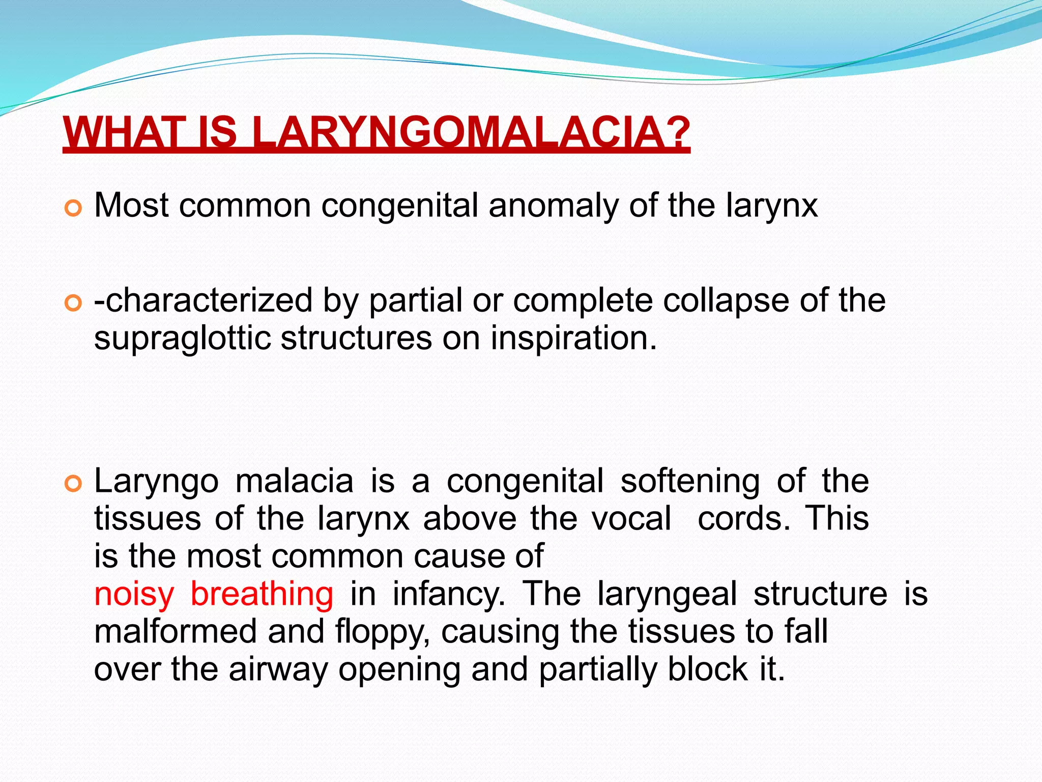
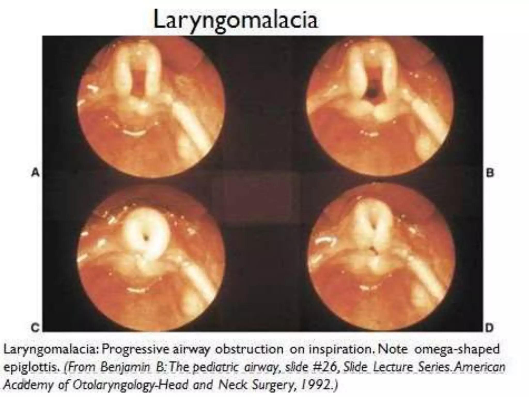
Congenital disorders of the
Larynx
LARYNGOMALACIA
BY : DR RIZWAN AHMED
[MD PEDIATRICS]
PART 1 ―LARYNGOMALACIA
WHAT IS LARYNGOMALACIA?
 Most common congenital anomaly of the larynx
 -characterized by partial or complete collapse of the
supraglottic structures on inspiration.
 Laryngo malacia is a congenital softening of the
tissues of the larynx above the vocal cords. This
is the most common cause of
noisy breathing in infancy. The laryngeal structure is
malformed and floppy, causing the tissues to fall
over the airway opening and partially block it.
LARYNGOMALACIA..
 Etiology: Unkown
• Theories:
• 1)GERD (gastroesophageal reflux disease)
• 2) Immature neuromuscular control
 Epiglottis is derived from the 3rd and 4th branchial
arches. Overgrowth of the 3rd results in elongation of
the structure and the observed laryngomalacia
Congenital disorders of the Larynx
Organogenesis
At birth the larynx is located high in the larynx between
levels C1- C4
By age 2 the larynx begins to descend inferiorly
By age 6 the larynx reaches the adult position between
levels C4 through C7
CONGENITAL DISORDERS OF THE LARYNX
EMBRYOGNESIS :
 The larynx develops from the endodermal lining
and the adjacent mesenchyme of the foregut
between the fourth and sixth branchial arches.
 At 20 days' gestation, the foregut is first identifiable
with a ventral laryngotracheal groove
 The laryngotracheal groove continues to deepen
until its lateral edges fuse. By day 26, this tube
descends caudally, where the trachea becomes
separated from the esophagus by the
tracheoesophageal septum with a persistent slitlike
opening into the pharynx.
CLINICAL DIAGNOSIS:
 It is considered a benign, transient cause of
inspiratory stridor and is characterized by collapse
of arytenoids, epiglottis and aryepiglottic folds
during the inspiratory phase.
 . The clinical diagnosis comprises of identifying
three anatomic abnormalities
 (1) flaccid epiglottis prolapsing
 backwards during inspiration;
 (2) poorly supported arytenoids that prolapse
forward during inspiration;
 (3) short aryepiglottic folds.
NORMAL VS LARYNGO MALACIAC VIEW
CAUSES:
 It can be due to the relaxation or a lack of muscle tone in
the upper airway.
 Insufficient or delayed calcium deposition
 The part of the nervous system that gives tone to the
airway is most likely underdeveloped.
 Gastroesophageal reflux (GER) may play an etiological
role in laryngomalacia
 Immature neuromuscular control may be responsible for
the arytenoid prolapse observed in laryngomalacia
TYPES:
 Type I: Inward collapse of aryepiglottic folds
 Type II: Long tubular epiglottis infolding on itself (Omega
shaped)
 Type III: Anterior and medial collapse of corniculate and
cuneiform cartilages
 Type IV: Posterior displacement of epiglottis against
posterior pharyngeal wall or inferior collapse towards
vocal folds
 Type V: Short aryepiglottic folds
DEGREE OF LARYNGOMALACIA
 Laryngomalacia can be mild, moderate or severe. 99%
of infants with laryngomalacia have mild or moderate
laryngomalacia.
 Mild laryngomalacia:
 noisy breathing or inspiratory stridor.
 no significant airway obstruction, feeding difficulties or
other symptoms associated with laryngomalacia.
 The noisy breathing is annoying to caregivers, but does
not cause other health care problems.
 Infants with mild laryngomalacia usually outgrow the
stridor by 12 – 18 months of age
MODERATE LARYNGOMALACIA
 noisy breathing or inspiratory stridor
 Feeding difficulties without poor weight gain
 (vomiting or spitting up)
 Choking on food
 Gastroesophageal reflux)
 Mild to moderate chest and/or neck retractions
(chest and/or neck sinking in with breathing), but no
severe distres
SEVERE LARYNGOMALACIA
 Feeding difficulties with poor weight gain
 (chest and/or neck sinking in with breathing)
 Significant cyanosis (turning blue)
 Life threatening apnea (pauses in breathing)
 Heart or lung problems from chronic oxygen
deprivation (low oxygen)
 Airway symptoms severe enough to cause multiple
visits to an emergency department or hospital
 Only 1% of infants with laryngomalacia have severe
laryngomalacia. Infants with severe laryngomalacia
usually need surgery.
SYMPTOMS
 Noisy breathing (stridor) — An audible wheeze when the baby
inhales . It is often worse when the baby is agitated, feeding,
crying or sleeping on his back.
 High pitched sound
 Difficulty feeding
 Poor weight gain
 Choking while feeding
 Apnea
 Pulling in neck and chest with each breath
 Cyanosis
 Gastro esophageal reflux (vomiting and regurgitation)
 Aspiration
TREATMENT
 Surgical management
 -Restoration of an adequate airway can be achieved
 by performing an endoscopic aryepiglottoplasty (sometimes
 termed a supraglottoplasty; Jani et al. 1991).
 Supraglottoplasty
 1) Using cup forceps and microscissors (or alternatively the
carbon dioxide laser), each aryepiglottic fold is first divided to
release it from the edge of the epiglottis,
 2) the redundant mucosa and submucosal tissue are then
excised from over the arytenoids, together if necessary with
part or all of the cuneiform cartilages. The stridor is usually
improved immediately following
laryngomalacia.pptx
laryngomalacia.pptx

laryngomalacia.pptx

  • 1.
    Congenital disorders ofthe Larynx LARYNGOMALACIA BY : DR RIZWAN AHMED [MD PEDIATRICS]
  • 2.
  • 3.
    WHAT IS LARYNGOMALACIA? Most common congenital anomaly of the larynx  -characterized by partial or complete collapse of the supraglottic structures on inspiration.  Laryngo malacia is a congenital softening of the tissues of the larynx above the vocal cords. This is the most common cause of noisy breathing in infancy. The laryngeal structure is malformed and floppy, causing the tissues to fall over the airway opening and partially block it.
  • 4.
    LARYNGOMALACIA..  Etiology: Unkown •Theories: • 1)GERD (gastroesophageal reflux disease) • 2) Immature neuromuscular control  Epiglottis is derived from the 3rd and 4th branchial arches. Overgrowth of the 3rd results in elongation of the structure and the observed laryngomalacia
  • 5.
    Congenital disorders ofthe Larynx Organogenesis At birth the larynx is located high in the larynx between levels C1- C4 By age 2 the larynx begins to descend inferiorly By age 6 the larynx reaches the adult position between levels C4 through C7
  • 7.
    CONGENITAL DISORDERS OFTHE LARYNX EMBRYOGNESIS :  The larynx develops from the endodermal lining and the adjacent mesenchyme of the foregut between the fourth and sixth branchial arches.  At 20 days' gestation, the foregut is first identifiable with a ventral laryngotracheal groove  The laryngotracheal groove continues to deepen until its lateral edges fuse. By day 26, this tube descends caudally, where the trachea becomes separated from the esophagus by the tracheoesophageal septum with a persistent slitlike opening into the pharynx.
  • 9.
    CLINICAL DIAGNOSIS:  Itis considered a benign, transient cause of inspiratory stridor and is characterized by collapse of arytenoids, epiglottis and aryepiglottic folds during the inspiratory phase.  . The clinical diagnosis comprises of identifying three anatomic abnormalities  (1) flaccid epiglottis prolapsing  backwards during inspiration;  (2) poorly supported arytenoids that prolapse forward during inspiration;  (3) short aryepiglottic folds.
  • 10.
    NORMAL VS LARYNGOMALACIAC VIEW
  • 11.
    CAUSES:  It canbe due to the relaxation or a lack of muscle tone in the upper airway.  Insufficient or delayed calcium deposition  The part of the nervous system that gives tone to the airway is most likely underdeveloped.  Gastroesophageal reflux (GER) may play an etiological role in laryngomalacia  Immature neuromuscular control may be responsible for the arytenoid prolapse observed in laryngomalacia
  • 12.
    TYPES:  Type I:Inward collapse of aryepiglottic folds  Type II: Long tubular epiglottis infolding on itself (Omega shaped)  Type III: Anterior and medial collapse of corniculate and cuneiform cartilages  Type IV: Posterior displacement of epiglottis against posterior pharyngeal wall or inferior collapse towards vocal folds  Type V: Short aryepiglottic folds
  • 18.
    DEGREE OF LARYNGOMALACIA Laryngomalacia can be mild, moderate or severe. 99% of infants with laryngomalacia have mild or moderate laryngomalacia.  Mild laryngomalacia:  noisy breathing or inspiratory stridor.  no significant airway obstruction, feeding difficulties or other symptoms associated with laryngomalacia.  The noisy breathing is annoying to caregivers, but does not cause other health care problems.  Infants with mild laryngomalacia usually outgrow the stridor by 12 – 18 months of age
  • 19.
    MODERATE LARYNGOMALACIA  noisybreathing or inspiratory stridor  Feeding difficulties without poor weight gain  (vomiting or spitting up)  Choking on food  Gastroesophageal reflux)  Mild to moderate chest and/or neck retractions (chest and/or neck sinking in with breathing), but no severe distres
  • 20.
    SEVERE LARYNGOMALACIA  Feedingdifficulties with poor weight gain  (chest and/or neck sinking in with breathing)  Significant cyanosis (turning blue)  Life threatening apnea (pauses in breathing)  Heart or lung problems from chronic oxygen deprivation (low oxygen)  Airway symptoms severe enough to cause multiple visits to an emergency department or hospital  Only 1% of infants with laryngomalacia have severe laryngomalacia. Infants with severe laryngomalacia usually need surgery.
  • 21.
  • 22.
     Noisy breathing(stridor) — An audible wheeze when the baby inhales . It is often worse when the baby is agitated, feeding, crying or sleeping on his back.  High pitched sound  Difficulty feeding  Poor weight gain  Choking while feeding  Apnea  Pulling in neck and chest with each breath  Cyanosis  Gastro esophageal reflux (vomiting and regurgitation)  Aspiration
  • 24.
    TREATMENT  Surgical management -Restoration of an adequate airway can be achieved  by performing an endoscopic aryepiglottoplasty (sometimes  termed a supraglottoplasty; Jani et al. 1991).  Supraglottoplasty  1) Using cup forceps and microscissors (or alternatively the carbon dioxide laser), each aryepiglottic fold is first divided to release it from the edge of the epiglottis,  2) the redundant mucosa and submucosal tissue are then excised from over the arytenoids, together if necessary with part or all of the cuneiform cartilages. The stridor is usually improved immediately following