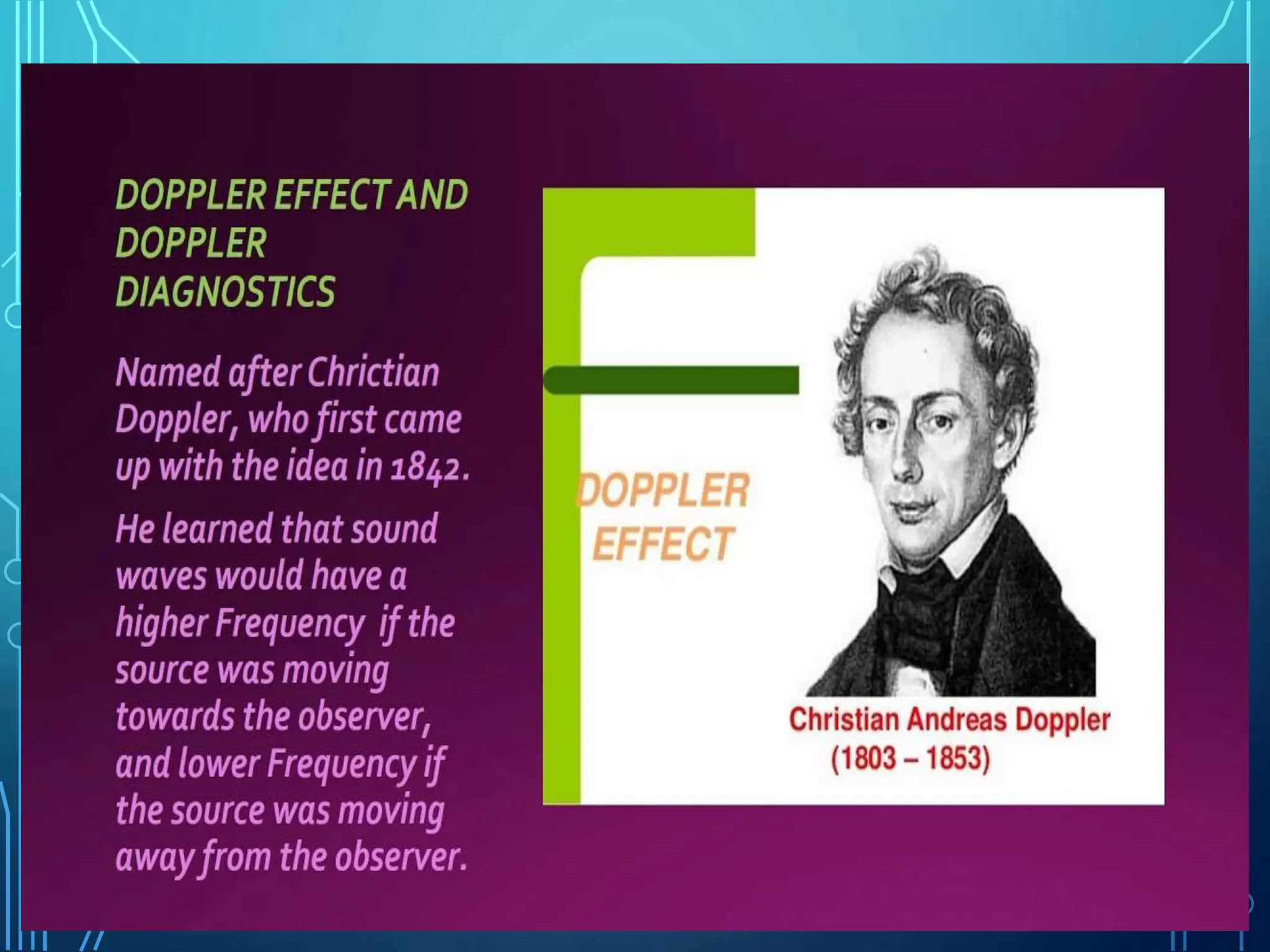
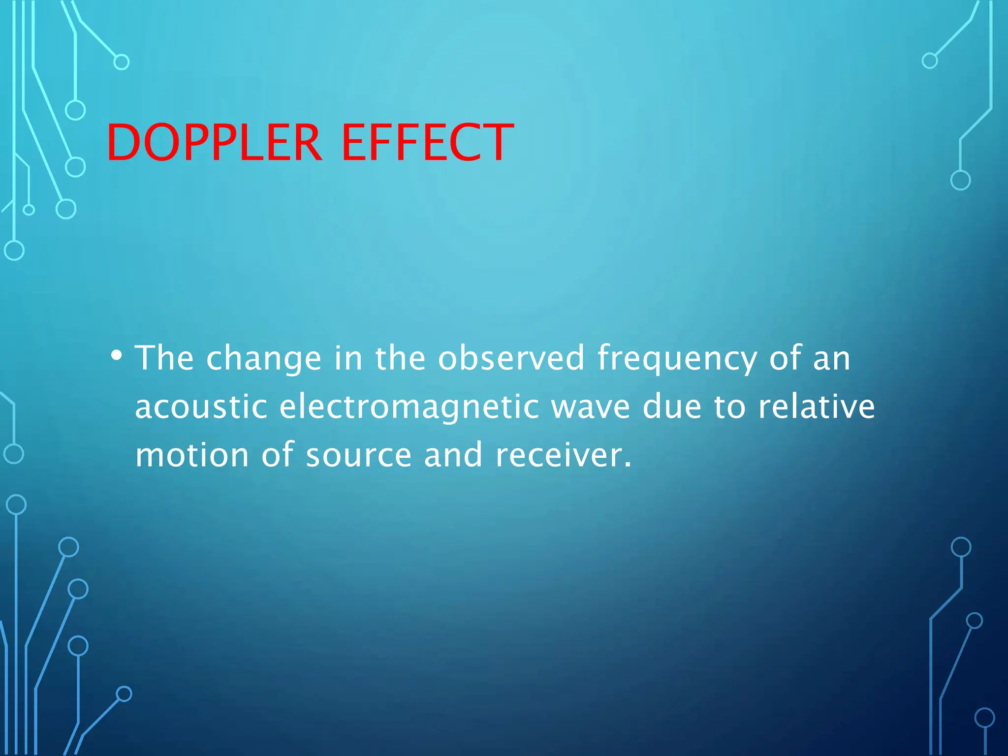
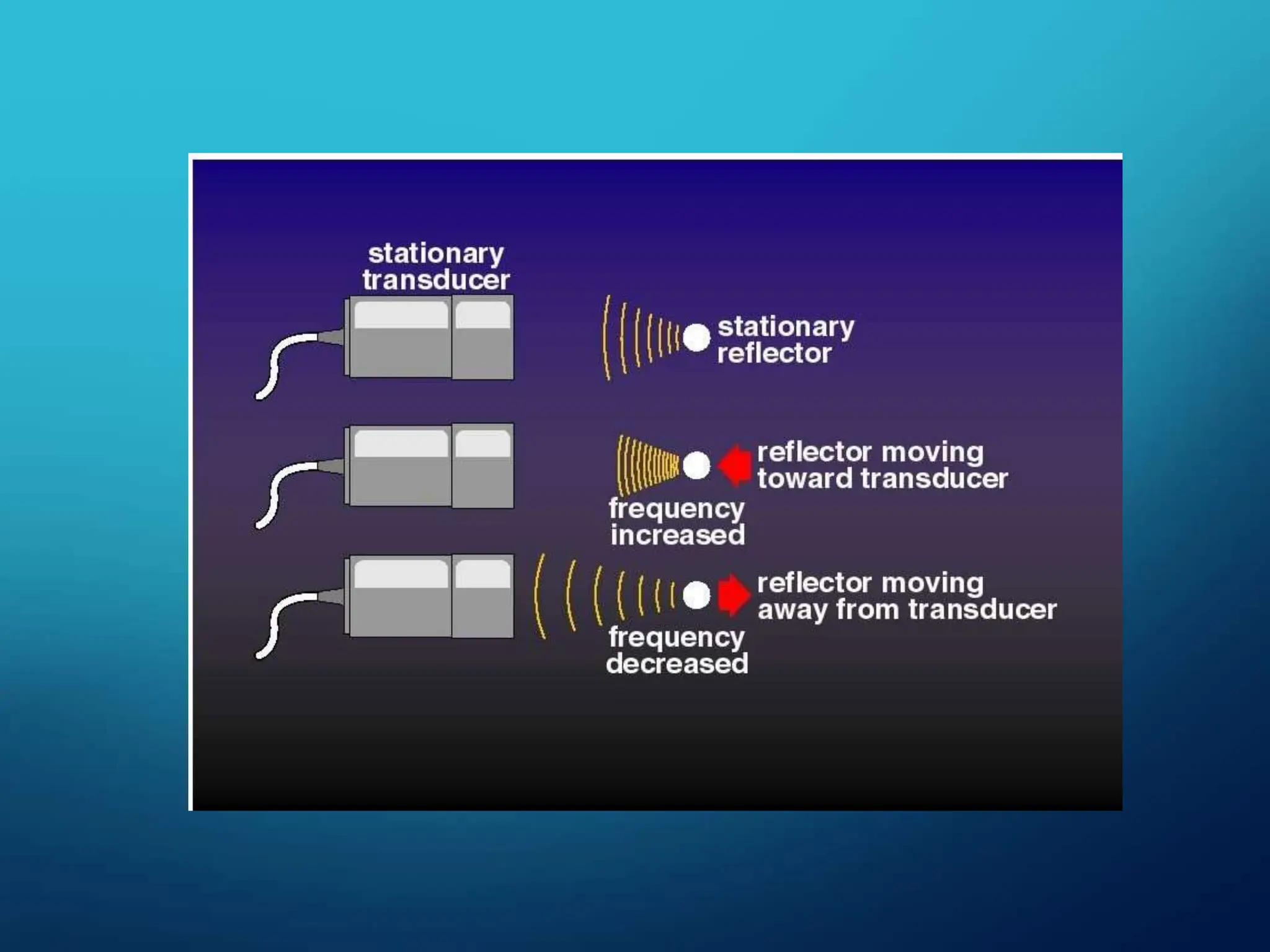
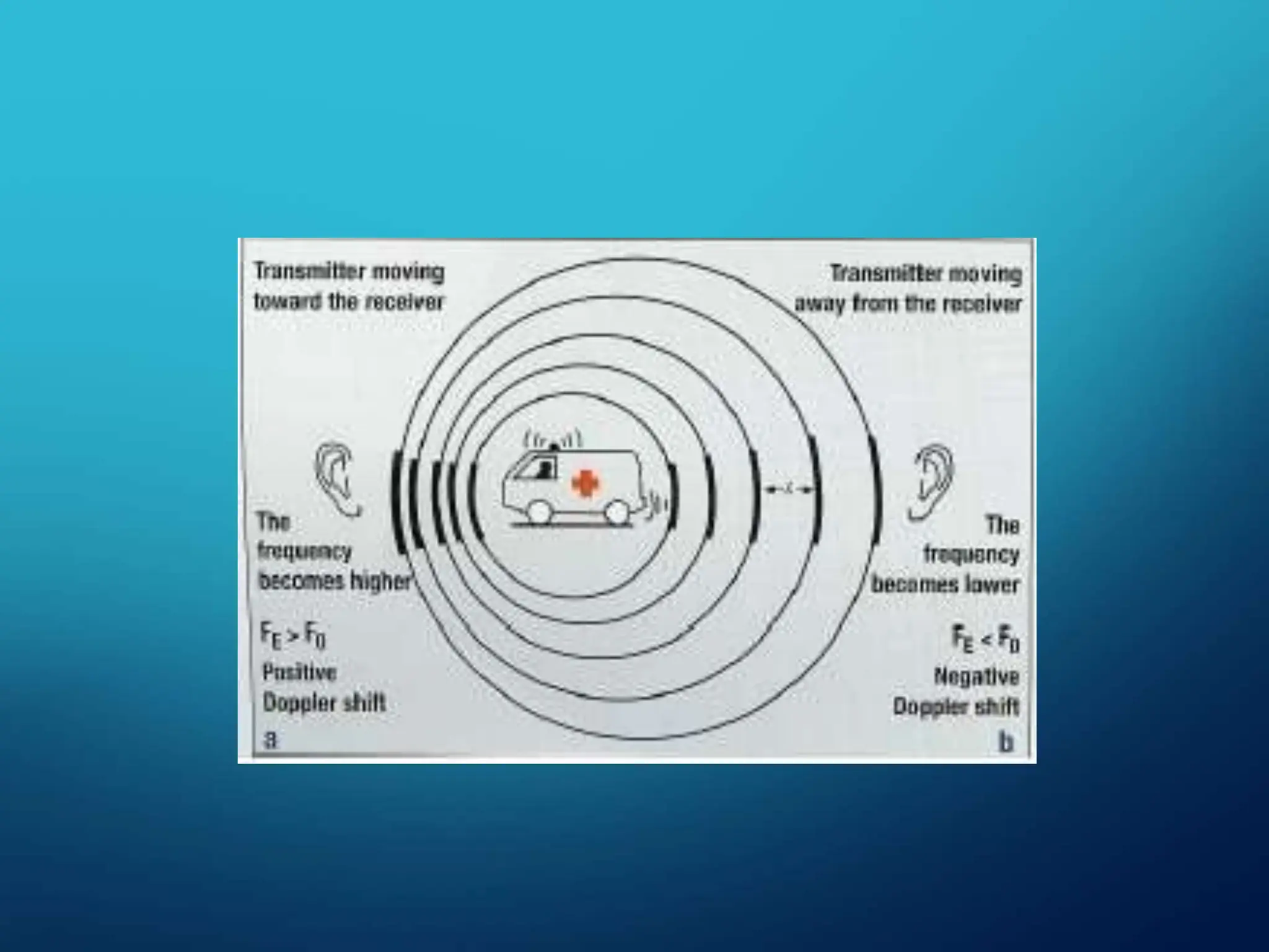
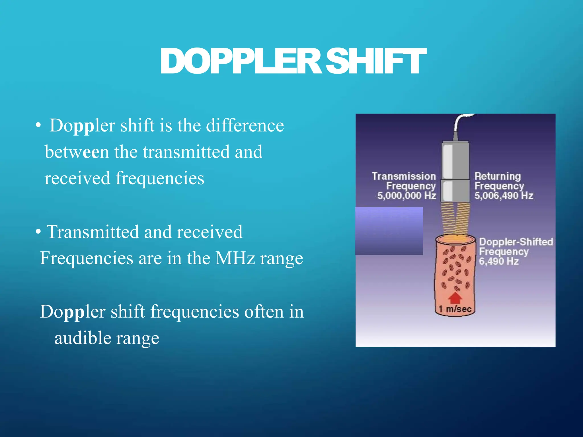
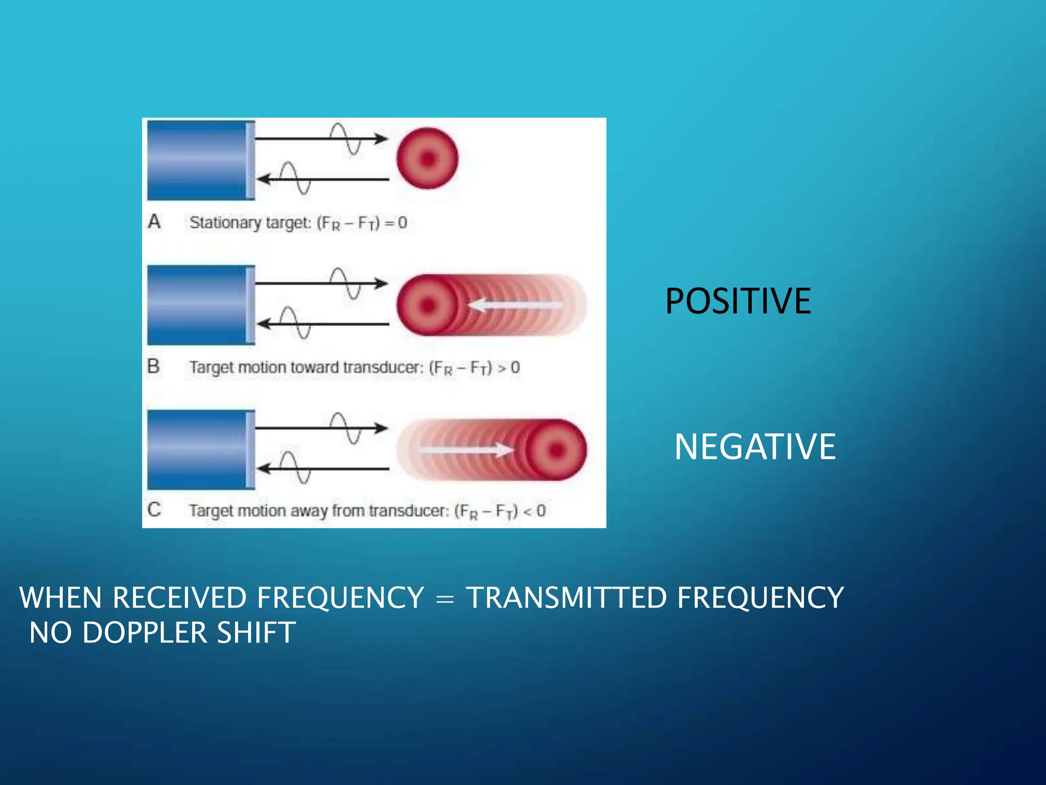
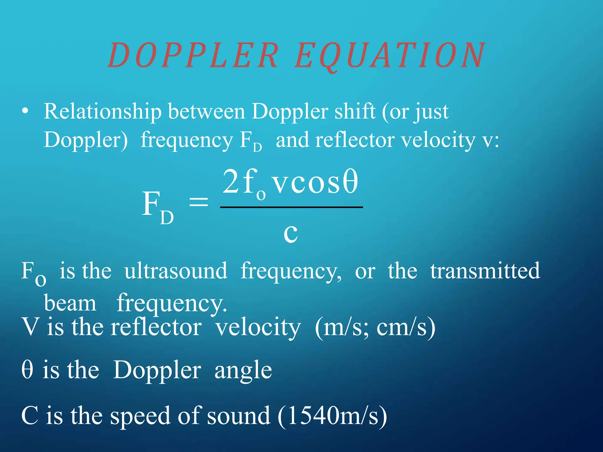
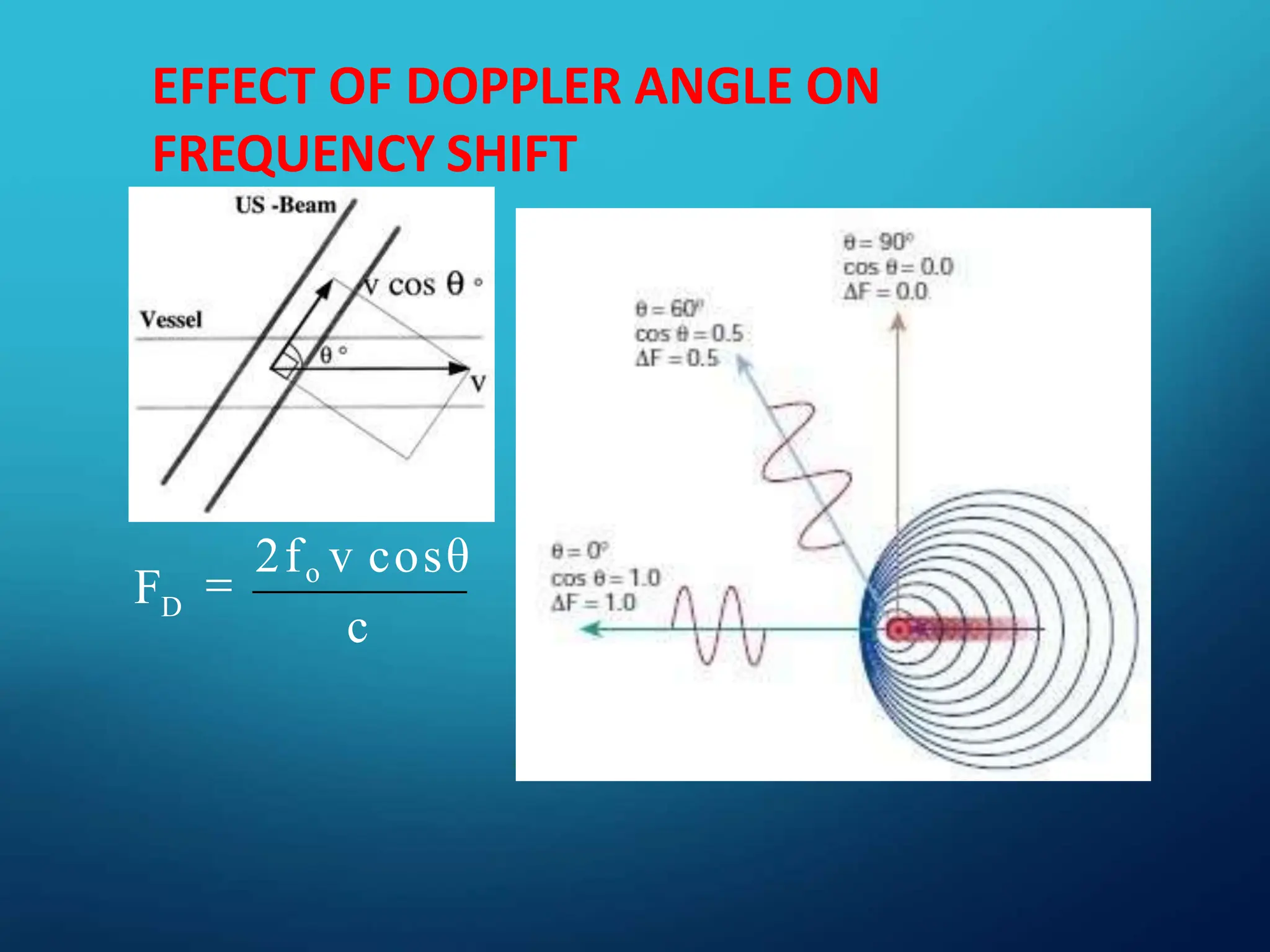
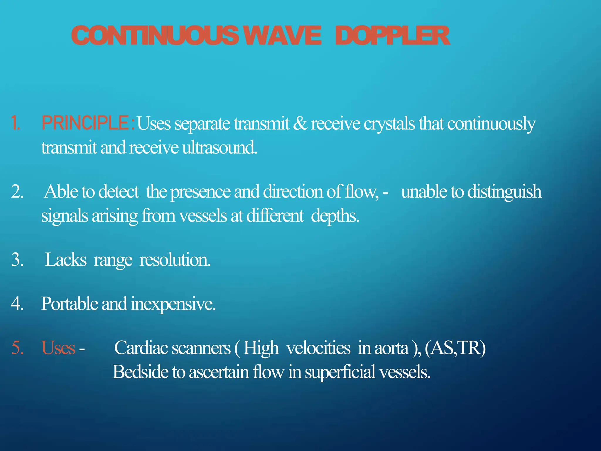
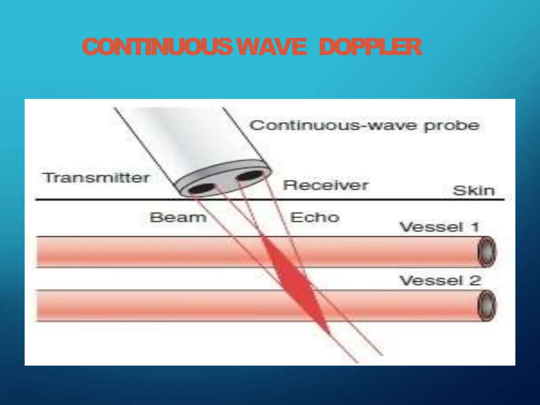
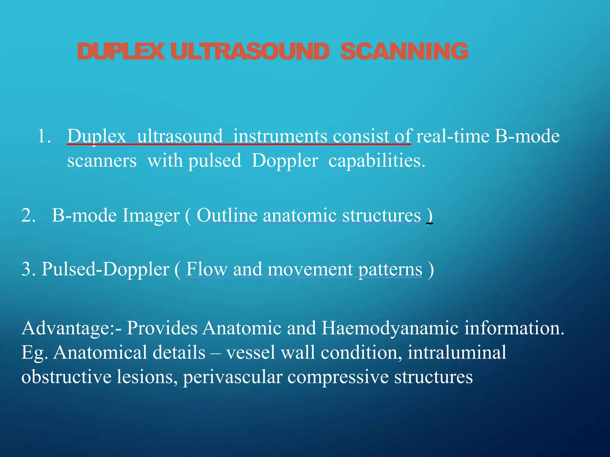
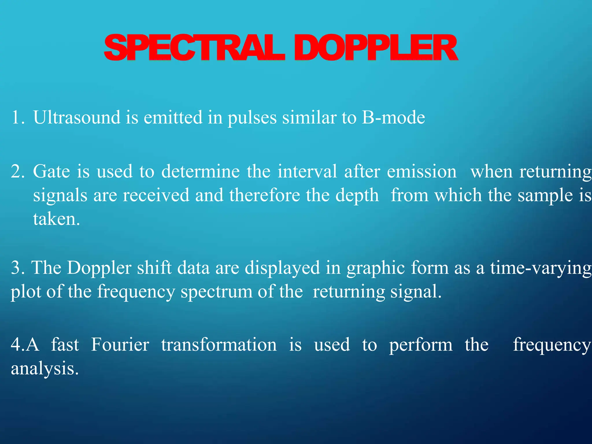
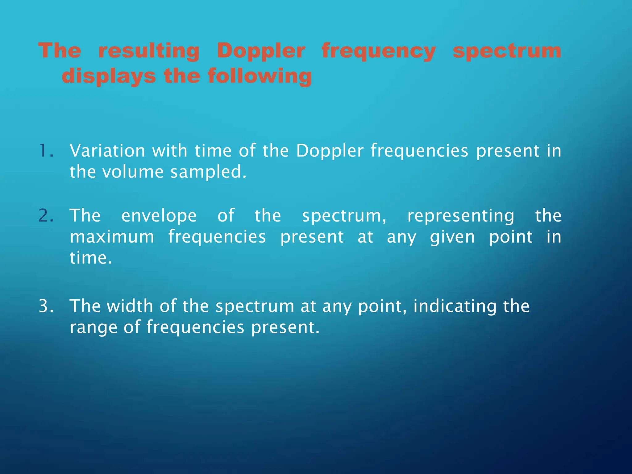
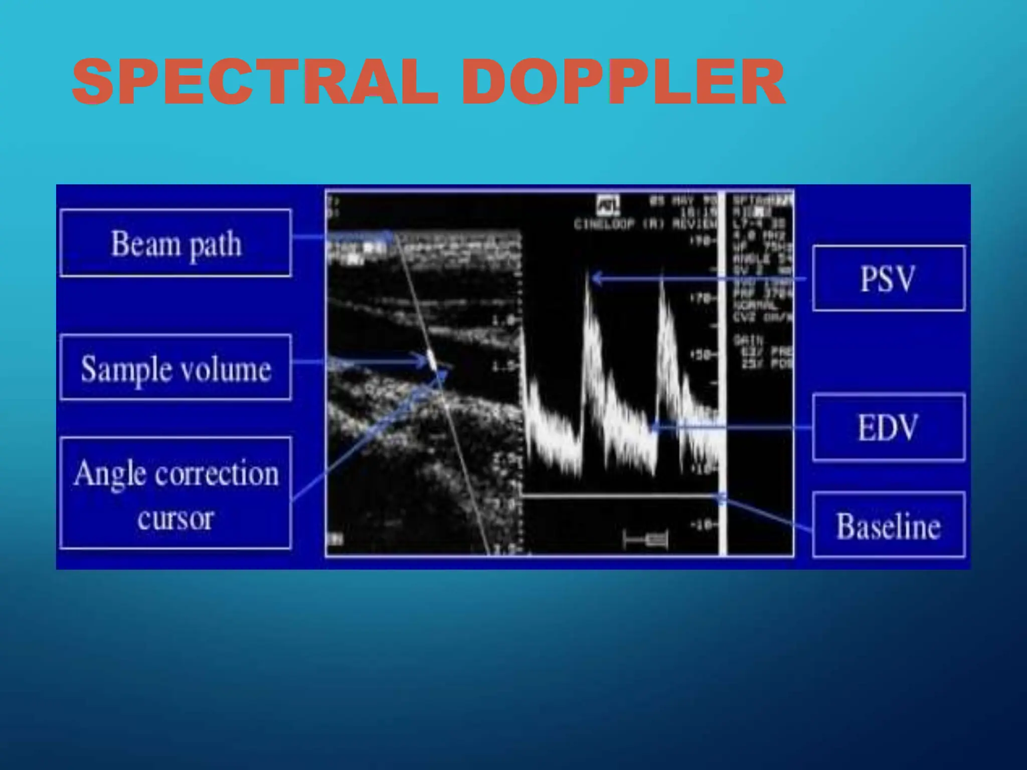
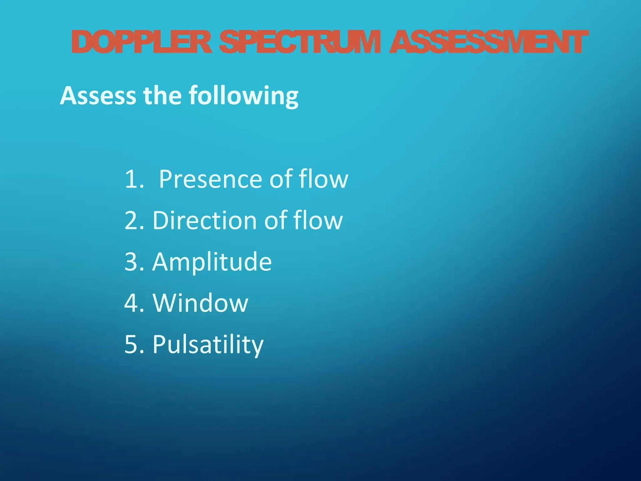
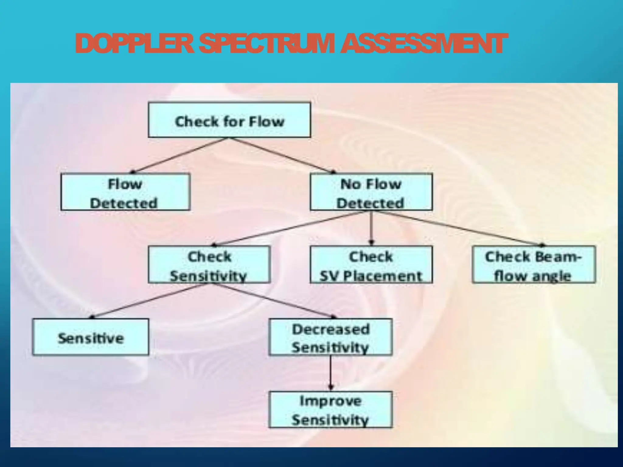
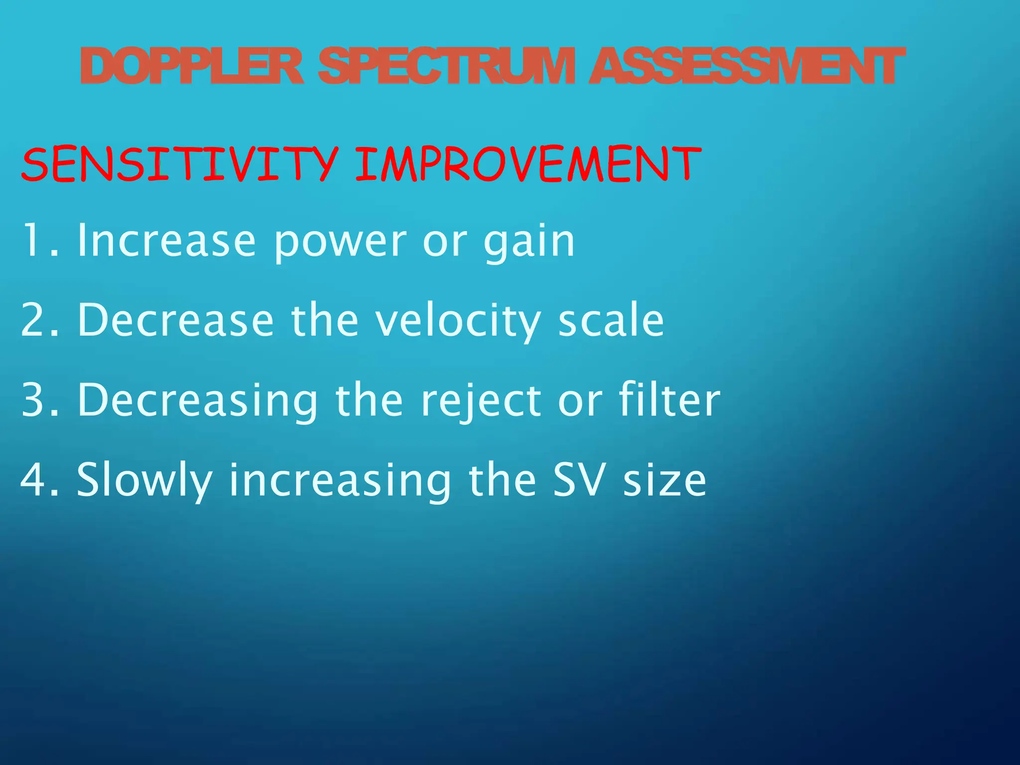
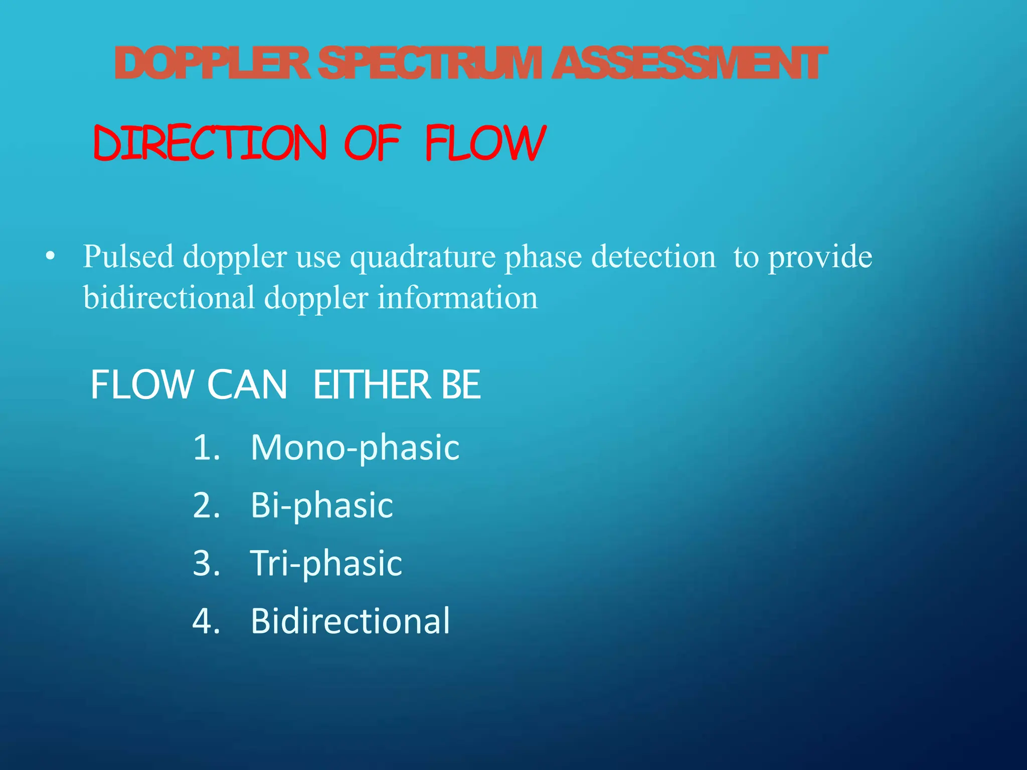
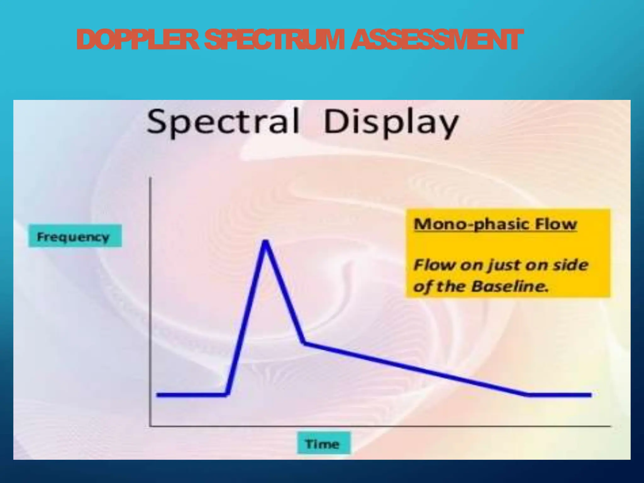
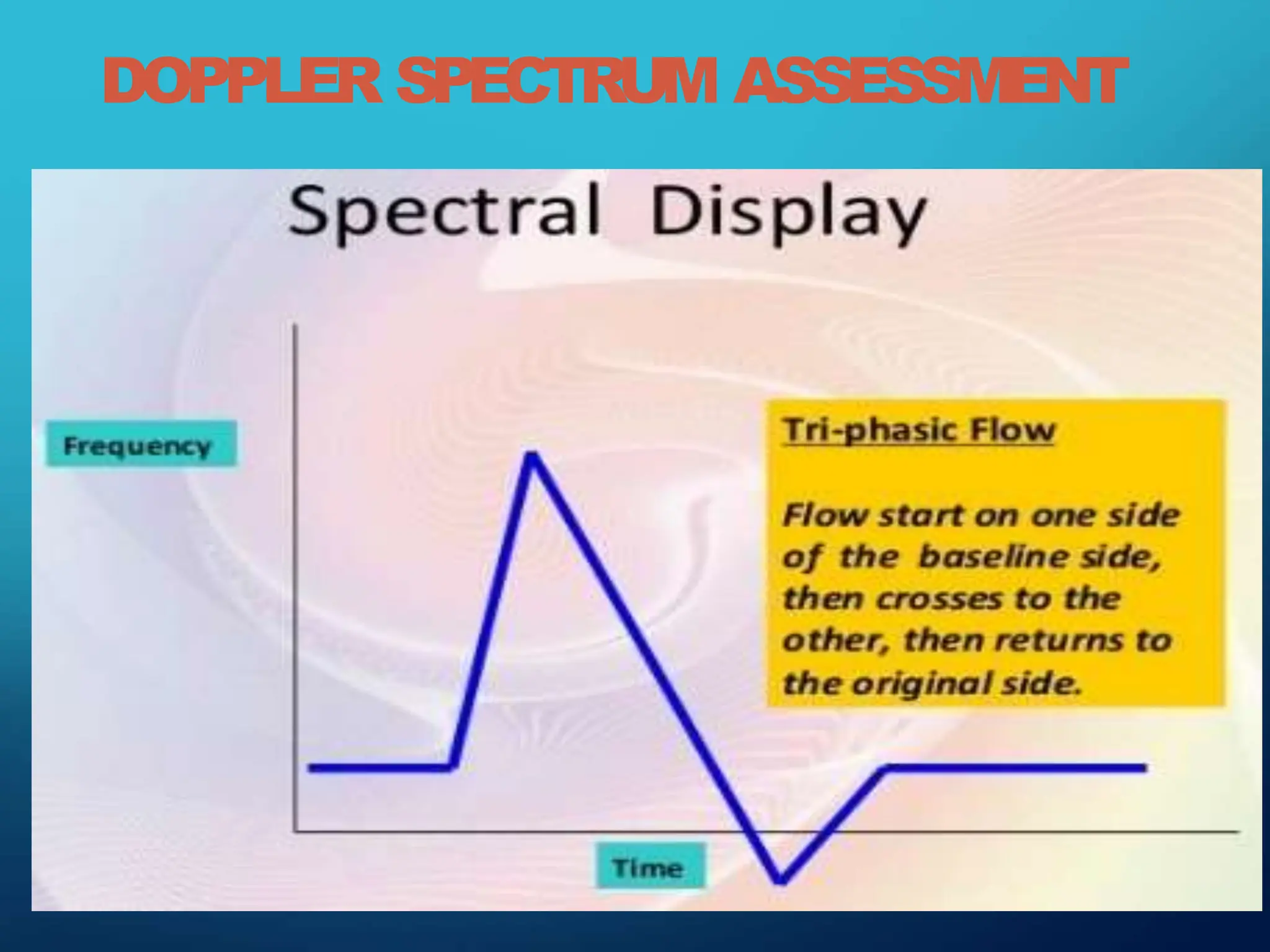
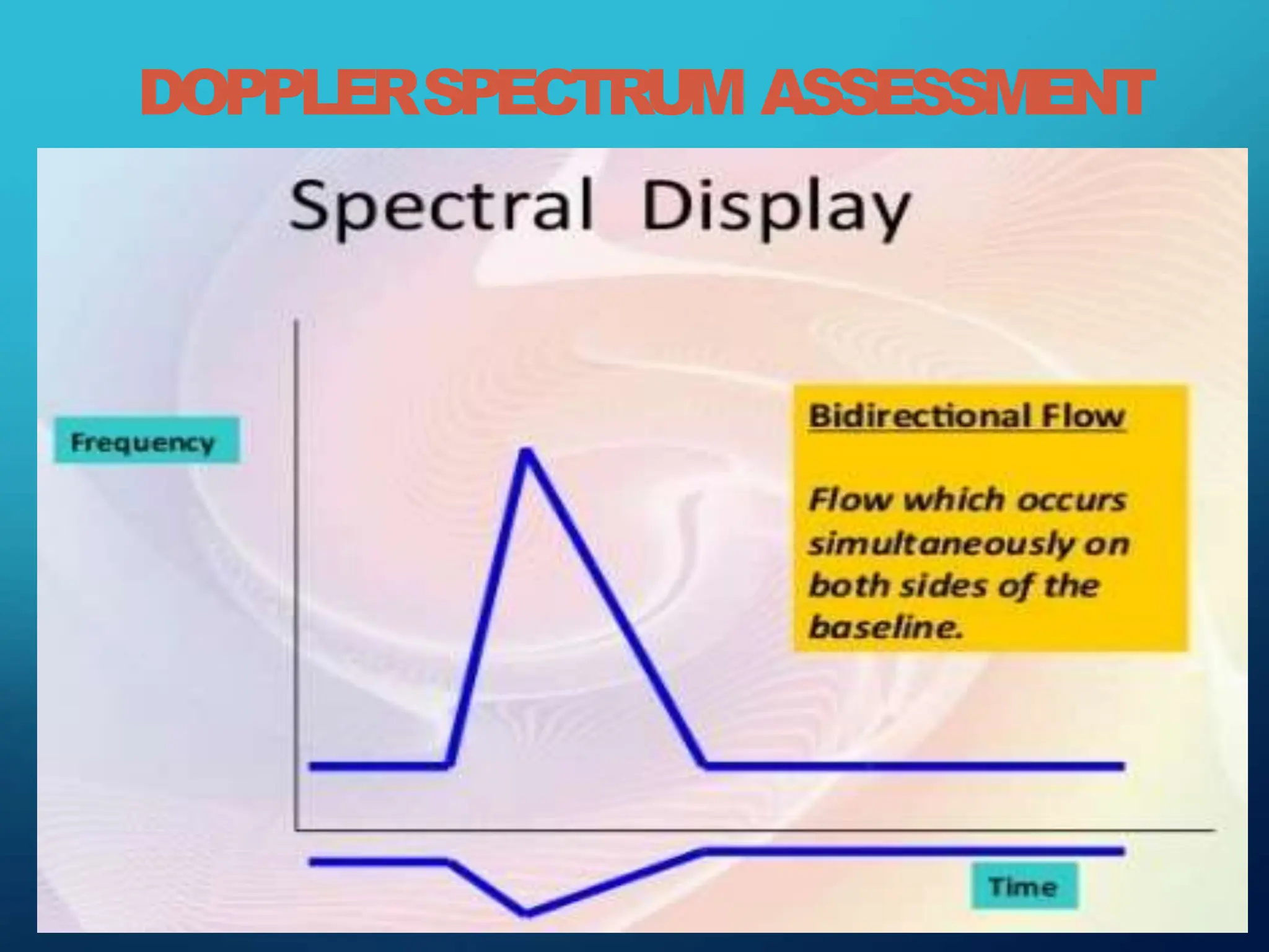
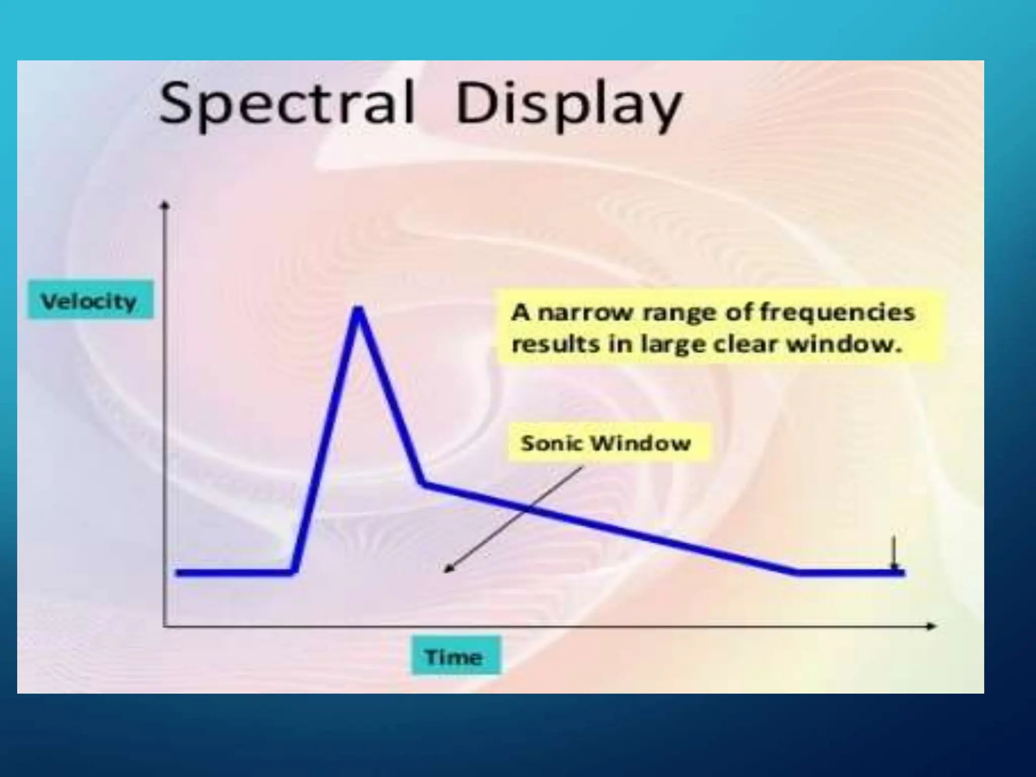
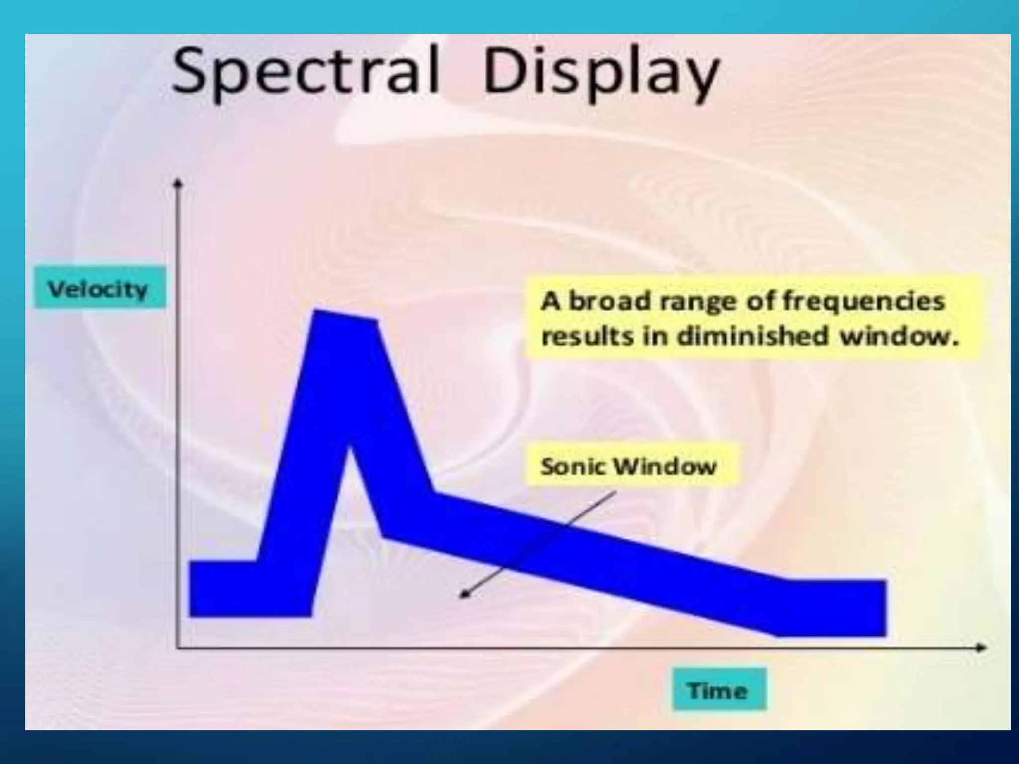
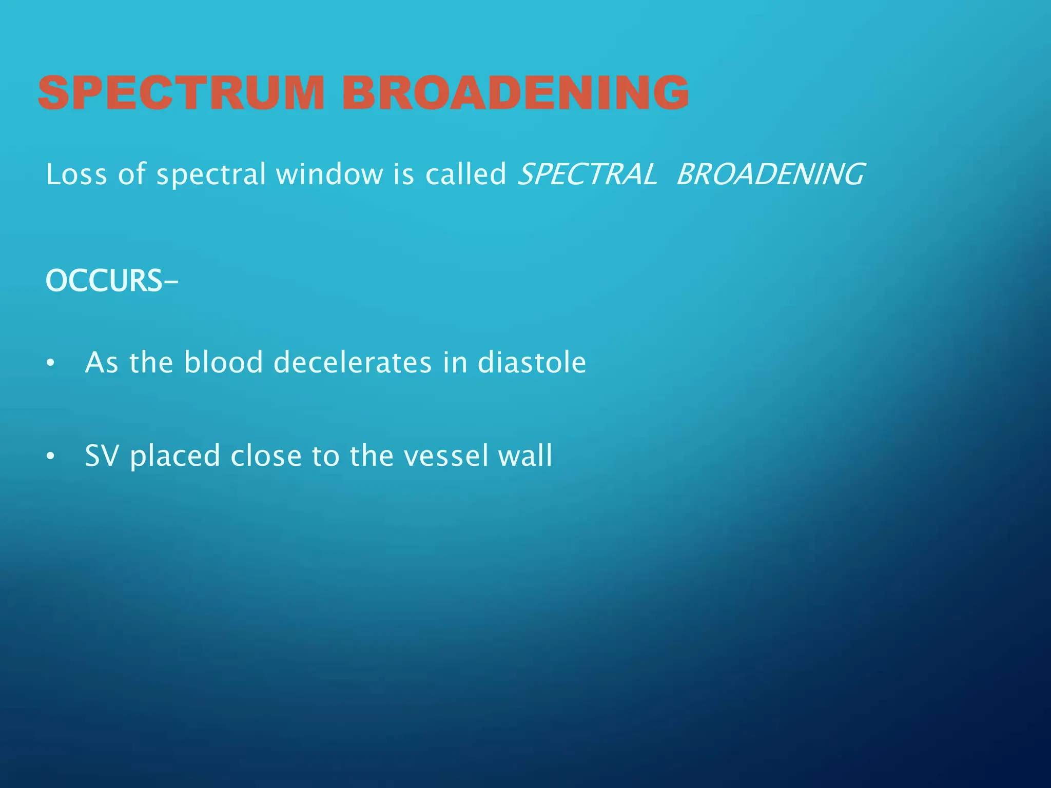
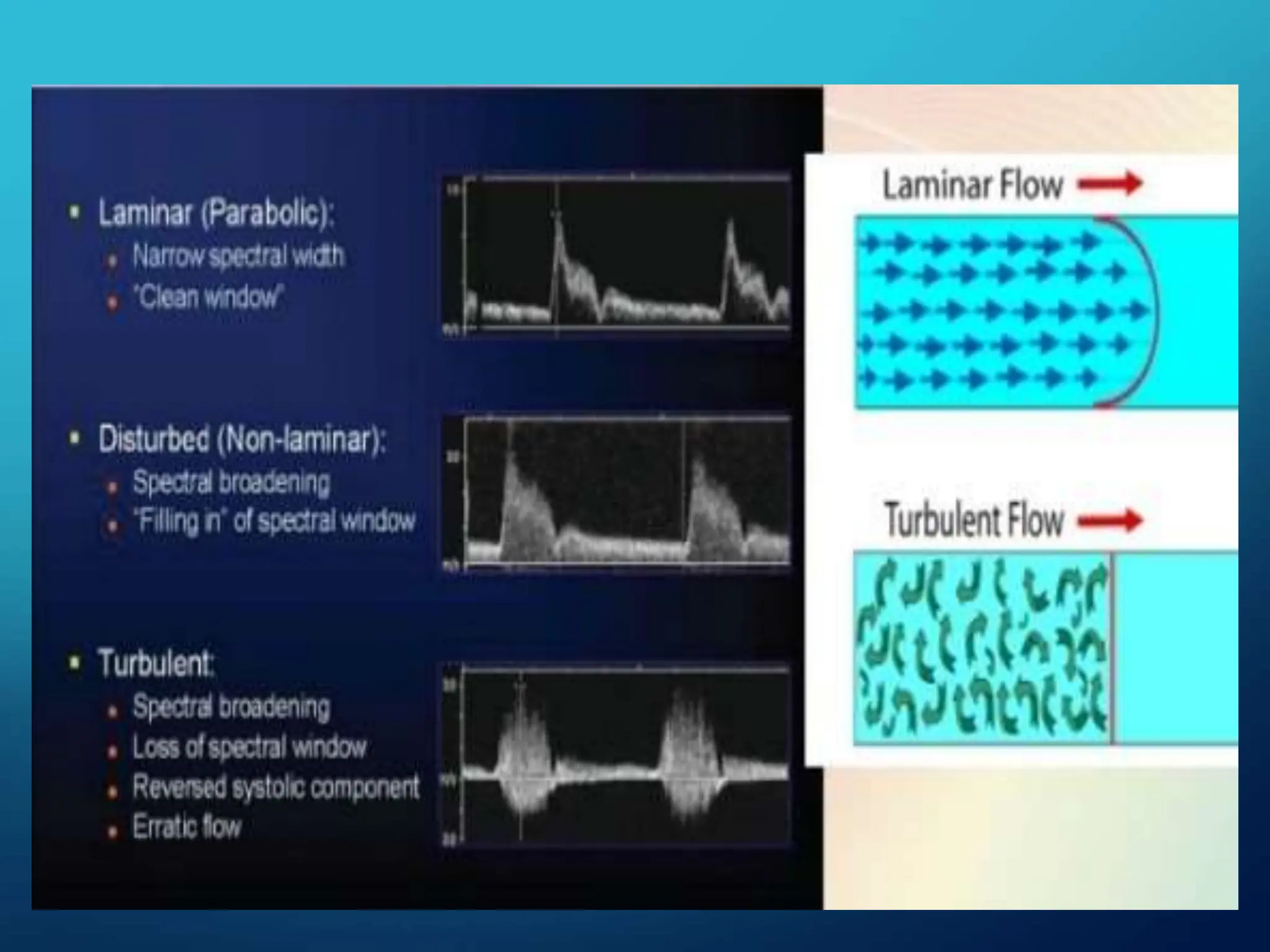
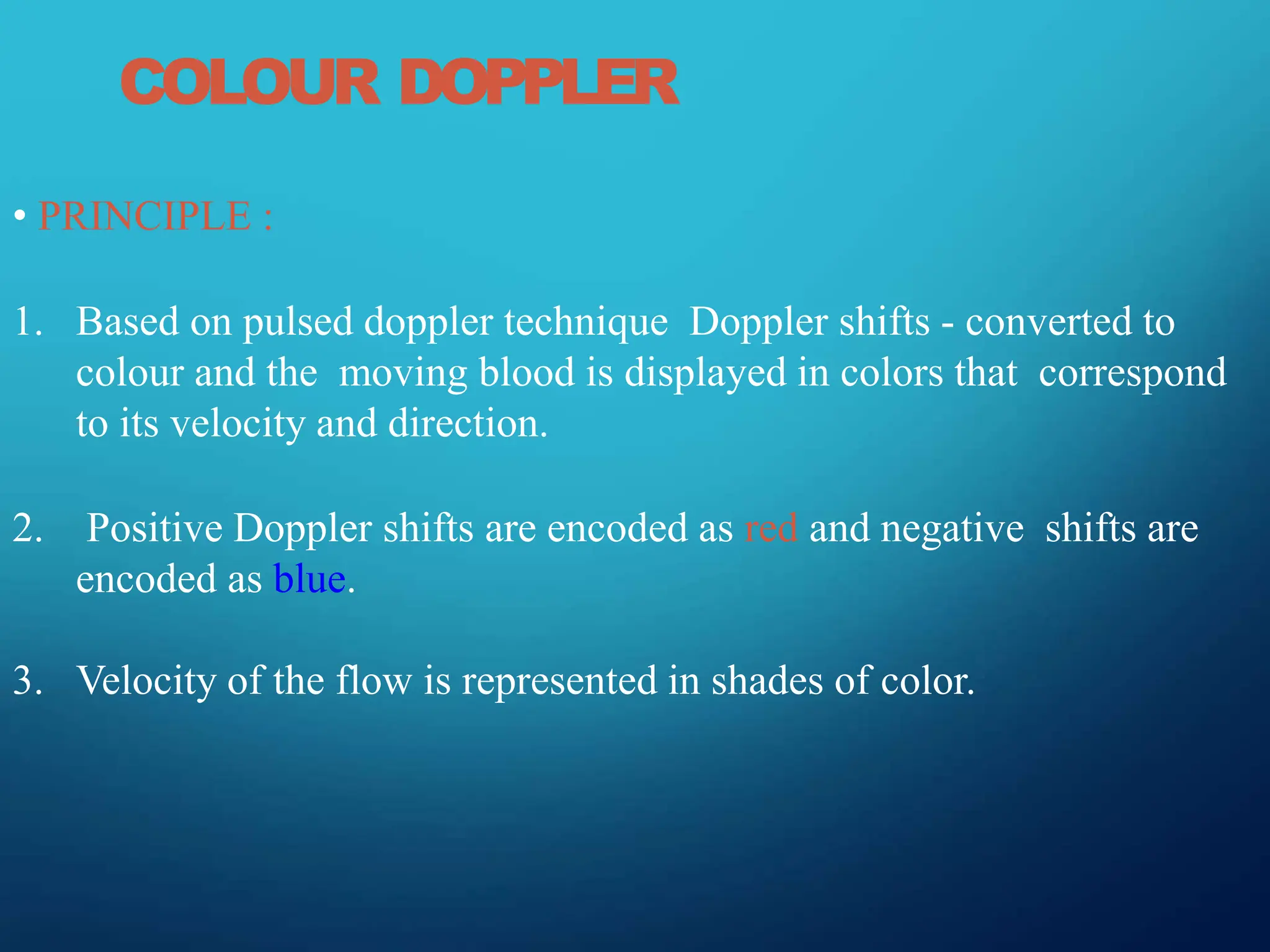
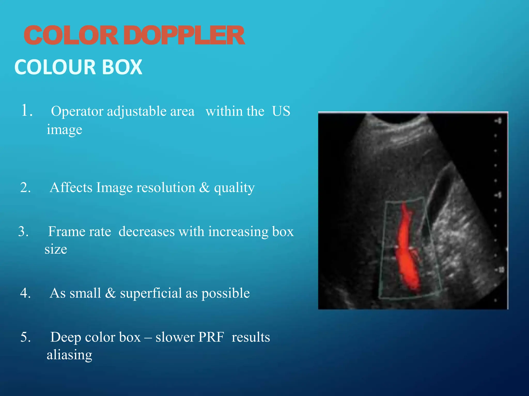
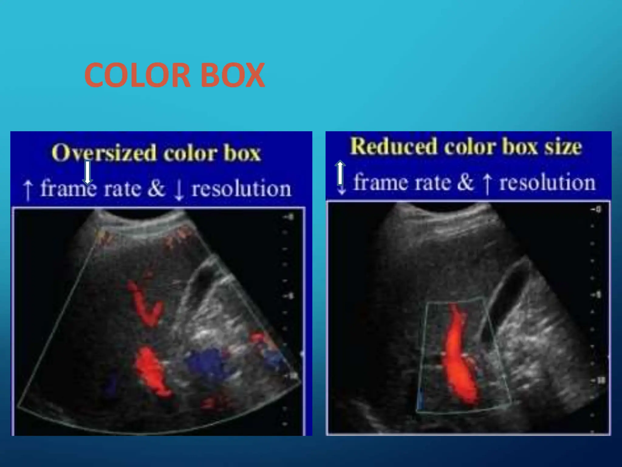
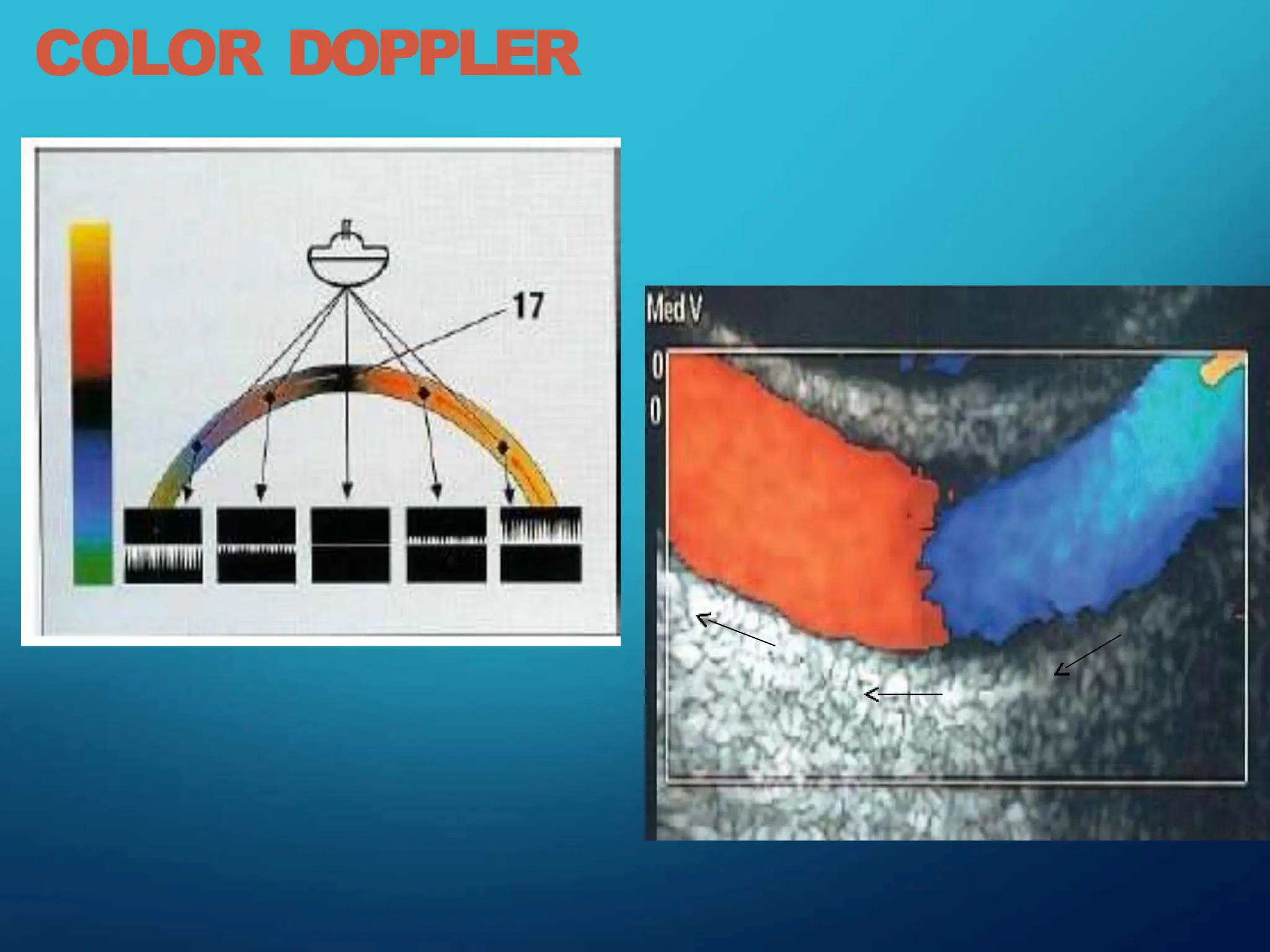
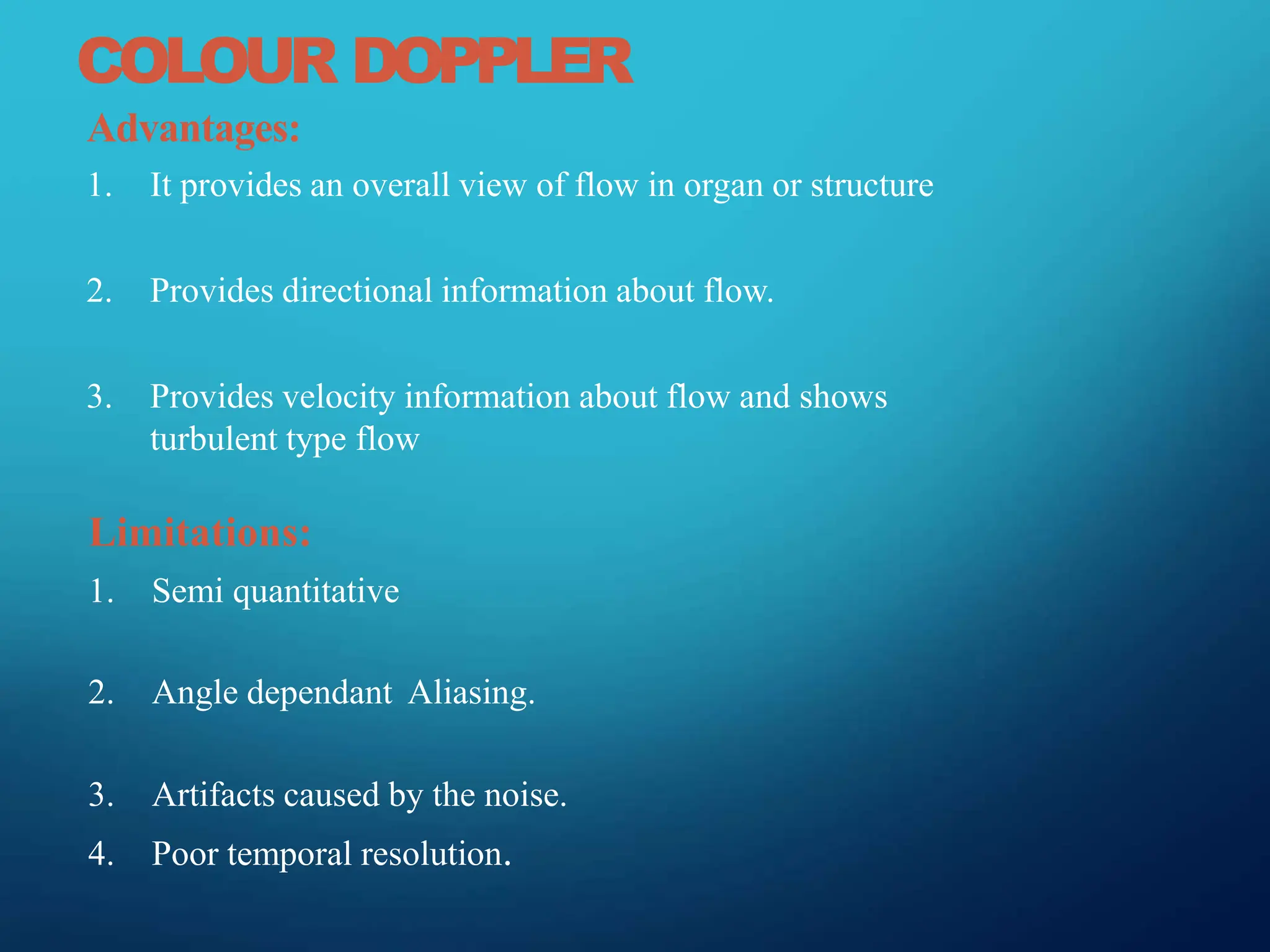
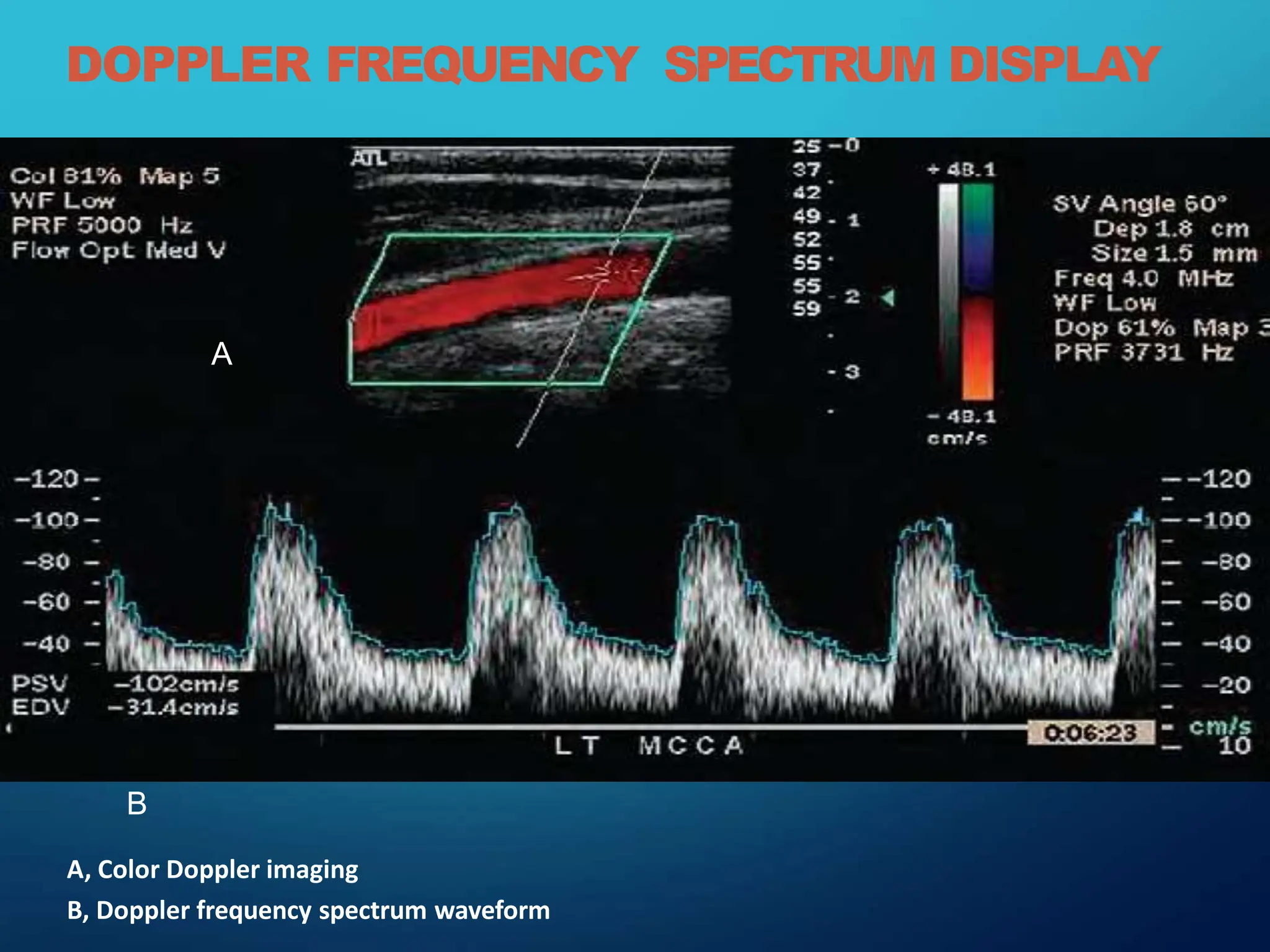
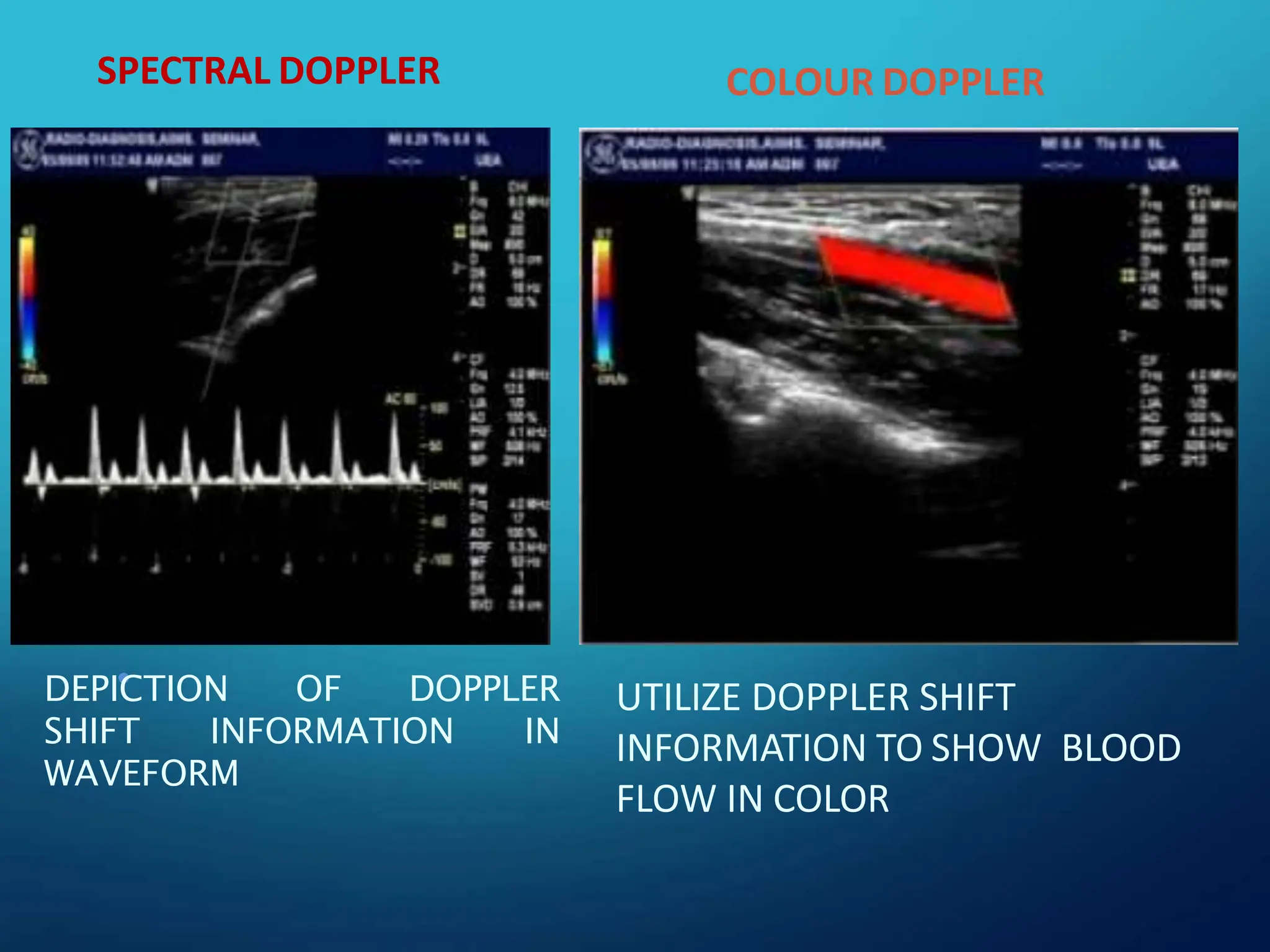
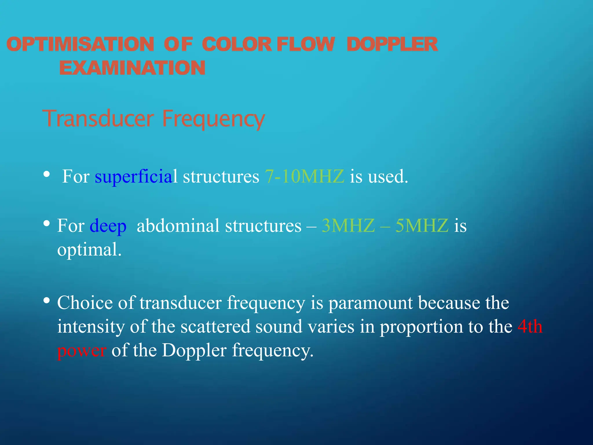
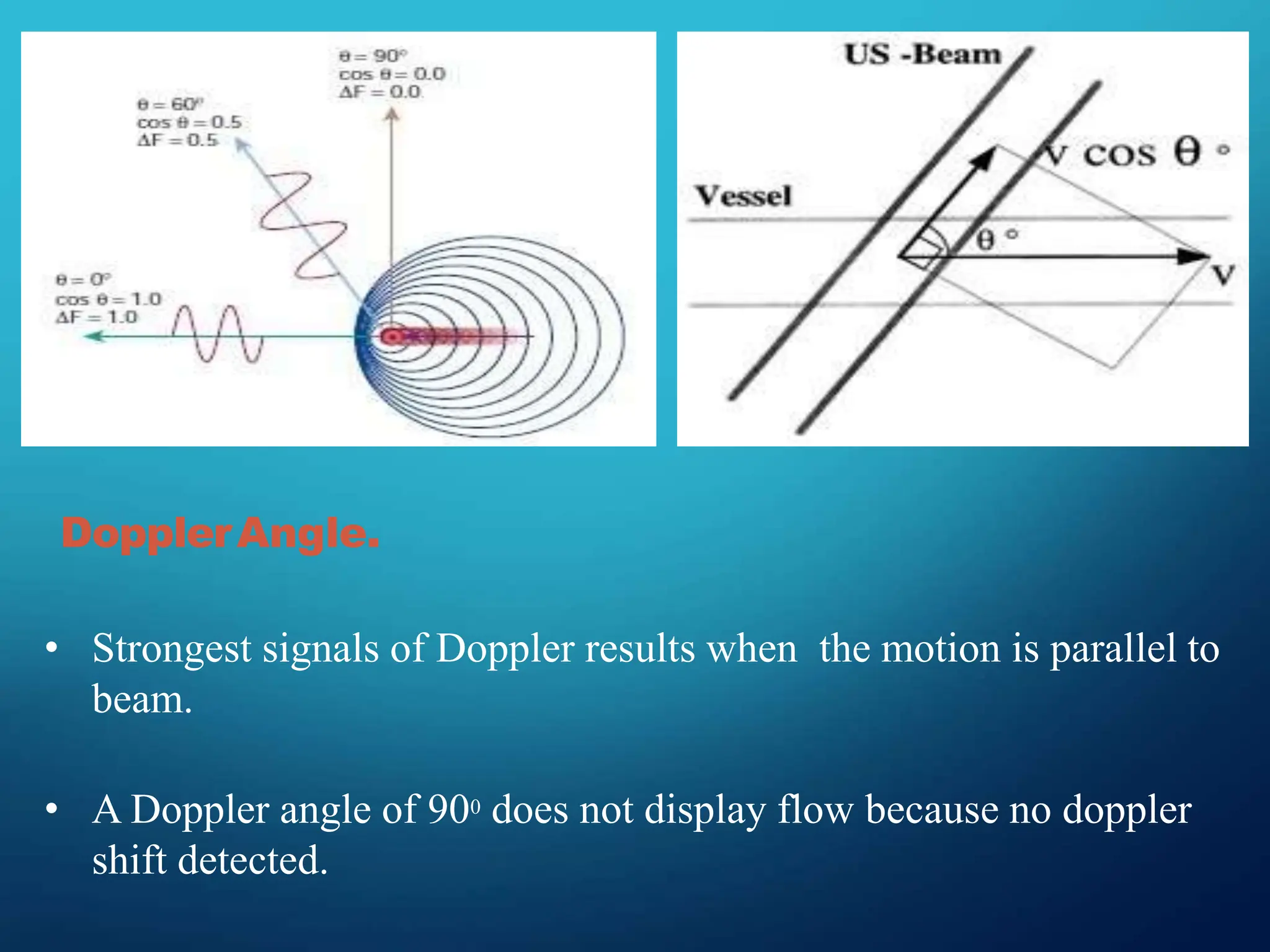
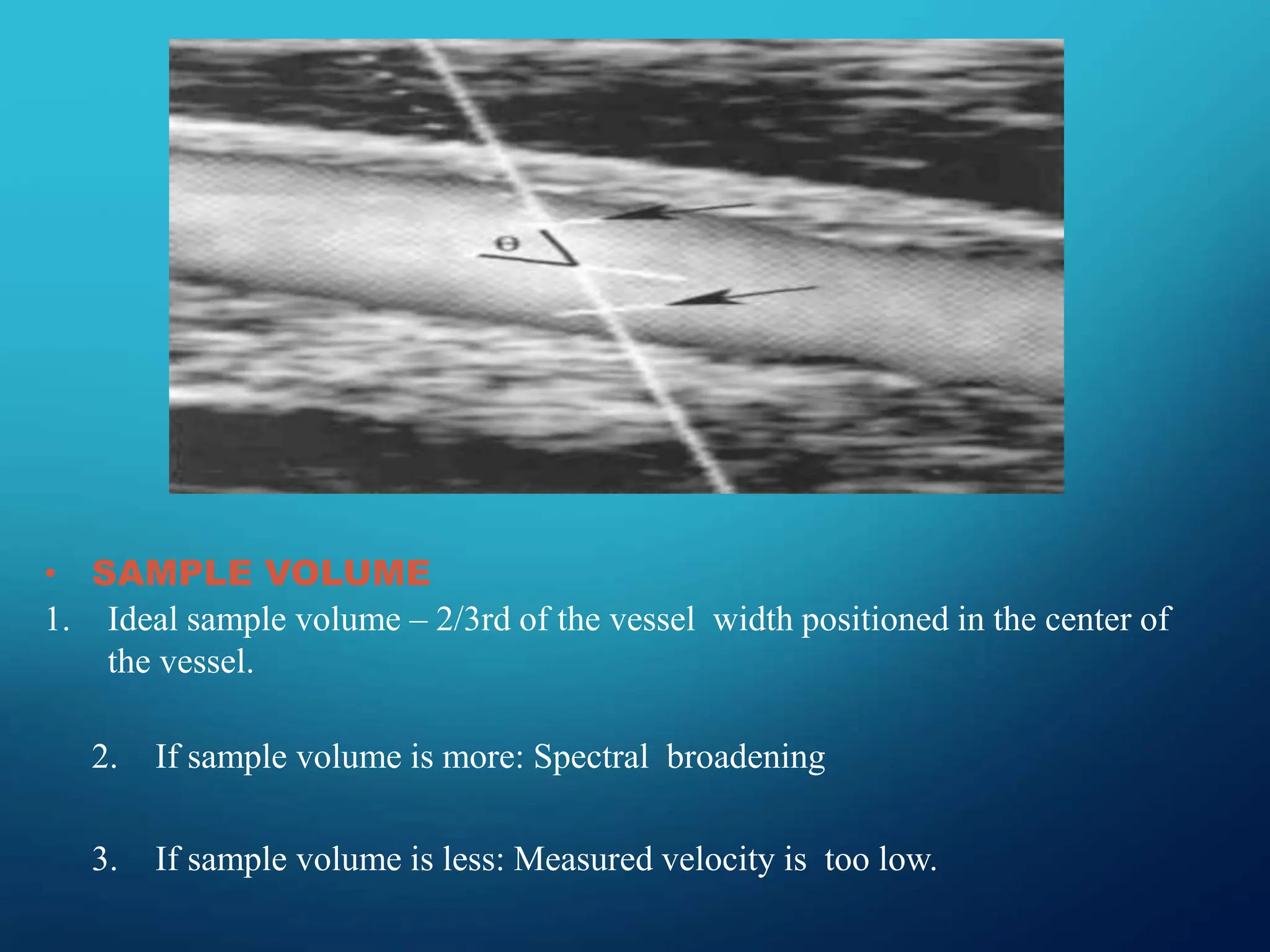
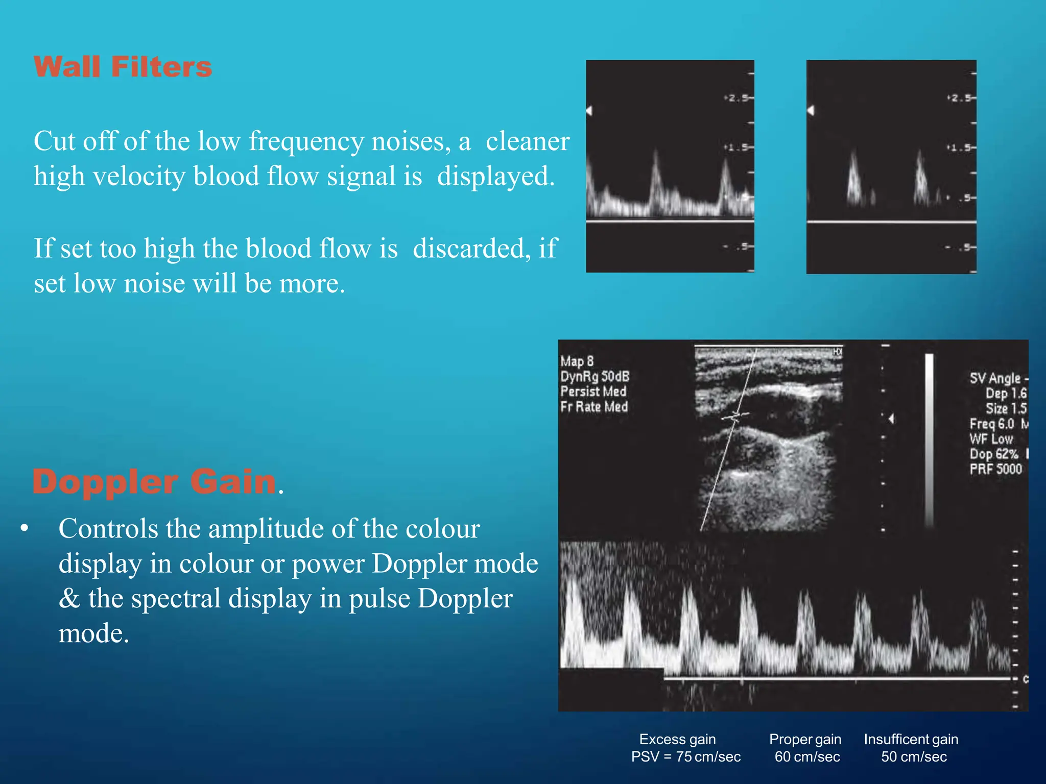
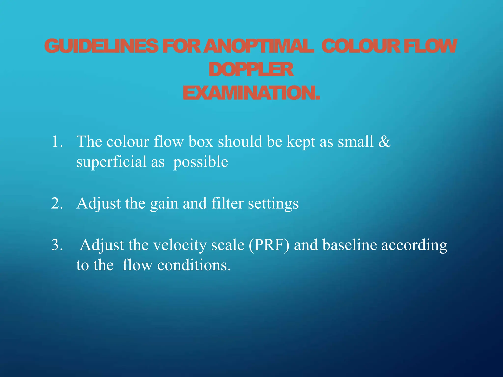
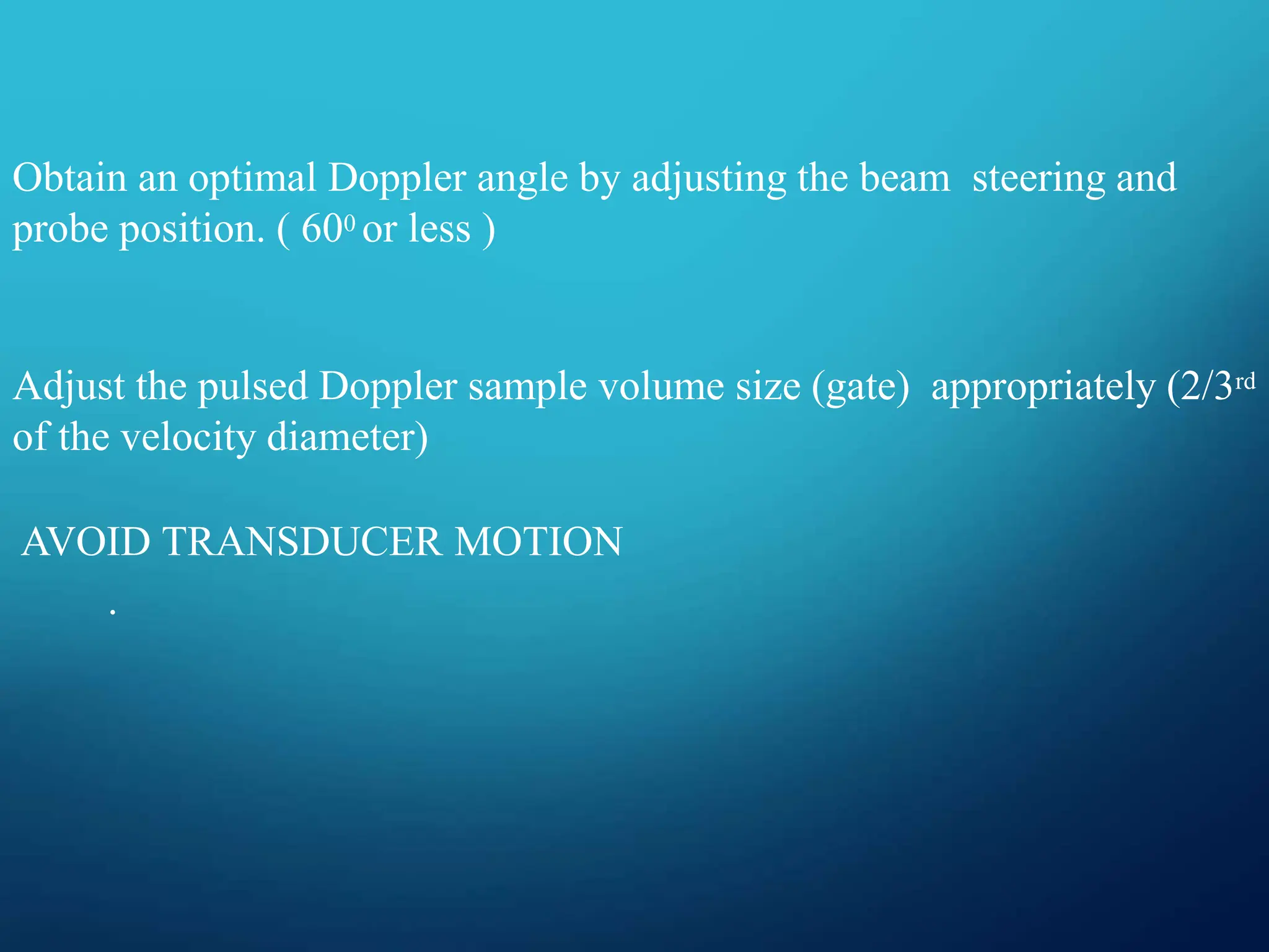
DOPPLER
PHYSICS
Dr. AAFRIN
DOPPLER EFFECT
• The change in the observed frequency of an
acoustic electromagnetic wave due to relative
motion of source and receiver.
• Doppler shift is the difference
between the transmitted and
received frequencies
• Transmitted and received
Frequencies are in the MHz range
Doppler shift frequencies often in
audible range
DOPPLERSHIFT
POSITIVE
NEGATIVE
WHEN RECEIVED FREQUENCY = TRANSMITTED FREQUENCY
NO DOPPLER SHIFT
• Relationship between Doppler shift (or just
Doppler) frequency FD and reflector velocity v:
Fo is the ultrasound frequency, or the transmitted
beam frequency.
V is the reflector velocity (m/s; cm/s)
θ is the Doppler angle
C is the speed of sound (1540m/s)
DOPPLER EQUATION
c
FD

2fo vcosθ
EFFECT OF DOPPLER ANGLE ON
FREQUENCY SHIFT
c
FD

2fo v cosθ
DOPPLERULTRASOUND
MODES ORINSTRUMENTATIONS
1. ContinuouswaveDoppler
2. PulsedwaveDoppler
3. Spectral doppler
4. Colour doppler
5. Powerdoppler
CONTINUOUSWAVE DOPPLER
1. PRINCIPLE:Usesseparatetransmit&receivecrystalsthatcontinuously
transmitandreceiveultrasound.
2. Abletodetect thepresenceanddirectionofflow,- unabletodistinguish
signalsarisingfromvesselsatdifferent depths.
3. Lacks range resolution.
4. Portableandinexpensive.
5. Uses- Cardiacscanners(High velocities inaorta),(AS,TR)
Bedsidetoascertainflowinsuperficialvessels.
CONTINUOUSWAVE DOPPLER
•
1. PRINCIPLE : Uses brief pulses of ultrasound energy using only
one crystal.
2. The echo delay time ( Te ) can be converted into distance and the
DEPTH of echo source can be determined.
1. Advantage:- Range Resolution.
2. No mixture of signals like CW Doppler.
PULSED-WAVE(PW)DOPPLER
PULSEDWAVEDOPPLER
PULSE REPETITION FREQUENCY ( PRF) - No. of pulses transmitted
per second
PRINCIPLE : With increasing scanning depth PRF decreases as more time is
needed for the echoes to return
1. At a minimum, the PRF must be at least twice the frequency of the
Doppler signal to construct the signal successfully.
2. This sets upper limit to the flow velocities to be accurately recorded.
3. Use higher PRF setting for high flow velocities, low PRF for slow venous flow
A (PRF = 700 Hz) B (PRF = 4500 Hz)
DIFFERENCE BETWEEN CW
DOPPLER & PW DOPPLER
CW DOPPLER
Contains receiver and
transmitting crystals.
Advantage: Can measure
very high velocities.
Limitation : Range
Ambiguity.
Cannot be used for
imaging
No anatomic information.
PW DOPPLER
• Produced by single crystal
• Only Pulsed sound can
create images.
• Advantage: Measures
depth to detect exact
location from where
signals receive
• Limitation: Cannot
measure very high
velocities.
Continuous wave (CW) Doppler Pulsed wave (PW) Doppler
DUPLEX ULTRASOUND SCANNING
1. Duplex ultrasound instruments consist of real-time B-mode
scanners with pulsed Doppler capabilities.
2. B-mode Imager ( Outline anatomic structures )
3. Pulsed-Doppler ( Flow and movement patterns )
Advantage:- Provides Anatomic and Haemodyanamic information.
Eg. Anatomical details – vessel wall condition, intraluminal
obstructive lesions, perivascular compressive structures
SPECTRAL DOPPLER
1. Ultrasound is emitted in pulses similar to B-mode
2. Gate is used to determine the interval after emission when returning
signals are received and therefore the depth from which the sample is
taken.
3. The Doppler shift data are displayed in graphic form as a time-varying
plot of the frequency spectrum of the returning signal.
4.A fast Fourier transformation is used to perform the frequency
analysis.
The resulting Doppler frequency spectrum
displays the following
1. Variation with time of the Doppler frequencies present in
the volume sampled.
2. The envelope of the spectrum, representing the
maximum frequencies present at any given point in
time.
3. The width of the spectrum at any point, indicating the
range of frequencies present.
SPECTRAL DOPPLER
DOPPLER SPECTRUM ASSESSMENT
Assess the following
1. Presence of flow
2. Direction of flow
3. Amplitude
4. Window
5. Pulsatility
DOPPLERSPECTRUMASSESSMENT
DOPPLER SPECTRUM ASSESSMENT
SENSITIVITY IMPROVEMENT
1. Increase power or gain
2. Decrease the velocity scale
3. Decreasing the reject or filter
4. Slowly increasing the SV size
DOPPLERSPECTRUMASSESSMENT
DIRECTION OF FLOW
• Pulsed doppler use quadrature phase detection to provide
bidirectional doppler information
FLOW CAN EITHER BE
1. Mono-phasic
2. Bi-phasic
3. Tri-phasic
4. Bidirectional
DOPPLERSPECTRUMASSESSMENT
DOPPLER SPECTRUM ASSESSMENT
DOPPLERSPECTRUM ASSESSMENT
1. Received doppler shift consists of a range of
frequencies.
2. Narrow range of frequencies will result in a
narrow display line.
3. The clear underneath the spectrum is called the
window
DOPPLER SPECTRUM ASSESSMENT
SPECTRUM BROADENING
Loss of spectral window is called SPECTRAL BROADENING
OCCURS-
• As the blood decelerates in diastole
• SV placed close to the vessel wall
SPECTRAL BROADENING
1. Tortous vessels
2. Low flow states
3. Excessive gain/dynamic range
COLOUR DOPPLER
• PRINCIPLE :
1. Based on pulsed doppler technique Doppler shifts - converted to
colour and the moving blood is displayed in colors that correspond
to its velocity and direction.
2. Positive Doppler shifts are encoded as red and negative shifts are
encoded as blue.
3. Velocity of the flow is represented in shades of color.
COLORDOPPLER.
B
A
COLOUR BOX
1. Operator adjustable area within the US
image
2. Affects Image resolution & quality
3. Frame rate decreases with increasing box
size
4. As small & superficial as possible
5. Deep color box – slower PRF results
aliasing
COLORDOPPLER
COLOR BOX
COLOR DOPPLER
COLOUR DOPPLER
Advantages:
1. It provides an overall view of flow in organ or structure
2. Provides directional information about flow.
3. Provides velocity information about flow and shows
turbulent type flow
Limitations:
1. Semi quantitative
2. Angle dependant Aliasing.
3. Artifacts caused by the noise.
4. Poor temporal resolution.
DOPPLER FREQUENCY SPECTRUM DISPLAY
B
A, Color Doppler imaging
B, Doppler frequency spectrum waveform
A
SPECTRAL DOPPLER
•
DEPICTION OF DOPPLER
SHIFT INFORMATION IN
WAVEFORM
COLOUR DOPPLER
UTILIZE DOPPLER SHIFT
INFORMATION TO SHOW BLOOD
FLOW IN COLOR
SPECTRAL DOPPLER
ADVANTAGES
1. Depicts quantitaive flow at
one site
2. Allows calculations of
velocity and indices
3. Good temporal resolution
COLOR DOPPLER
• ADVANTAGES
1. Overall view of flow
2. Directional information
about flow
3. Average velocity
information about flow
POWERDOPPLER
1. In this mode, colour is assigned to the power /strength/ energy of the Doppler
signal rather than the Doppler frequency shift.
2. Also known as Energy Doppler / Amplitude Doppler.
3. Flow is usually displayed with one colour
USES:
1. To detect very slow flow, flow in small vessels, or where transducer
angling is awkward.
2. Power Doppler can be used in conjunction with contrast agents of
varying brightness
POWERDOPPLER
POWER DOPPLER
Advantages
1. Angle independent
2. No aliasing
3. Improved signal to noise ratio
4. More sensitive to detect low flow
5. Better able to define
boundaries
Limitations
1. No directional information
2. Poor temporal resolution (due to
relatively low PRF ).
3. Due to the low PRF, power
Doppler is particularly sensitive
to flash artefact
OPTIMISATION OF COLOR FLOW DOPPLER
EXAMINATION
Transducer Frequency
• For superficial structures 7-10MHZ is used.
• For deep abdominal structures – 3MHZ – 5MHZ is
optimal.
• Choice of transducer frequency is paramount because the
intensity of the scattered sound varies in proportion to the 4th
power of the Doppler frequency.
DopplerAngle.
• Strongest signals of Doppler results when the motion is parallel to
beam.
• A Doppler angle of 900 does not display flow because no doppler
shift detected.
• SAMPLE VOLUME
1. Ideal sample volume – 2/3rd of the vessel width positioned in the center of
the vessel.
2. If sample volume is more: Spectral broadening
3. If sample volume is less: Measured velocity is too low.
Wall Filters
Cut off of the low frequency noises, a cleaner
high velocity blood flow signal is displayed.
If set too high the blood flow is discarded, if
set low noise will be more.
Doppler Gain.
• Controls the amplitude of the colour
display in colour or power Doppler mode
& the spectral display in pulse Doppler
mode.
Excess gain
PSV = 75 cm/sec
Proper gain
60 cm/sec
Insufficent gain
50 cm/sec
GUIDELINESFORANOPTIMAL COLOURFLOW
DOPPLER
EXAMINATION.
1. The colour flow box should be kept as small &
superficial as possible
2. Adjust the gain and filter settings
3. Adjust the velocity scale (PRF) and baseline according
to the flow conditions.
Obtain an optimal Doppler angle by adjusting the beam steering and
probe position. ( 600 or less )
Adjust the pulsed Doppler sample volume size (gate) appropriately (2/3rd
of the velocity diameter)
AVOID TRANSDUCER MOTION
.
THANK YOU.

Exploring Doppler Effect Physics: Frequency Shifts in Sound and Light Waves Due to Relative Motion"

  • 1.
  • 3.
    DOPPLER EFFECT • Thechange in the observed frequency of an acoustic electromagnetic wave due to relative motion of source and receiver.
  • 6.
    • Doppler shiftis the difference between the transmitted and received frequencies • Transmitted and received Frequencies are in the MHz range Doppler shift frequencies often in audible range DOPPLERSHIFT
  • 7.
    POSITIVE NEGATIVE WHEN RECEIVED FREQUENCY= TRANSMITTED FREQUENCY NO DOPPLER SHIFT
  • 8.
    • Relationship betweenDoppler shift (or just Doppler) frequency FD and reflector velocity v: Fo is the ultrasound frequency, or the transmitted beam frequency. V is the reflector velocity (m/s; cm/s) θ is the Doppler angle C is the speed of sound (1540m/s) DOPPLER EQUATION c FD  2fo vcosθ
  • 9.
    EFFECT OF DOPPLERANGLE ON FREQUENCY SHIFT c FD  2fo v cosθ
  • 10.
    DOPPLERULTRASOUND MODES ORINSTRUMENTATIONS 1. ContinuouswaveDoppler 2.PulsedwaveDoppler 3. Spectral doppler 4. Colour doppler 5. Powerdoppler
  • 11.
    CONTINUOUSWAVE DOPPLER 1. PRINCIPLE:Usesseparatetransmit&receivecrystalsthatcontinuously transmitandreceiveultrasound. 2.Abletodetect thepresenceanddirectionofflow,- unabletodistinguish signalsarisingfromvesselsatdifferent depths. 3. Lacks range resolution. 4. Portableandinexpensive. 5. Uses- Cardiacscanners(High velocities inaorta),(AS,TR) Bedsidetoascertainflowinsuperficialvessels.
  • 12.
  • 13.
    • 1. PRINCIPLE :Uses brief pulses of ultrasound energy using only one crystal. 2. The echo delay time ( Te ) can be converted into distance and the DEPTH of echo source can be determined. 1. Advantage:- Range Resolution. 2. No mixture of signals like CW Doppler. PULSED-WAVE(PW)DOPPLER
  • 14.
    PULSEDWAVEDOPPLER PULSE REPETITION FREQUENCY( PRF) - No. of pulses transmitted per second PRINCIPLE : With increasing scanning depth PRF decreases as more time is needed for the echoes to return 1. At a minimum, the PRF must be at least twice the frequency of the Doppler signal to construct the signal successfully. 2. This sets upper limit to the flow velocities to be accurately recorded. 3. Use higher PRF setting for high flow velocities, low PRF for slow venous flow
  • 15.
    A (PRF =700 Hz) B (PRF = 4500 Hz)
  • 16.
    DIFFERENCE BETWEEN CW DOPPLER& PW DOPPLER CW DOPPLER Contains receiver and transmitting crystals. Advantage: Can measure very high velocities. Limitation : Range Ambiguity. Cannot be used for imaging No anatomic information. PW DOPPLER • Produced by single crystal • Only Pulsed sound can create images. • Advantage: Measures depth to detect exact location from where signals receive • Limitation: Cannot measure very high velocities.
  • 17.
    Continuous wave (CW)Doppler Pulsed wave (PW) Doppler
  • 18.
    DUPLEX ULTRASOUND SCANNING 1.Duplex ultrasound instruments consist of real-time B-mode scanners with pulsed Doppler capabilities. 2. B-mode Imager ( Outline anatomic structures ) 3. Pulsed-Doppler ( Flow and movement patterns ) Advantage:- Provides Anatomic and Haemodyanamic information. Eg. Anatomical details – vessel wall condition, intraluminal obstructive lesions, perivascular compressive structures
  • 19.
    SPECTRAL DOPPLER 1. Ultrasoundis emitted in pulses similar to B-mode 2. Gate is used to determine the interval after emission when returning signals are received and therefore the depth from which the sample is taken. 3. The Doppler shift data are displayed in graphic form as a time-varying plot of the frequency spectrum of the returning signal. 4.A fast Fourier transformation is used to perform the frequency analysis.
  • 20.
    The resulting Dopplerfrequency spectrum displays the following 1. Variation with time of the Doppler frequencies present in the volume sampled. 2. The envelope of the spectrum, representing the maximum frequencies present at any given point in time. 3. The width of the spectrum at any point, indicating the range of frequencies present.
  • 21.
  • 22.
    DOPPLER SPECTRUM ASSESSMENT Assessthe following 1. Presence of flow 2. Direction of flow 3. Amplitude 4. Window 5. Pulsatility
  • 23.
  • 24.
    DOPPLER SPECTRUM ASSESSMENT SENSITIVITYIMPROVEMENT 1. Increase power or gain 2. Decrease the velocity scale 3. Decreasing the reject or filter 4. Slowly increasing the SV size
  • 25.
    DOPPLERSPECTRUMASSESSMENT DIRECTION OF FLOW •Pulsed doppler use quadrature phase detection to provide bidirectional doppler information FLOW CAN EITHER BE 1. Mono-phasic 2. Bi-phasic 3. Tri-phasic 4. Bidirectional
  • 26.
  • 27.
  • 28.
  • 29.
  • 30.
    1. Received dopplershift consists of a range of frequencies. 2. Narrow range of frequencies will result in a narrow display line. 3. The clear underneath the spectrum is called the window DOPPLER SPECTRUM ASSESSMENT
  • 33.
    SPECTRUM BROADENING Loss ofspectral window is called SPECTRAL BROADENING OCCURS- • As the blood decelerates in diastole • SV placed close to the vessel wall
  • 34.
    SPECTRAL BROADENING 1. Tortousvessels 2. Low flow states 3. Excessive gain/dynamic range
  • 36.
    COLOUR DOPPLER • PRINCIPLE: 1. Based on pulsed doppler technique Doppler shifts - converted to colour and the moving blood is displayed in colors that correspond to its velocity and direction. 2. Positive Doppler shifts are encoded as red and negative shifts are encoded as blue. 3. Velocity of the flow is represented in shades of color.
  • 37.
  • 38.
    COLOUR BOX 1. Operatoradjustable area within the US image 2. Affects Image resolution & quality 3. Frame rate decreases with increasing box size 4. As small & superficial as possible 5. Deep color box – slower PRF results aliasing COLORDOPPLER
  • 39.
  • 40.
  • 41.
    COLOUR DOPPLER Advantages: 1. Itprovides an overall view of flow in organ or structure 2. Provides directional information about flow. 3. Provides velocity information about flow and shows turbulent type flow Limitations: 1. Semi quantitative 2. Angle dependant Aliasing. 3. Artifacts caused by the noise. 4. Poor temporal resolution.
  • 42.
    DOPPLER FREQUENCY SPECTRUMDISPLAY B A, Color Doppler imaging B, Doppler frequency spectrum waveform A
  • 43.
    SPECTRAL DOPPLER • DEPICTION OFDOPPLER SHIFT INFORMATION IN WAVEFORM COLOUR DOPPLER UTILIZE DOPPLER SHIFT INFORMATION TO SHOW BLOOD FLOW IN COLOR
  • 44.
    SPECTRAL DOPPLER ADVANTAGES 1. Depictsquantitaive flow at one site 2. Allows calculations of velocity and indices 3. Good temporal resolution COLOR DOPPLER • ADVANTAGES 1. Overall view of flow 2. Directional information about flow 3. Average velocity information about flow
  • 45.
    POWERDOPPLER 1. In thismode, colour is assigned to the power /strength/ energy of the Doppler signal rather than the Doppler frequency shift. 2. Also known as Energy Doppler / Amplitude Doppler. 3. Flow is usually displayed with one colour USES: 1. To detect very slow flow, flow in small vessels, or where transducer angling is awkward. 2. Power Doppler can be used in conjunction with contrast agents of varying brightness
  • 46.
  • 47.
    POWER DOPPLER Advantages 1. Angleindependent 2. No aliasing 3. Improved signal to noise ratio 4. More sensitive to detect low flow 5. Better able to define boundaries Limitations 1. No directional information 2. Poor temporal resolution (due to relatively low PRF ). 3. Due to the low PRF, power Doppler is particularly sensitive to flash artefact
  • 48.
    OPTIMISATION OF COLORFLOW DOPPLER EXAMINATION Transducer Frequency • For superficial structures 7-10MHZ is used. • For deep abdominal structures – 3MHZ – 5MHZ is optimal. • Choice of transducer frequency is paramount because the intensity of the scattered sound varies in proportion to the 4th power of the Doppler frequency.
  • 49.
    DopplerAngle. • Strongest signalsof Doppler results when the motion is parallel to beam. • A Doppler angle of 900 does not display flow because no doppler shift detected.
  • 50.
    • SAMPLE VOLUME 1.Ideal sample volume – 2/3rd of the vessel width positioned in the center of the vessel. 2. If sample volume is more: Spectral broadening 3. If sample volume is less: Measured velocity is too low.
  • 51.
    Wall Filters Cut offof the low frequency noises, a cleaner high velocity blood flow signal is displayed. If set too high the blood flow is discarded, if set low noise will be more. Doppler Gain. • Controls the amplitude of the colour display in colour or power Doppler mode & the spectral display in pulse Doppler mode. Excess gain PSV = 75 cm/sec Proper gain 60 cm/sec Insufficent gain 50 cm/sec
  • 52.
    GUIDELINESFORANOPTIMAL COLOURFLOW DOPPLER EXAMINATION. 1. Thecolour flow box should be kept as small & superficial as possible 2. Adjust the gain and filter settings 3. Adjust the velocity scale (PRF) and baseline according to the flow conditions.
  • 53.
    Obtain an optimalDoppler angle by adjusting the beam steering and probe position. ( 600 or less ) Adjust the pulsed Doppler sample volume size (gate) appropriately (2/3rd of the velocity diameter) AVOID TRANSDUCER MOTION .
  • 54.