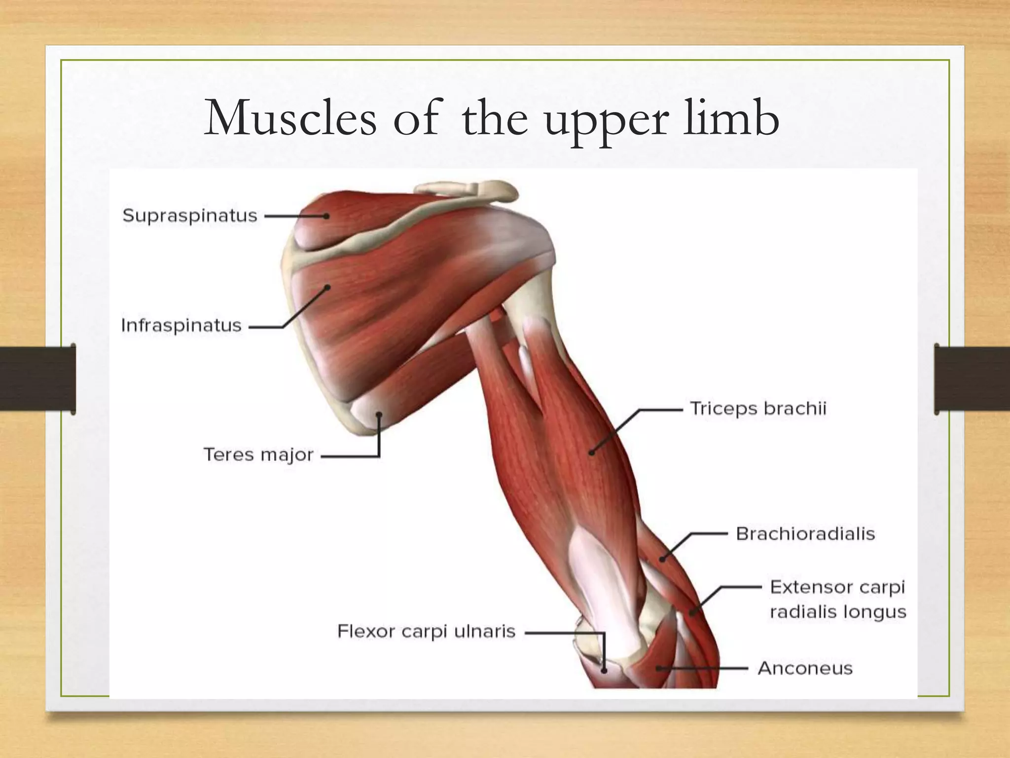
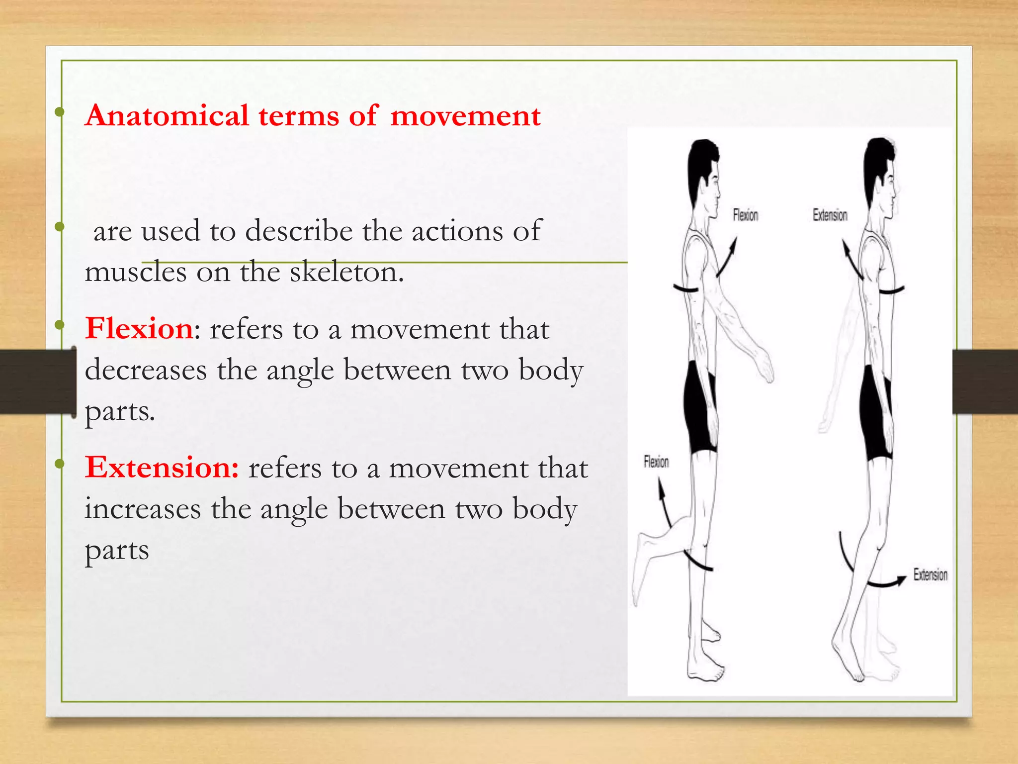
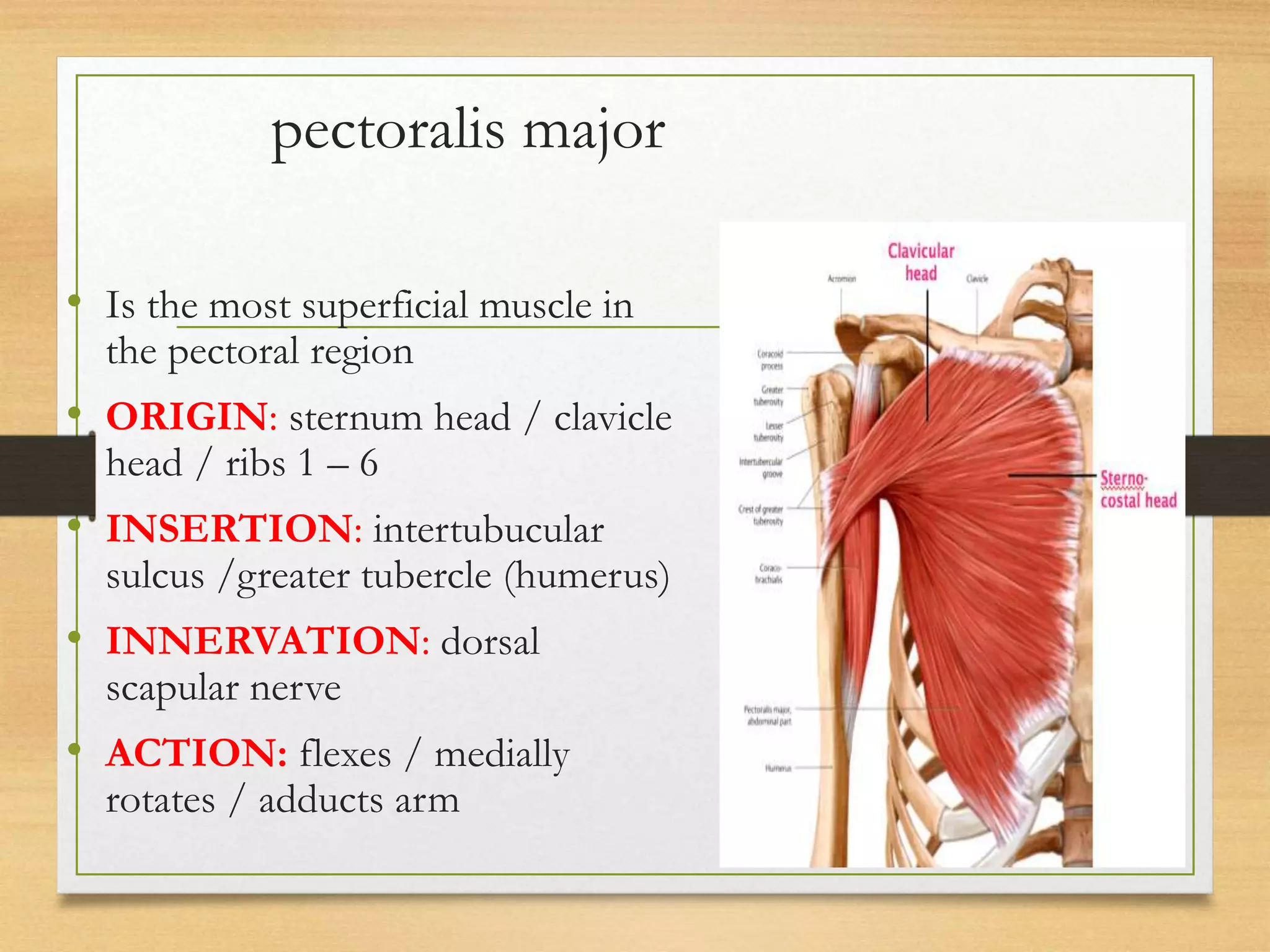
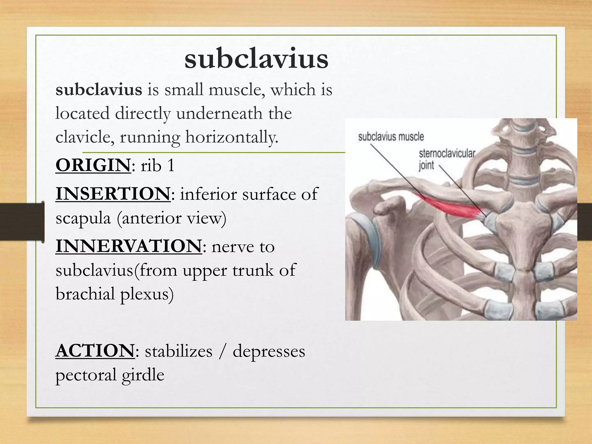
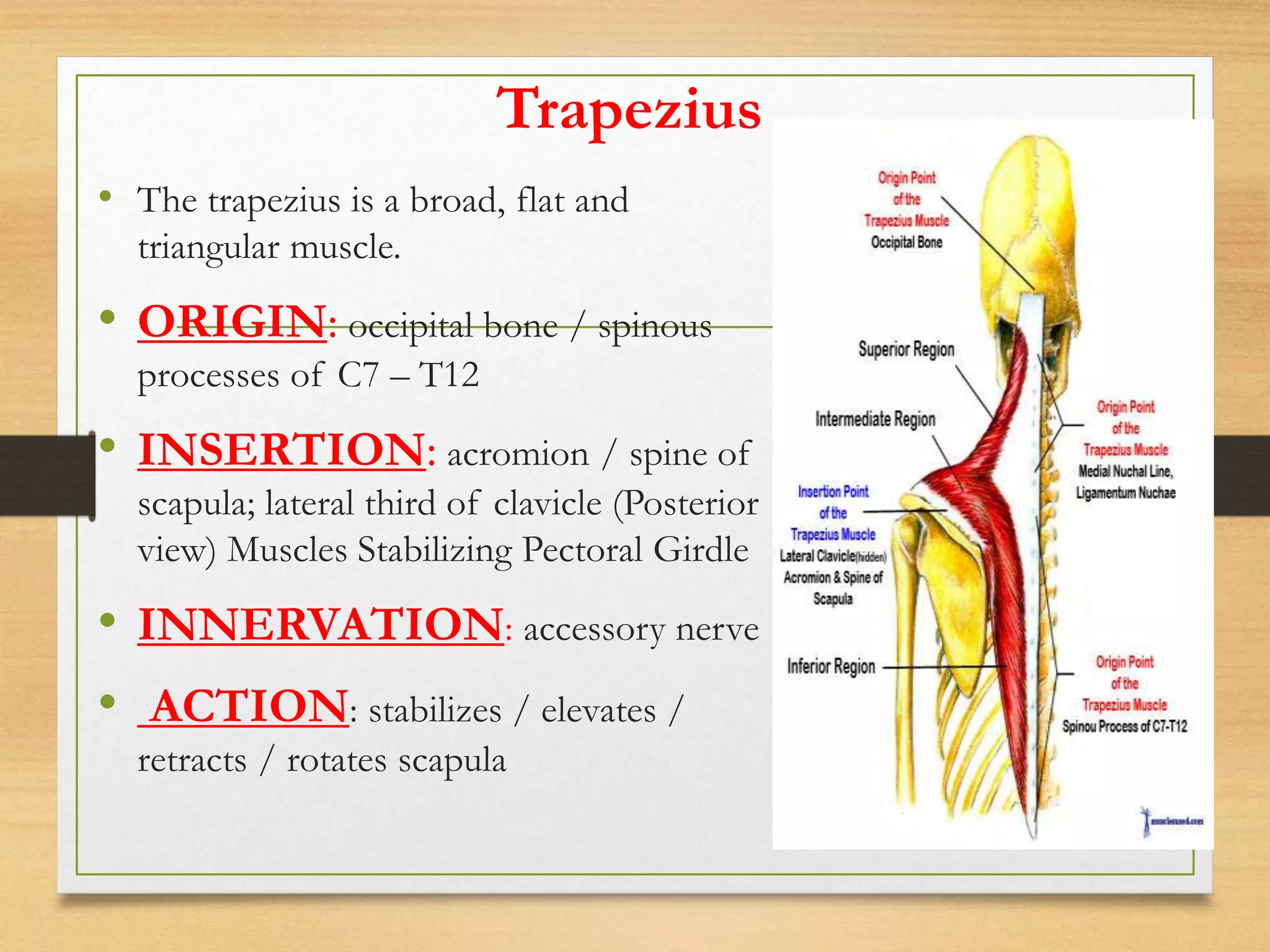
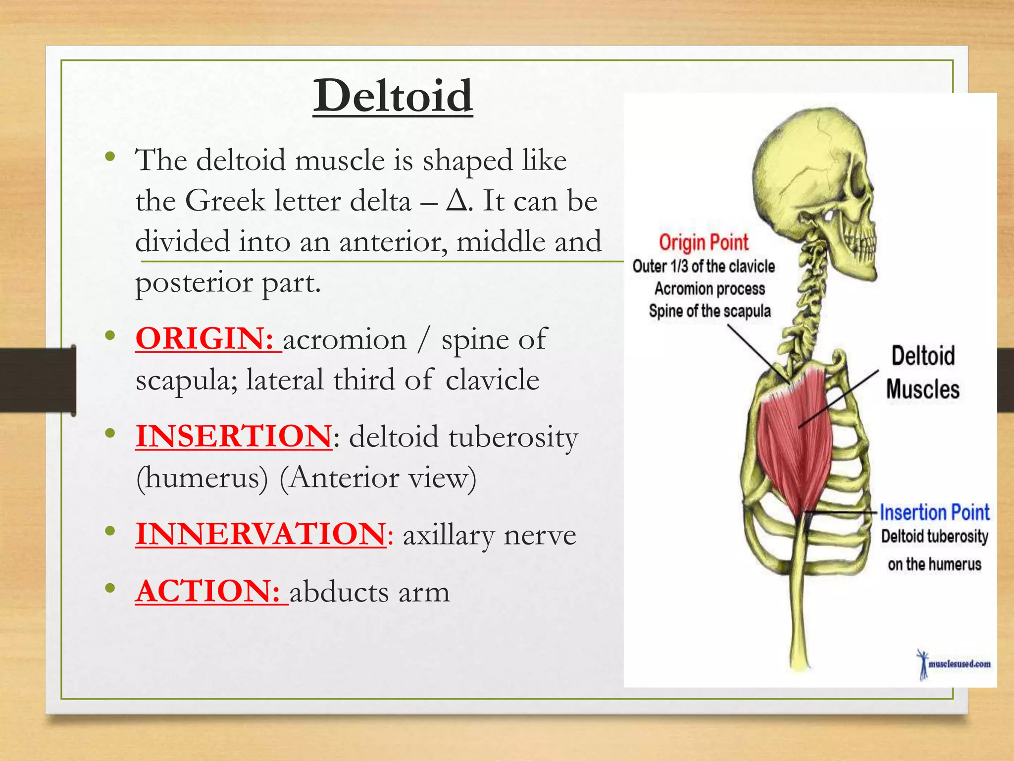
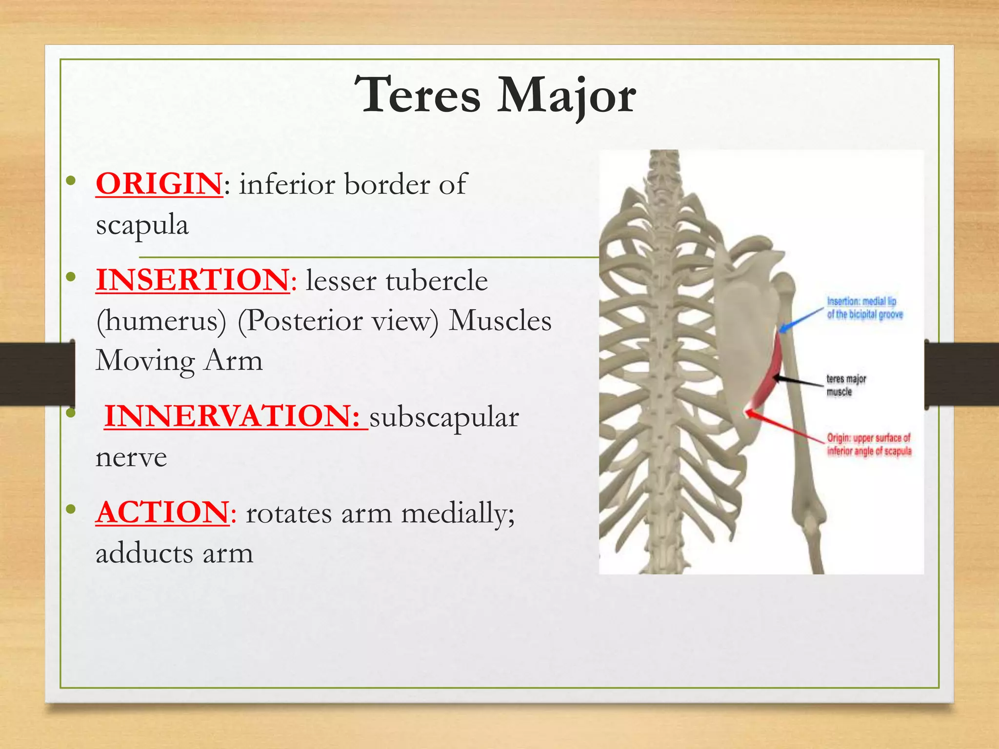
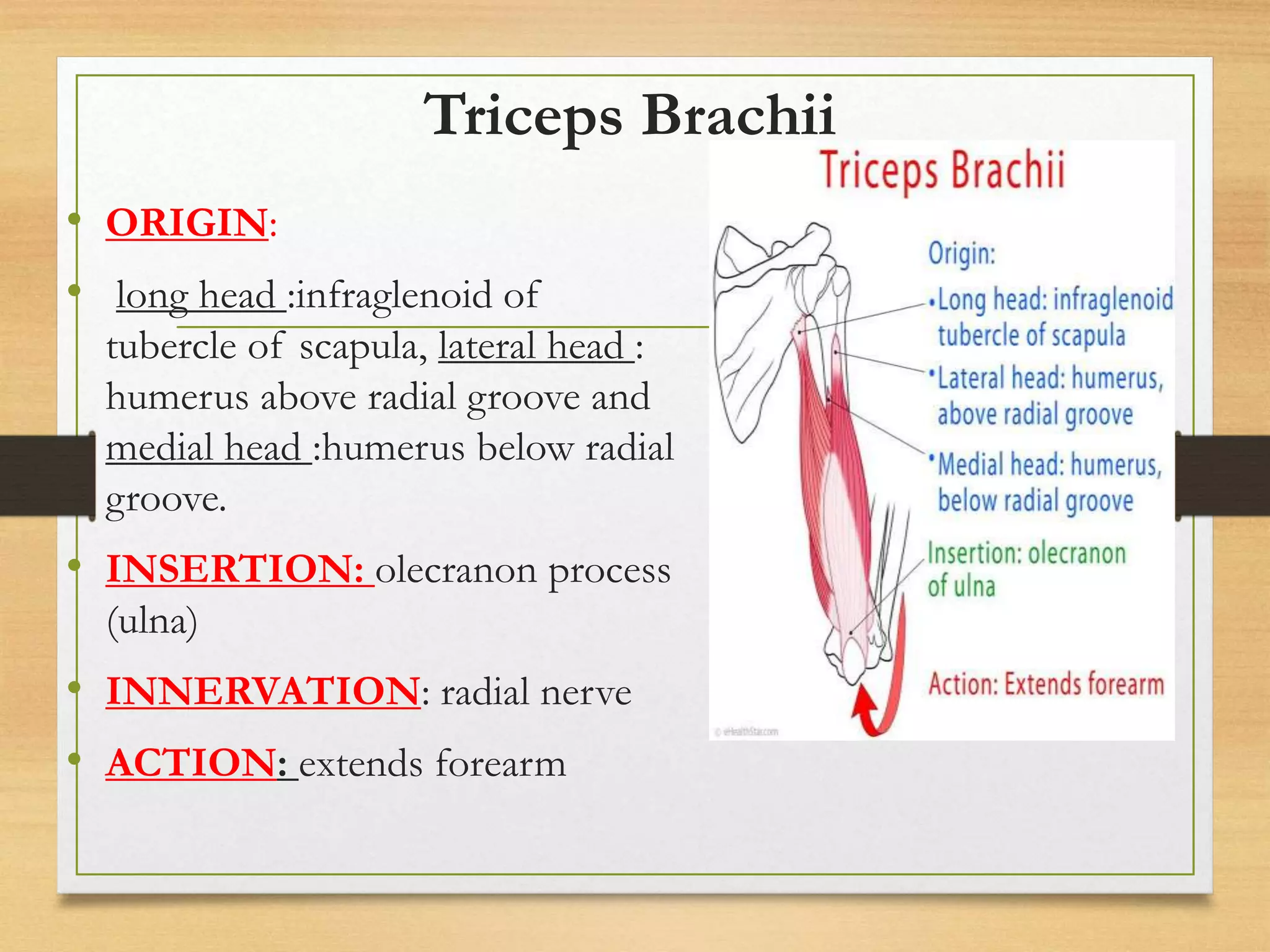
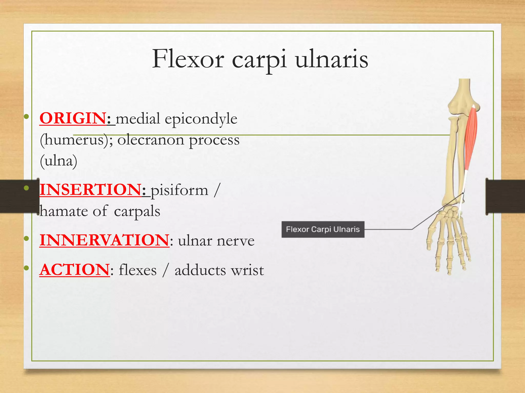
Human anatomy
Dr. Ahmed H. Ahmed
Muscles of the upper limb
introduction
A muscle is made of strong tissue that can
contract in an orderly way.
•the muscular system is made of different types
of muscles and has several functions.
•All muscle tissues are made of cells that
contract.
Function of muscle
Movement:
 These muscles make your skeleton move.
When muscles contract, they move bones
Stability:
Muscles that are attached to your bones
support your body and help you keep your
balance.
Cont.
Protection:
•Muscles protect your body.
• They cover most of your skeleton.
•Muscles also cover most of the organs inside your
body.
Temperature regulation:
• The muscular system helps your body keep
your internal temperature within a certain
range.
Conti.
Posture
• when someone is sitting or Skeletal muscles help
keep the body in the correct position standing.
Respiration
• Breathing involves the use of the diaphragm muscle.
Cont…
• muscles are also:
Contractible (they can shorten in length)
Extensible (they can extend or stretch)
Elastic (they can return to their original shape)
Conti.
• Skeletal muscle is composed of skeletal
muscle tissue and also contains nervous
tissue, blood vessels and connective tissue
• Half of the body’s weight is muscle tissue
• Skeletal muscle = 40% in males, 32% in females
• Cardiac muscle = 10%
(b) Cardiac muscle (c) Visceral smooth muscle
(a) Skeletal muscle
Three Types of Muscular Tissue
Location Function Appearance Control
Skeletal
skeleton
movement,
heat, posture
striated, multi-
nucleated (eccentric),
fibers parallel
voluntary
Cardiac
heart
pump blood
continuously
striated, one central
nucleus
involuntary
Visceral
(smooth muscle) G.I. tract,
uterus, eye,
blood vessels
Peristalsis,
blood pressure,
pupil size,
erects hairs
no striations, one
central nucleus
involuntary
Three Types of Muscular Tissue
Tendons
• Tendons attach muscles to bones.
•Tendons work with muscles and keep
your joints in place when your body
moves.
•Tendons also help hold your body in a
correct posture, or shape.
Origin ,insertion and action
• Origin: is the muscle attachment site that
does NOT move during contraction.
• Insertion: is the muscle attachment that
does move during contraction.
• Innervation: The supply or distribution
of nerve fibres to any part of the body.
• During a muscle action,
the INSERTION is pulled towards
the ORIGIN.
• Anatomical terms of movement
• are used to describe the actions of
muscles on the skeleton.
• Flexion: refers to a movement that
decreases the angle between two body
parts.
• Extension: refers to a movement that
increases the angle between two body
parts
Conti.
• Abduction: is a movement away from
the midline – just as abducting
someone is to take them away.
• Adduction: is a movement towards the
midline. Adduction of the hip squeezes
the legs together.
• Medial rotation: is a rotational
movement towards the midline.
• Lateral rotation: is a rotating
movement away from the midline.
The muscles of upper limb can be divided into
6 different regions
1. Muscles of the Pectoral region
2. Muscles of the shoulder region
3. Muscles of the upper arm
4. Muscles in the anterior compartment of the forearm
5. Muscles in the posterior compartment of the forearm
6. Muscles of the hand
Muscles of the Pectoral region
The pectoral region is located on the anterior chest wall.
It contains four muscles that exert a force on the upper
limb;
the pectoralis major,
 pectoralis minor,
 serratus anterior
and subclavius.
pectoralis major
• Is the most superficial muscle in
the pectoral region
• ORIGIN: sternum head / clavicle
head / ribs 1 – 6
• INSERTION: intertubucular
sulcus /greater tubercle (humerus)
• INNERVATION: dorsal
scapular nerve
• ACTION: flexes / medially
rotates / adducts arm
Pectoralis minor
• ORIGIN: anterior surface of ribs 3 – 5
• INSERTION: coracoid process (scapula)
• INNERVATION: pectoral nerves
• ACTION: protracts / retarction scapula
serratus anterior
• ORIGIN: ribs 1 - 8
• INSERTION: medial border of
scapula (Lateral view)
• INNERVATION: long thoracic
nerve
• ACTION: rotates scapula laterally
subclavius
subclavius is small muscle, which is
located directly underneath the
clavicle, running horizontally.
ORIGIN: rib 1
INSERTION: inferior surface of
scapula (anterior view)
INNERVATION: nerve to
subclavius(from upper trunk of
brachial plexus)
ACTION: stabilizes / depresses
pectoral girdle
Muscles of the shoulder region
• The muscles of the shoulder are associated with movements of
the upper limb. , and can be divided into two groups:
• Extrinsic – originate from the trunck, and attach to the bones
of the shoulder (clavicle, scapula or humerus).
• Intrinsic – originate from the scapula and clavicle, and attach
to the humerus.
There are two superficial extrinsic muscles – the trapezius and
latissimus dorsi.
There are three deep muscles – the levator scapulae and the
two rhomboids.
Trapezius
• The trapezius is a broad, flat and
triangular muscle.
• ORIGIN: occipital bone / spinous
processes of C7 – T12
• INSERTION: acromion / spine of
scapula; lateral third of clavicle (Posterior
view) Muscles Stabilizing Pectoral Girdle
• INNERVATION: accessory nerve
• ACTION: stabilizes / elevates /
retracts / rotates scapula
Latissimus Dorsi
• ORIGIN: spinous processes of T7
– L5; ribs 9 – 12; iliac crest of oscoxae
• INSERTION: intertubercular
groove (humerus) Muscles Moving Arm
• INNERVATION: thoracodorsal
nerve
• ACTION: extends / adducts
arm/medial rotation
Deep layer
• There are three muscles in this group –
the levator scapulae
and the two rhomboids.
Levator Scapulae
The levator scapulae is a small strap-like
muscle.
ORIGIN: transverse processes of C1
– C4
INSERTION: upper medial border
of scapula (Posterior view)
INNERVATION: dorsal scapular
nerve
ACTION: elevates / adducts scapula
Rhomboids (major / minor)
• ORIGIN: spinous processes
of C7 – T5
• INSERTION: medial border
of scapula (Posterior view)
Rhomboids (major / minor)
Muscles Stabilizing Pectoral
Girdle
• INNERVATION: dorsal
scapular nerve
• ACTION: adducts / rotates
scapula
Intrinsic muscles of shoulder
• The intrinsic muscles (also known as the
scapulohumeral group) originate from the scapula
and clavicle, and attach to the humerus.
• There are six muscles in this group – the deltoid,
teres major, and the four rotator cuff muscles
(supraspinatus, infraspinatus, subscapularis and
teres minor).
Deltoid
• The deltoid muscle is shaped like
the Greek letter delta – Δ. It can be
divided into an anterior, middle and
posterior part.
• ORIGIN: acromion / spine of
scapula; lateral third of clavicle
• INSERTION: deltoid tuberosity
(humerus) (Anterior view)
• INNERVATION: axillary nerve
• ACTION: abducts arm
Teres Major
• ORIGIN: inferior border of
scapula
• INSERTION: lesser tubercle
(humerus) (Posterior view) Muscles
Moving Arm
• INNERVATION: subscapular
nerve
• ACTION: rotates arm medially;
adducts arm
Teres Minor
• ORIGIN: lateral border of
scapula
• INSERTION: greater tubercle
(humerus) (Posterior view)
Muscles Moving Arm
• INNERVATION: subscapular
nerve
• ACTION: externally rotates the
arm
Rotator Cuff Muscles
• The rotator cuff muscles are a group of four muscles
that originate from the scapula and attach to the
humeral head.
• Supraspinatus
• Infraspinatus
• Subscapularis
• Teres Minor
Infraspinatus
• ORIGIN: infraspinous fossa
(scapula)
• INSERTION: greater tubercle
(humerus) (Posterior view)
• INNERVATION:
suprascapular nerve
• ACTION: rotates arm laterally
Supraspinatus
• ORIGIN: supraspinous fossa
(scapula)
• INSERTION: greater tubercle
(humerus) (Posterior view)
Muscles Moving Arm
• INNERVATION:
suprascapular nerve
• ACTION: abducts arm
Subscapularis
• ORIGIN: subscapular fossa
(scapula)
• INSERTION: lesser tubercle
(humerus)
• INNERVATION: subscapular
nerve
• ACTION: rotates arm medially
Muscles of the Upper Arm
The upper arm is located between the shoulder joint
and elbow joint.
It contains four muscles – three in the anterior
compartment (biceps brachii, brachialis,
coracobrachialis), and one in the posterior
compartment (triceps brachii).
biceps brachii
• The biceps brachii is a two-
headed(long and short) muscle.
• ORIGIN: short head coracoid
process / supraglenoid tubercle
(scapula)
• INSERTION: radial tuberosity
(radius)
• INNERVATION:
musculocutaneous nerve
• ACTION: flexes forearm
brachialis
• ORIGIN: anterior face of
distal humerus
• INSERTION: coronoid
process (ulna)
• INNERVATION:
musculocutaneous nerve
• ACTION: flexes forearm
coracobrachialis
• ORIGIN: coracoid process
(scapula)
• INSERTION: medial shaft of
humerus (Anterior view)
Muscles Moving Arm
• INNERVATION:
musculocutaneous nerve
• ACTION: flexes / adducts arm
Triceps Brachii
• ORIGIN:
• long head :infraglenoid of
tubercle of scapula, lateral head :
humerus above radial groove and
medial head :humerus below radial
groove.
• INSERTION: olecranon process
(ulna)
• INNERVATION: radial nerve
• ACTION: extends forearm
Muscles in the Anterior Compartment of
the Forearm
• There are many muscles in the forearm. In the
anterior compartment, they are split into three
categories; superficial, intermediate and deep.
• In general, muscles in the anterior compartment of
the forearm perform flexion at the wrist and fingers.
Superficial Compartment
The superficial muscles in the anterior compartment are the
flexor carpi ulnaris,
palmaris longus,
flexor carpi radialis and
 pronator teres.
They all originate from a common tendon, which arises from
the medial epicondyle of the humurus
flexor carpi radialis
• ORIGIN: medial epicondyle (humerus)
• INSERTION: metacarpals 2 – 3
• INNERVATION: median nerve
• ACTION: flexes / abducts wrist
Flexor carpi ulnaris
• ORIGIN: medial epicondyle
(humerus); olecranon process
(ulna)
• INSERTION: pisiform /
hamate of carpals
• INNERVATION: ulnar nerve
• ACTION: flexes / adducts wrist
Intermediate Compartment
• The flexor digitorum superficialis is the only
muscle of the intermediate compartment.
• It can sometimes be classed as a superficial muscle, it
lies between the deep and superficial muscle layers.
flexor digitorum superficialis
• ORIGIN: medial epicondyle
(humerus); coronoid process (ulna);
shaft of radius
• INSERTION: middle phalanges 2 -
5
• INNERVATION: median nerve
• ACTION: flexes wrist / fingers
Deep Compartment
There are three muscles in the deep anterior
forearm;
• flexor digitorum profundus,
• flexor pollicis longus, and
• pronator quadratus.
flexor digitorum profundus
• ORIGIN: coronoid process /
anteriolmedial surface (ulna)
INSERTION: distal
phalanges 2 - 5
• INNERVATION: ulnar /
median nerves
• ACTION: flexes wrist /
fingers
flexor pollicis longus
• ORIGIN: anterior surface of
radius
• INSERTION: distal phalanx
of thumb
• INNERVATION: median
nerve
• ACTION: flexes thumb
pronator quadratus
• ORIGIN: distal surface of
anterior ulna
• INSERTION: distal surface of
anterior radius
• INNERVATION: median
nerve
• ACTION: pronates( Rotation)
forearm
Muscles in the Posterior
Compartment of the Forearm
• The muscles in the posterior compartment of the
forearm are commonly known as the extensor
muscles.
• The general function of these muscles is to produce
extension at the wrist and fingers.
• The muscles in this compartment can be divided into
two layers; deep and superficial.
Superficial Muscles
The superficial layer of the posterior forearm contains six
muscles.
Brachioradialis.
Anconeus
extensor carpi radialis brevis,
extensor digitorum,
extensor carpi ulnaris and
extensor digiti minimi.
thanks

L9 muscles of upper limb [Autosaved].pptx

  • 1.
  • 2.
    Muscles of theupper limb
  • 3.
    introduction A muscle ismade of strong tissue that can contract in an orderly way. •the muscular system is made of different types of muscles and has several functions. •All muscle tissues are made of cells that contract.
  • 4.
    Function of muscle Movement: These muscles make your skeleton move. When muscles contract, they move bones Stability: Muscles that are attached to your bones support your body and help you keep your balance.
  • 5.
    Cont. Protection: •Muscles protect yourbody. • They cover most of your skeleton. •Muscles also cover most of the organs inside your body. Temperature regulation: • The muscular system helps your body keep your internal temperature within a certain range.
  • 6.
    Conti. Posture • when someoneis sitting or Skeletal muscles help keep the body in the correct position standing. Respiration • Breathing involves the use of the diaphragm muscle.
  • 7.
    Cont… • muscles arealso: Contractible (they can shorten in length) Extensible (they can extend or stretch) Elastic (they can return to their original shape)
  • 8.
    Conti. • Skeletal muscleis composed of skeletal muscle tissue and also contains nervous tissue, blood vessels and connective tissue • Half of the body’s weight is muscle tissue • Skeletal muscle = 40% in males, 32% in females • Cardiac muscle = 10%
  • 9.
    (b) Cardiac muscle(c) Visceral smooth muscle (a) Skeletal muscle Three Types of Muscular Tissue
  • 10.
    Location Function AppearanceControl Skeletal skeleton movement, heat, posture striated, multi- nucleated (eccentric), fibers parallel voluntary Cardiac heart pump blood continuously striated, one central nucleus involuntary Visceral (smooth muscle) G.I. tract, uterus, eye, blood vessels Peristalsis, blood pressure, pupil size, erects hairs no striations, one central nucleus involuntary Three Types of Muscular Tissue
  • 11.
    Tendons • Tendons attachmuscles to bones. •Tendons work with muscles and keep your joints in place when your body moves. •Tendons also help hold your body in a correct posture, or shape.
  • 12.
    Origin ,insertion andaction • Origin: is the muscle attachment site that does NOT move during contraction. • Insertion: is the muscle attachment that does move during contraction. • Innervation: The supply or distribution of nerve fibres to any part of the body. • During a muscle action, the INSERTION is pulled towards the ORIGIN.
  • 13.
    • Anatomical termsof movement • are used to describe the actions of muscles on the skeleton. • Flexion: refers to a movement that decreases the angle between two body parts. • Extension: refers to a movement that increases the angle between two body parts
  • 14.
    Conti. • Abduction: isa movement away from the midline – just as abducting someone is to take them away. • Adduction: is a movement towards the midline. Adduction of the hip squeezes the legs together. • Medial rotation: is a rotational movement towards the midline. • Lateral rotation: is a rotating movement away from the midline.
  • 15.
    The muscles ofupper limb can be divided into 6 different regions 1. Muscles of the Pectoral region 2. Muscles of the shoulder region 3. Muscles of the upper arm 4. Muscles in the anterior compartment of the forearm 5. Muscles in the posterior compartment of the forearm 6. Muscles of the hand
  • 16.
    Muscles of thePectoral region The pectoral region is located on the anterior chest wall. It contains four muscles that exert a force on the upper limb; the pectoralis major,  pectoralis minor,  serratus anterior and subclavius.
  • 17.
    pectoralis major • Isthe most superficial muscle in the pectoral region • ORIGIN: sternum head / clavicle head / ribs 1 – 6 • INSERTION: intertubucular sulcus /greater tubercle (humerus) • INNERVATION: dorsal scapular nerve • ACTION: flexes / medially rotates / adducts arm
  • 18.
    Pectoralis minor • ORIGIN:anterior surface of ribs 3 – 5 • INSERTION: coracoid process (scapula) • INNERVATION: pectoral nerves • ACTION: protracts / retarction scapula
  • 19.
    serratus anterior • ORIGIN:ribs 1 - 8 • INSERTION: medial border of scapula (Lateral view) • INNERVATION: long thoracic nerve • ACTION: rotates scapula laterally
  • 20.
    subclavius subclavius is smallmuscle, which is located directly underneath the clavicle, running horizontally. ORIGIN: rib 1 INSERTION: inferior surface of scapula (anterior view) INNERVATION: nerve to subclavius(from upper trunk of brachial plexus) ACTION: stabilizes / depresses pectoral girdle
  • 21.
    Muscles of theshoulder region • The muscles of the shoulder are associated with movements of the upper limb. , and can be divided into two groups: • Extrinsic – originate from the trunck, and attach to the bones of the shoulder (clavicle, scapula or humerus). • Intrinsic – originate from the scapula and clavicle, and attach to the humerus. There are two superficial extrinsic muscles – the trapezius and latissimus dorsi. There are three deep muscles – the levator scapulae and the two rhomboids.
  • 22.
    Trapezius • The trapeziusis a broad, flat and triangular muscle. • ORIGIN: occipital bone / spinous processes of C7 – T12 • INSERTION: acromion / spine of scapula; lateral third of clavicle (Posterior view) Muscles Stabilizing Pectoral Girdle • INNERVATION: accessory nerve • ACTION: stabilizes / elevates / retracts / rotates scapula
  • 23.
    Latissimus Dorsi • ORIGIN:spinous processes of T7 – L5; ribs 9 – 12; iliac crest of oscoxae • INSERTION: intertubercular groove (humerus) Muscles Moving Arm • INNERVATION: thoracodorsal nerve • ACTION: extends / adducts arm/medial rotation
  • 24.
    Deep layer • Thereare three muscles in this group – the levator scapulae and the two rhomboids.
  • 25.
    Levator Scapulae The levatorscapulae is a small strap-like muscle. ORIGIN: transverse processes of C1 – C4 INSERTION: upper medial border of scapula (Posterior view) INNERVATION: dorsal scapular nerve ACTION: elevates / adducts scapula
  • 26.
    Rhomboids (major /minor) • ORIGIN: spinous processes of C7 – T5 • INSERTION: medial border of scapula (Posterior view) Rhomboids (major / minor) Muscles Stabilizing Pectoral Girdle • INNERVATION: dorsal scapular nerve • ACTION: adducts / rotates scapula
  • 27.
    Intrinsic muscles ofshoulder • The intrinsic muscles (also known as the scapulohumeral group) originate from the scapula and clavicle, and attach to the humerus. • There are six muscles in this group – the deltoid, teres major, and the four rotator cuff muscles (supraspinatus, infraspinatus, subscapularis and teres minor).
  • 28.
    Deltoid • The deltoidmuscle is shaped like the Greek letter delta – Δ. It can be divided into an anterior, middle and posterior part. • ORIGIN: acromion / spine of scapula; lateral third of clavicle • INSERTION: deltoid tuberosity (humerus) (Anterior view) • INNERVATION: axillary nerve • ACTION: abducts arm
  • 29.
    Teres Major • ORIGIN:inferior border of scapula • INSERTION: lesser tubercle (humerus) (Posterior view) Muscles Moving Arm • INNERVATION: subscapular nerve • ACTION: rotates arm medially; adducts arm
  • 30.
    Teres Minor • ORIGIN:lateral border of scapula • INSERTION: greater tubercle (humerus) (Posterior view) Muscles Moving Arm • INNERVATION: subscapular nerve • ACTION: externally rotates the arm
  • 31.
    Rotator Cuff Muscles •The rotator cuff muscles are a group of four muscles that originate from the scapula and attach to the humeral head. • Supraspinatus • Infraspinatus • Subscapularis • Teres Minor
  • 32.
    Infraspinatus • ORIGIN: infraspinousfossa (scapula) • INSERTION: greater tubercle (humerus) (Posterior view) • INNERVATION: suprascapular nerve • ACTION: rotates arm laterally
  • 33.
    Supraspinatus • ORIGIN: supraspinousfossa (scapula) • INSERTION: greater tubercle (humerus) (Posterior view) Muscles Moving Arm • INNERVATION: suprascapular nerve • ACTION: abducts arm
  • 34.
    Subscapularis • ORIGIN: subscapularfossa (scapula) • INSERTION: lesser tubercle (humerus) • INNERVATION: subscapular nerve • ACTION: rotates arm medially
  • 35.
    Muscles of theUpper Arm The upper arm is located between the shoulder joint and elbow joint. It contains four muscles – three in the anterior compartment (biceps brachii, brachialis, coracobrachialis), and one in the posterior compartment (triceps brachii).
  • 36.
    biceps brachii • Thebiceps brachii is a two- headed(long and short) muscle. • ORIGIN: short head coracoid process / supraglenoid tubercle (scapula) • INSERTION: radial tuberosity (radius) • INNERVATION: musculocutaneous nerve • ACTION: flexes forearm
  • 37.
    brachialis • ORIGIN: anteriorface of distal humerus • INSERTION: coronoid process (ulna) • INNERVATION: musculocutaneous nerve • ACTION: flexes forearm
  • 38.
    coracobrachialis • ORIGIN: coracoidprocess (scapula) • INSERTION: medial shaft of humerus (Anterior view) Muscles Moving Arm • INNERVATION: musculocutaneous nerve • ACTION: flexes / adducts arm
  • 39.
    Triceps Brachii • ORIGIN: •long head :infraglenoid of tubercle of scapula, lateral head : humerus above radial groove and medial head :humerus below radial groove. • INSERTION: olecranon process (ulna) • INNERVATION: radial nerve • ACTION: extends forearm
  • 40.
    Muscles in theAnterior Compartment of the Forearm • There are many muscles in the forearm. In the anterior compartment, they are split into three categories; superficial, intermediate and deep. • In general, muscles in the anterior compartment of the forearm perform flexion at the wrist and fingers.
  • 41.
    Superficial Compartment The superficialmuscles in the anterior compartment are the flexor carpi ulnaris, palmaris longus, flexor carpi radialis and  pronator teres. They all originate from a common tendon, which arises from the medial epicondyle of the humurus
  • 42.
    flexor carpi radialis •ORIGIN: medial epicondyle (humerus) • INSERTION: metacarpals 2 – 3 • INNERVATION: median nerve • ACTION: flexes / abducts wrist
  • 43.
    Flexor carpi ulnaris •ORIGIN: medial epicondyle (humerus); olecranon process (ulna) • INSERTION: pisiform / hamate of carpals • INNERVATION: ulnar nerve • ACTION: flexes / adducts wrist
  • 44.
    Intermediate Compartment • Theflexor digitorum superficialis is the only muscle of the intermediate compartment. • It can sometimes be classed as a superficial muscle, it lies between the deep and superficial muscle layers.
  • 45.
    flexor digitorum superficialis •ORIGIN: medial epicondyle (humerus); coronoid process (ulna); shaft of radius • INSERTION: middle phalanges 2 - 5 • INNERVATION: median nerve • ACTION: flexes wrist / fingers
  • 46.
    Deep Compartment There arethree muscles in the deep anterior forearm; • flexor digitorum profundus, • flexor pollicis longus, and • pronator quadratus.
  • 47.
    flexor digitorum profundus •ORIGIN: coronoid process / anteriolmedial surface (ulna) INSERTION: distal phalanges 2 - 5 • INNERVATION: ulnar / median nerves • ACTION: flexes wrist / fingers
  • 48.
    flexor pollicis longus •ORIGIN: anterior surface of radius • INSERTION: distal phalanx of thumb • INNERVATION: median nerve • ACTION: flexes thumb
  • 49.
    pronator quadratus • ORIGIN:distal surface of anterior ulna • INSERTION: distal surface of anterior radius • INNERVATION: median nerve • ACTION: pronates( Rotation) forearm
  • 50.
    Muscles in thePosterior Compartment of the Forearm • The muscles in the posterior compartment of the forearm are commonly known as the extensor muscles. • The general function of these muscles is to produce extension at the wrist and fingers. • The muscles in this compartment can be divided into two layers; deep and superficial.
  • 51.
    Superficial Muscles The superficiallayer of the posterior forearm contains six muscles. Brachioradialis. Anconeus extensor carpi radialis brevis, extensor digitorum, extensor carpi ulnaris and extensor digiti minimi.
  • 52.