Development of face
Dr.abdisamad osman shiekh
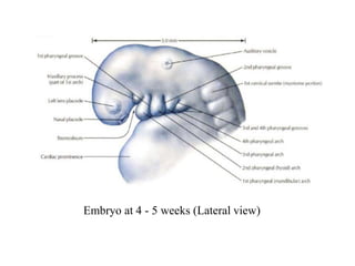
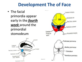
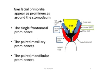
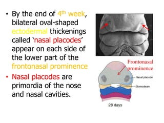
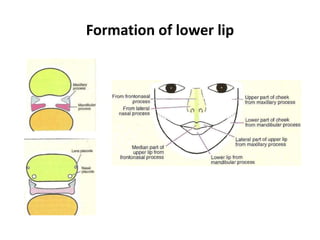
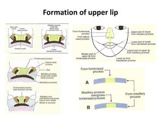
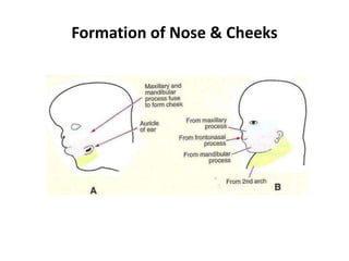
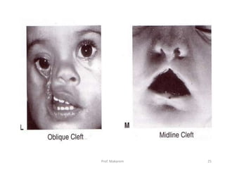
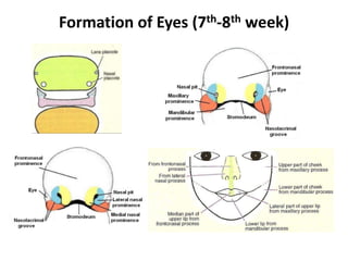
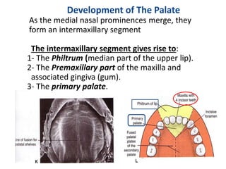
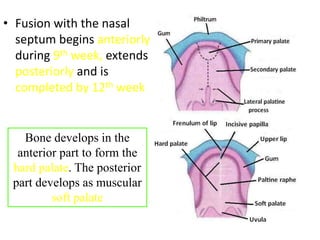
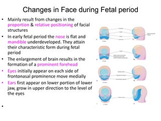
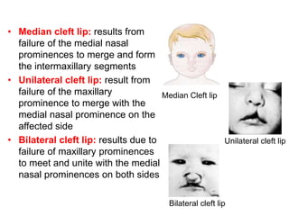
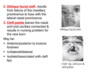
• The development of the face occurs mainly
between 5 – 8 weeks
• The lower jaw (mandible) is the first to form (4th
week)
• The facial proportions develop during the fetal
period (9th week to birth)
• During infancy & childhood, following the
development of teeth and paranasal sinuses, the
facial skeleton increases in size and contribute to
the definitive shape of the face
Embryo at 4 - 5 weeks (Lateral view)
Development The of Face
• The facial
primordia appear
early in the fourth
week around the
primordial
stomodeum
md
md
FN
S
4
Prof. Makarem
MX
MX
Five facial primordia
appear as prominences
around the stomodeum
• The single frontonasal
prominence
• The paired maxillary
prominences
• The paired mandibular
prominences
FNP
5
Prof. Makarem
• The single frontonasal
prominence ventral to
the forebrain
• The paired maxillary
prominences develop
from the cranial part of
first branchial arch
• The paired mandibular
prominences develop
from the caudal part of
first branchial arch
Lateral view
• The mesoderm of the
five prominences is
continuous with each
other
• There is no internal
division
corresponding to the
grooves demarcating
the prominences
externally
Stomodeum
• An ectoderm lined depression
• Separated from the primitive pharynx by the
buccopharyngeal (oropharyngeal) membrane
• The membrane later breaks down and stomodeum
opens into the pharynx
Forms the
vestibule of the
oral cavity
• By the end of 4th week,
bilateral oval-shaped
ectodermal thickenings
called ‘nasal placodes’
appear on each side of
the lower part of the
frontonasal prominence
• Nasal placodes are
primordia of the nose
and nasal cavities.
Frontonasal
prominence
• Mesenchymal cells
proliferate at the margin of
the placodes and produce
horse-shoe shaped
swellings around these.
• The sides of these swellings
are called ‘medial’ and
‘lateral’ nasal prominences
• The placodes now lie in the
floor of a depression called
‘nasal pits’
Each lateral nasal prominence is separated from
the maxillary swelling by nasolacrimal groove
Formation of lower lip
• The lower jaw and lower
lips are the first parts of
the face to form
• They result from merging
of the medial ends of the
mandibular prominences
in the median plane
• Median cleft lower lip is
a very rare condition M
12
Prof. Makarem
Formation of upper lip
• The maxillary prominences
continue to increase in size
and:
• Laterally, merge with the
mandibular prominences to
form the cheek
• Medially, compress the
medial nasal prominences
toward the midline and
finally fuses with these to
form the upper lip.
The upper lip is formed by the two medial nasal
prominences & the two maxillary prominences
• Between the 7th and 8th weeks, the two medial
nasal prominences merge with each other and
with the maxillary and lateral nasal prominences
Merging of the medial nasal and maxillary
prominences results in continuity of the upper
jaw and upper lip and separation of the nasal
pits from the stomodeum
15
Prof. Makarem
Formation of Nose & Cheeks
• With the formation of
the medial and lateral
nasal prominences, the
nasal placodes lie in the
floor of depressions
called the nasal pits
• By the end of 6th week,
nasal pits deepen and
form nasal sacs
• Each nasal sac grows
dorsocaudally, ventral to
the developing brain
• Initially the nasal
sacs are separated
from the oral cavity
by oronasal
membrane.
• The oronasal
membrane ruptures
by the 7th week,
communicating the
primitive nasal
cavities with the oral
cavity
• These communications
are called the primitive
choanae and are
located posterior to the
primary palate
• After the development
of the secondary palate,
the choanae change
their position and
become located at the
junction of nasal cavity
and the pharynx
• The superior, middle
and inferior conchae
develop on the lateral
wall of each nasal
cavity
• The ectodermal
epithelium in the roof
of each nasal cavity
becomes specialized
as the olfactory
epithelium
• The olfactory cells
of the olfactory
epithelium give
origin to olfactory
nerve fibers that
grow into the
olfactory bulb
• The paranasal sinuses
develop as diverticulae
of the walls of the nasal
cavity
• Maxillary sinuses and
few anterior & posterior
ethmoidal air cells
develop in fetal life
• Frontal and sphenoidal
sinuses develop after
birth
E
M
From a 3 months old fetus, showing
ethmoid & maxillary sinuses
Nasolacrimal duct
• Develops from a rod-like thickening of the ectoderm in the
floor of the nasolacrimal groove
• This solid cord of cells separates from the surface ectoderm
and lies in the underlying mesenchyme
• The cord gets canalized to form the nasolacrimal duct
• The cranial end of the duct expands to form the lacrimal sac
• The caudal end opens into the inferior meatus of the nasal
cavity
• The duct is usually becomes completely patent only after
birth
• Failure of complete canalization of the duct leads to atresia
of the duct (seen in about 6% of newborn infants)
Prof. Makarem 25
Formation of External Ear (6th week)
Development of the External Ear
• By the end of the fifth week, the
primordia of the auricles of the ears have
begun to develop
• Six auricular hillocks form around the first
pharyngeal groove (cleft).
• Three on each side of the 1st pharyngeal
groove (cleft).
• These are the primordia of the auricle
and external acoustic meatus.
• Initially the ear located in the neck.
• As the mandible develops the ears ascend
to the level of the eye.
Prof. Makarem 27
Formation of Eyes (7th-8th week)
Development of Palate (Palatogenesis)
Prof. Makarem 30
Development of The Palate
As the medial nasal prominences merge, they
form an intermaxillary segment
The intermaxillary segment gives rise to:
1- The Philtrum (median part of the upper lip).
2- The Premaxillary part of the maxilla and
associated gingiva (gum).
3- The primary palate.
The palate develops from two primordia:
• The Primary palate
• The Secondary palate
• Begins at the end of the 5th week
• Gets completed by the end of the 12th week
• The most critical period for the development
of palate is from the end of 6th week to the
beginning of 9th week
Palatogenesis
The Primary Palate
• Begins to develop:
 Early in the 6th week
 From the deep part of
the intermaxillary
segment, as median
palatine process
• Lies behind the
premaxillary part of the
maxilla
• Fuses with the
developing secondary
palate
The primary palate represents only a small
part lying anterior to the incisive fossa, of the
adult hard palate
Hard palate
Primary
palate
Soft palate
Secondary
palate
The Secondary Palate
• Is the primordia of hard
and soft palate
posterior to the incisive
fossa
• Begins to develop:
 Early in the 6th week
 From the internal
aspect of the
maxillary processes,
as lateral palatine
process
• In the beginning, the
lateral palatine
processes project
inferomedially on each
side of the tongue
• With the development
of the jaws, the tongue
moves inferiorly.
• During 7th & 8th weeks,
the lateral palatine
processes elongate and
ascend to a horizontal
position above the
tongue
Tongue
• Gradually the lateral
palatine processes:
 Grow medially and
fuse in the median
plane
 Also fuse with the:
• Posterior part of
the primary palate
&
• The nasal septum
• Fusion with the nasal
septum begins anteriorly
during 9th week, extends
posteriorly and is
completed by 12th week
Bone develops in the
anterior part to form the
hard palate. The posterior
part develops as muscular
soft palate
Changes in Face during Fetal period
• Mainly result from changes in the
proportion & relative positioning of facial
structures
• In early fetal period the nose is flat and
mandible underdeveloped. They attain
their characteristic form during fetal
period
• The enlargement of brain results in the
formation of a prominent forehead
• Eyes initially appear on each side of
frontonasal prominence move medially
• Ears first appear on lower portion of lower
jaw, grow in upper direction to the level of
the eyes
•
Anomalies related to Face, Nose
& Palate
Facial clefts
Failure of the embryonic facial
prominences to fuse properly
• May be unilateral or bilateral
• May involve:
 Lips only: Cleft lip
 Palate only: Cleft palate
 Lip & palate: Cleft lip & palate
 Region of nasolacrimal
groove: Facial clefts
Lead to
difficulty in
breathing
feeding
sucking
swallowing
&
speech
• Median cleft lip: results from
failure of the medial nasal
prominences to merge and form
the intermaxillary segments
• Unilateral cleft lip: result from
failure of the maxillary
prominence to merge with the
medial nasal prominence on the
affected side
• Bilateral cleft lip: results due to
failure of maxillary prominences
to meet and unite with the medial
nasal prominences on both sides
Median Cleft lip
Unilateral cleft lip
Bilateral cleft lip
2. Oblique facial cleft: results
from failure of the maxillary
prominence to fuse with the
lateral nasal prominence
3. Cleft palate leaves the nasal
and oral cavities connected &
results in nursing problem for
the new born
May be:
 Anterior/posterior to incisive
foramen
 Unilateral/bilateral
 Isolated/associated with cleft
lips
Cleft lip, cleft jaw &
cleft palate
Oblique facial cleft
Cleft lip coupled with clefts of the anterior
palate or entire palate.
What matters most is how you see yourself …
Thank U
&
Good luck

03-Development of face.pptx

  • 1.
  • 2.
    • The developmentof the face occurs mainly between 5 – 8 weeks • The lower jaw (mandible) is the first to form (4th week) • The facial proportions develop during the fetal period (9th week to birth) • During infancy & childhood, following the development of teeth and paranasal sinuses, the facial skeleton increases in size and contribute to the definitive shape of the face
  • 3.
    Embryo at 4- 5 weeks (Lateral view)
  • 4.
    Development The ofFace • The facial primordia appear early in the fourth week around the primordial stomodeum md md FN S 4 Prof. Makarem MX MX
  • 5.
    Five facial primordia appearas prominences around the stomodeum • The single frontonasal prominence • The paired maxillary prominences • The paired mandibular prominences FNP 5 Prof. Makarem
  • 6.
    • The singlefrontonasal prominence ventral to the forebrain • The paired maxillary prominences develop from the cranial part of first branchial arch • The paired mandibular prominences develop from the caudal part of first branchial arch Lateral view
  • 7.
    • The mesodermof the five prominences is continuous with each other • There is no internal division corresponding to the grooves demarcating the prominences externally
  • 8.
    Stomodeum • An ectodermlined depression • Separated from the primitive pharynx by the buccopharyngeal (oropharyngeal) membrane • The membrane later breaks down and stomodeum opens into the pharynx Forms the vestibule of the oral cavity
  • 9.
    • By theend of 4th week, bilateral oval-shaped ectodermal thickenings called ‘nasal placodes’ appear on each side of the lower part of the frontonasal prominence • Nasal placodes are primordia of the nose and nasal cavities. Frontonasal prominence
  • 10.
    • Mesenchymal cells proliferateat the margin of the placodes and produce horse-shoe shaped swellings around these. • The sides of these swellings are called ‘medial’ and ‘lateral’ nasal prominences • The placodes now lie in the floor of a depression called ‘nasal pits’ Each lateral nasal prominence is separated from the maxillary swelling by nasolacrimal groove
  • 11.
  • 12.
    • The lowerjaw and lower lips are the first parts of the face to form • They result from merging of the medial ends of the mandibular prominences in the median plane • Median cleft lower lip is a very rare condition M 12 Prof. Makarem
  • 13.
  • 14.
    • The maxillaryprominences continue to increase in size and: • Laterally, merge with the mandibular prominences to form the cheek • Medially, compress the medial nasal prominences toward the midline and finally fuses with these to form the upper lip. The upper lip is formed by the two medial nasal prominences & the two maxillary prominences
  • 15.
    • Between the7th and 8th weeks, the two medial nasal prominences merge with each other and with the maxillary and lateral nasal prominences Merging of the medial nasal and maxillary prominences results in continuity of the upper jaw and upper lip and separation of the nasal pits from the stomodeum 15 Prof. Makarem
  • 16.
  • 17.
    • With theformation of the medial and lateral nasal prominences, the nasal placodes lie in the floor of depressions called the nasal pits • By the end of 6th week, nasal pits deepen and form nasal sacs • Each nasal sac grows dorsocaudally, ventral to the developing brain
  • 18.
    • Initially thenasal sacs are separated from the oral cavity by oronasal membrane. • The oronasal membrane ruptures by the 7th week, communicating the primitive nasal cavities with the oral cavity
  • 19.
    • These communications arecalled the primitive choanae and are located posterior to the primary palate • After the development of the secondary palate, the choanae change their position and become located at the junction of nasal cavity and the pharynx
  • 20.
    • The superior,middle and inferior conchae develop on the lateral wall of each nasal cavity • The ectodermal epithelium in the roof of each nasal cavity becomes specialized as the olfactory epithelium
  • 21.
    • The olfactorycells of the olfactory epithelium give origin to olfactory nerve fibers that grow into the olfactory bulb
  • 22.
    • The paranasalsinuses develop as diverticulae of the walls of the nasal cavity • Maxillary sinuses and few anterior & posterior ethmoidal air cells develop in fetal life • Frontal and sphenoidal sinuses develop after birth E M From a 3 months old fetus, showing ethmoid & maxillary sinuses
  • 23.
    Nasolacrimal duct • Developsfrom a rod-like thickening of the ectoderm in the floor of the nasolacrimal groove • This solid cord of cells separates from the surface ectoderm and lies in the underlying mesenchyme • The cord gets canalized to form the nasolacrimal duct • The cranial end of the duct expands to form the lacrimal sac • The caudal end opens into the inferior meatus of the nasal cavity • The duct is usually becomes completely patent only after birth • Failure of complete canalization of the duct leads to atresia of the duct (seen in about 6% of newborn infants)
  • 25.
  • 26.
    Formation of ExternalEar (6th week)
  • 27.
    Development of theExternal Ear • By the end of the fifth week, the primordia of the auricles of the ears have begun to develop • Six auricular hillocks form around the first pharyngeal groove (cleft). • Three on each side of the 1st pharyngeal groove (cleft). • These are the primordia of the auricle and external acoustic meatus. • Initially the ear located in the neck. • As the mandible develops the ears ascend to the level of the eye. Prof. Makarem 27
  • 28.
    Formation of Eyes(7th-8th week)
  • 29.
    Development of Palate(Palatogenesis)
  • 30.
    Prof. Makarem 30 Developmentof The Palate As the medial nasal prominences merge, they form an intermaxillary segment The intermaxillary segment gives rise to: 1- The Philtrum (median part of the upper lip). 2- The Premaxillary part of the maxilla and associated gingiva (gum). 3- The primary palate.
  • 31.
    The palate developsfrom two primordia: • The Primary palate • The Secondary palate • Begins at the end of the 5th week • Gets completed by the end of the 12th week • The most critical period for the development of palate is from the end of 6th week to the beginning of 9th week Palatogenesis
  • 32.
    The Primary Palate •Begins to develop:  Early in the 6th week  From the deep part of the intermaxillary segment, as median palatine process • Lies behind the premaxillary part of the maxilla • Fuses with the developing secondary palate
  • 33.
    The primary palaterepresents only a small part lying anterior to the incisive fossa, of the adult hard palate Hard palate Primary palate Soft palate Secondary palate
  • 34.
    The Secondary Palate •Is the primordia of hard and soft palate posterior to the incisive fossa • Begins to develop:  Early in the 6th week  From the internal aspect of the maxillary processes, as lateral palatine process
  • 35.
    • In thebeginning, the lateral palatine processes project inferomedially on each side of the tongue • With the development of the jaws, the tongue moves inferiorly. • During 7th & 8th weeks, the lateral palatine processes elongate and ascend to a horizontal position above the tongue Tongue
  • 36.
    • Gradually thelateral palatine processes:  Grow medially and fuse in the median plane  Also fuse with the: • Posterior part of the primary palate & • The nasal septum
  • 37.
    • Fusion withthe nasal septum begins anteriorly during 9th week, extends posteriorly and is completed by 12th week Bone develops in the anterior part to form the hard palate. The posterior part develops as muscular soft palate
  • 38.
    Changes in Faceduring Fetal period • Mainly result from changes in the proportion & relative positioning of facial structures • In early fetal period the nose is flat and mandible underdeveloped. They attain their characteristic form during fetal period • The enlargement of brain results in the formation of a prominent forehead • Eyes initially appear on each side of frontonasal prominence move medially • Ears first appear on lower portion of lower jaw, grow in upper direction to the level of the eyes •
  • 39.
    Anomalies related toFace, Nose & Palate
  • 40.
    Facial clefts Failure ofthe embryonic facial prominences to fuse properly • May be unilateral or bilateral • May involve:  Lips only: Cleft lip  Palate only: Cleft palate  Lip & palate: Cleft lip & palate  Region of nasolacrimal groove: Facial clefts Lead to difficulty in breathing feeding sucking swallowing & speech
  • 41.
    • Median cleftlip: results from failure of the medial nasal prominences to merge and form the intermaxillary segments • Unilateral cleft lip: result from failure of the maxillary prominence to merge with the medial nasal prominence on the affected side • Bilateral cleft lip: results due to failure of maxillary prominences to meet and unite with the medial nasal prominences on both sides Median Cleft lip Unilateral cleft lip Bilateral cleft lip
  • 42.
    2. Oblique facialcleft: results from failure of the maxillary prominence to fuse with the lateral nasal prominence 3. Cleft palate leaves the nasal and oral cavities connected & results in nursing problem for the new born May be:  Anterior/posterior to incisive foramen  Unilateral/bilateral  Isolated/associated with cleft lips Cleft lip, cleft jaw & cleft palate Oblique facial cleft
  • 43.
    Cleft lip coupledwith clefts of the anterior palate or entire palate.
  • 45.
    What matters mostis how you see yourself … Thank U & Good luck