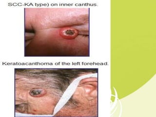
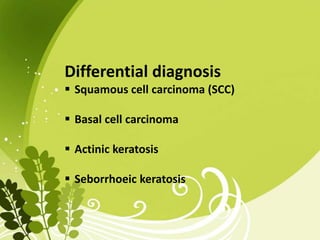
AMNA
HASSAN
ROLL no : 12
PRESENTED TO :
MAAM FAIZA
KERATOACANTHOMA
Introduction
• Benign epithelial neoplasm
• Rapidly growing skin cancer
• originates in the hair follicles epithelium
above the sebaceous glands
Epidemiology
• Incidence is estimated at 1 in 1,000.
• Peak incidence occurs in those aged over
60 years. Is rare in young adults.
• It is uncommon in darker-skinned
patients.
• Males are twice as often affected as
females.
• Sunlight and chemical carcinogens have been implicated.
• Trauma, genetic factors and immunocompromised status
have also been associated
• Industrial workers exposed to pitch and tar
• Long-term suppression of the immune system, such as organ
transplant recipients
• Long-term presence of scars, such as from a gasoline burn
• Chronic ulcers
• Presence of particular strains of the wart virus (human
papillomavirus)
• Previous skin cancer
Risk factors
Typically rapid growth over a few weeks to months,
followed by a slow spontaneous resolution over 4-6
months (but may take up to 1 year).
• Most occur on sun-exposed areas, e.g the face,
neck, and dorsum of hands and forearms.
• They are usually solitary and begin as firm, round,
skin-coloured or reddish papule
• rapidly progress to dome-shaped nodules with a
smooth shiny surface.
CLINICAL FEATURES
• A central crater of ulceration may develop, or a
keratin plug that may project like a horn.
• It leaves a residual scar if not excised.
• Occasionally presents as multiple tumors
The most common locations for keratoacanthoma
include:
• Center of the face
• Backs of hands
• Forearms
• Ears
• Scalp
• Lower legs, especially in women
SIGNS AND SYMPTOMS
Histopathology
• Cells appear mature and dyskeratosis
• Central plug of keratin
• Surface epithelium at the lateral edge of
tumor appears normal
• Sharply demarcated ,cup shaped buttress of
normal epidermis
• Epithelium exhibiting pseudo-carcinomatous
growth pattern
• At the lip of the central crater an acute
angle is formed between the overlying
epithelium and the periphery of the lesion
• The central crater is filled with keratin and
the base of the crater
• Connective tissue exhibits moderate to
marked infiltrate of chronic inflammatory
cells
Differential diagnosis
 Squamous cell carcinoma (SCC)
 Basal cell carcinoma
 Actinic keratosis
 Seborrhoeic keratosis
Investigations
Shave biopsy of keratoacanthoma is indistinguishable from
invasive squamous cell carcinoma (SCC).
Therefore, excisional or deep incisional biopsy is
required.
• Complete excision is the treatment of
choice for all skin neoplasms thought
to be keratoacanthoma
• Treatments that have produced some
success include
systemic -retinoids (eg isotretinoin),
intraLesional methotrexate, 5-
fluorouracil, bleomycin and
steroids, and topical imiquimod and 5-
fluorouracil
MANAGEMENT
• Freezing with liquid nitrogen (cryosurgery), in
which very cold liquid nitrogen is sprayed on
the keratoacanthoma, freezing it and
destroying it in the process.
• In most the lesions are treated before they
reach their maximum size of 2 to 2.5cm
• Keratoacanthomas are radiosensitive and
respond well to low doses of radiation.
• Radiation therapy may be useful in selected
patients with large tumors when resection
will result in cosmetic deformity
• for tumor's that have recurred following
attempted excision.
Radiotherapy..
Keratoacanthoma

Keratoacanthoma

  • 2.
    AMNA HASSAN ROLL no :12 PRESENTED TO : MAAM FAIZA
  • 3.
    KERATOACANTHOMA Introduction • Benign epithelialneoplasm • Rapidly growing skin cancer • originates in the hair follicles epithelium above the sebaceous glands
  • 5.
    Epidemiology • Incidence isestimated at 1 in 1,000. • Peak incidence occurs in those aged over 60 years. Is rare in young adults. • It is uncommon in darker-skinned patients. • Males are twice as often affected as females.
  • 6.
    • Sunlight andchemical carcinogens have been implicated. • Trauma, genetic factors and immunocompromised status have also been associated • Industrial workers exposed to pitch and tar • Long-term suppression of the immune system, such as organ transplant recipients • Long-term presence of scars, such as from a gasoline burn • Chronic ulcers • Presence of particular strains of the wart virus (human papillomavirus) • Previous skin cancer Risk factors
  • 7.
    Typically rapid growthover a few weeks to months, followed by a slow spontaneous resolution over 4-6 months (but may take up to 1 year). • Most occur on sun-exposed areas, e.g the face, neck, and dorsum of hands and forearms. • They are usually solitary and begin as firm, round, skin-coloured or reddish papule • rapidly progress to dome-shaped nodules with a smooth shiny surface. CLINICAL FEATURES
  • 8.
    • A centralcrater of ulceration may develop, or a keratin plug that may project like a horn. • It leaves a residual scar if not excised. • Occasionally presents as multiple tumors
  • 10.
    The most commonlocations for keratoacanthoma include: • Center of the face • Backs of hands • Forearms • Ears • Scalp • Lower legs, especially in women SIGNS AND SYMPTOMS
  • 11.
    Histopathology • Cells appearmature and dyskeratosis • Central plug of keratin • Surface epithelium at the lateral edge of tumor appears normal • Sharply demarcated ,cup shaped buttress of normal epidermis • Epithelium exhibiting pseudo-carcinomatous growth pattern
  • 12.
    • At thelip of the central crater an acute angle is formed between the overlying epithelium and the periphery of the lesion • The central crater is filled with keratin and the base of the crater • Connective tissue exhibits moderate to marked infiltrate of chronic inflammatory cells
  • 15.
    Differential diagnosis  Squamouscell carcinoma (SCC)  Basal cell carcinoma  Actinic keratosis  Seborrhoeic keratosis
  • 16.
    Investigations Shave biopsy ofkeratoacanthoma is indistinguishable from invasive squamous cell carcinoma (SCC). Therefore, excisional or deep incisional biopsy is required.
  • 17.
    • Complete excisionis the treatment of choice for all skin neoplasms thought to be keratoacanthoma • Treatments that have produced some success include systemic -retinoids (eg isotretinoin), intraLesional methotrexate, 5- fluorouracil, bleomycin and steroids, and topical imiquimod and 5- fluorouracil MANAGEMENT
  • 18.
    • Freezing withliquid nitrogen (cryosurgery), in which very cold liquid nitrogen is sprayed on the keratoacanthoma, freezing it and destroying it in the process. • In most the lesions are treated before they reach their maximum size of 2 to 2.5cm
  • 19.
    • Keratoacanthomas areradiosensitive and respond well to low doses of radiation. • Radiation therapy may be useful in selected patients with large tumors when resection will result in cosmetic deformity • for tumor's that have recurred following attempted excision. Radiotherapy..