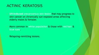
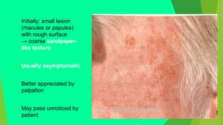
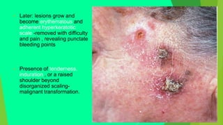
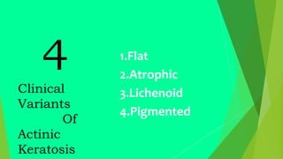
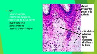
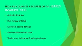
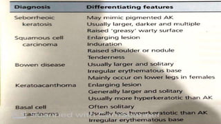
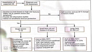
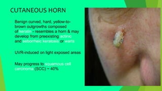
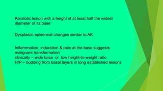
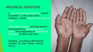
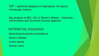
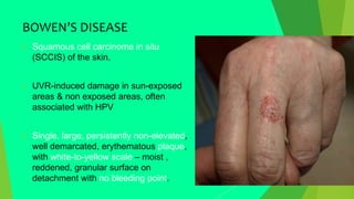
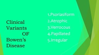
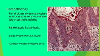
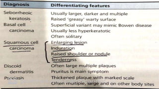
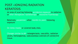
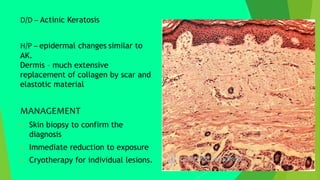
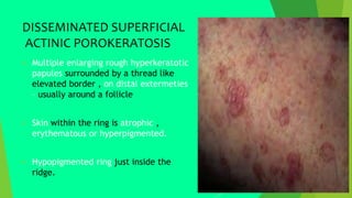
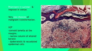
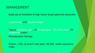
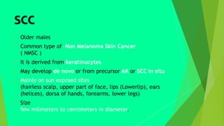
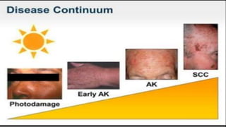
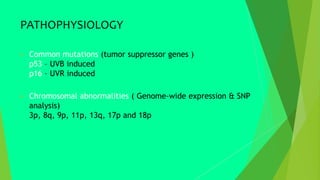
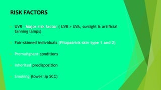
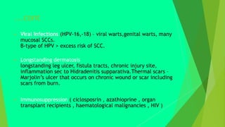
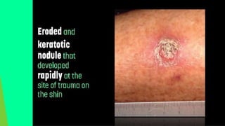
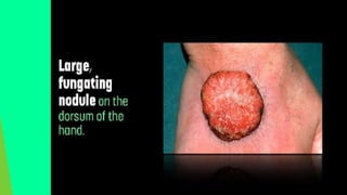
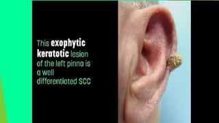
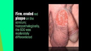
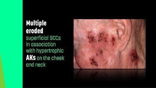
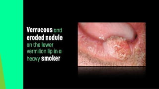
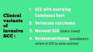
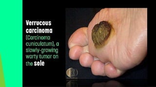
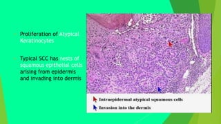
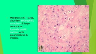
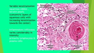
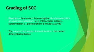
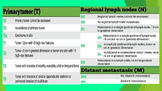
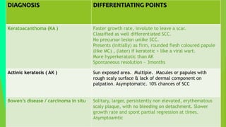
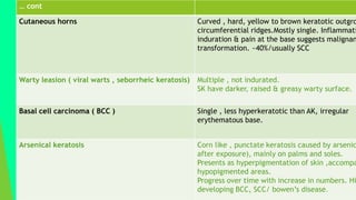
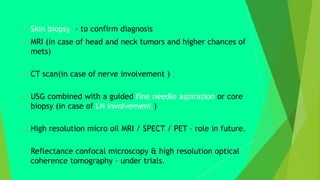
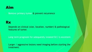
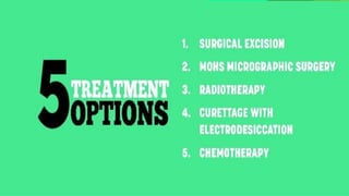
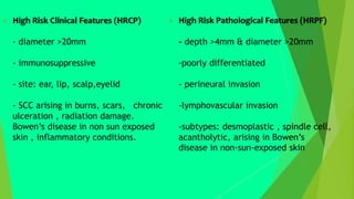
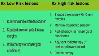
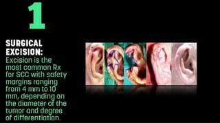
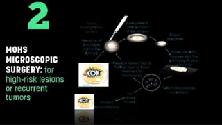
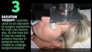
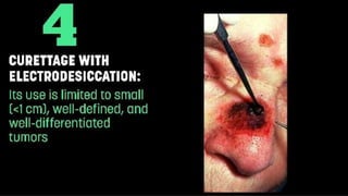
SQUAMOUS CELL
CARCINOMA
&
ITS PRECURSORS
PRECUROR
LESIONS
PREMALIGNANT / PRECURSOR LESIONS
 Actinic keratosis
 Cutaneous horn
 Arsenical keratosis
 Post ioninzing radiation keratosis
 Disseminated superficial actinic porokeratosis
 Bowen’s disease / carcinoma in situ
ACTINIC
KERATOSIS
ACTINIC KERATOSIS
 UV-induced precancerous skin lesion that may progress to
skin cancer on chronically sun exposed areas affecting
elderly males & females
 More common in white population & those with red hair &
blue eyes.
 Relapsing-remitting lesions.
 Initially: small lesion
(macules or papules)
with rough surface
→ coarse sandpaper-
like texture
 Usually asymptomatic
 Better appreciated by
palpation
 May pass unnoticed by
patient
 Later: lesions grow and
become erythematous and
adherent hyperkeratotic
scale -removed with difficulty
and pain , revealing punctate
bleeding points
 Presence of tenderness,
induration , or a raised
shoulder beyond
disorganized scaling-
malignant transformation.
4
Clinical
Variants
Of
Actinic
Keratosis
1.Flat
2.Atrophic
3.Lichenoid
4.Pigmented
 H/P
-solar elastosis ,
-epithelial dysplasia
restricted to basal layer
-hyperkeratosis
-parakeratosis
-absent granular layer
HIGH RISK CLINICAL FEATURES OF AK --EARLY
INVASIVE SCC
 Multiple thick Aks
 Past history of NMSC
 Extensive actinic damage
 Immunocompromised state
 Tenderness, induration & enlarging lesion
T
TREATMENT
Other Rx options
 Topical & oral retinoids
 Dermabrasions & chemical peels with trichloracetic acid
 Non ablative fractional resurfacing
CUTANEOUS HORN
CUTANEOUS HORN
• Benign curved, hard, yellow-to-
brown outgrowths composed
of keratin - resembles a horn & may
develop from preexisting actinic
and seborrheic keratosis or warts
• UVR-induced on light exposed areas
• May progress to squamous cell
carcinoma (SCC) ~ 40%
 Keratotic lesion with a height of at least half the widest
diameter of its base
 Dysplastic epidermal changes similar to AK
 Inflammation, induration & pain at the base suggests
malignant transformation
clinically – wide base or low height-to-weight ratio
H/P – budding from basal layers in long established lesions
DIFFERENTIAL DIAGNOSIS
 Viral warts
 Actinic keratosis
 Seborrheic keratosis
 Keratoacanthoma
 Invasive SCC
MANAGEMENT
 Clinical appearance
 H/P of base to rule out malignancy
 Surgical excision – to obtain pathology & rule out
malignany
ARSENICAL
KERATOSIS
ARSENICAL KERATOSIS
 A corn like punctate keratosis caused
by arsenic / a few years after
exposure, mainly affecting the palms &
soles.
 Hyperpigmentation –raindrop pattern ,
accompanied by hypopigmenated
areas, and progression to generalized
thickness of palms and soles .
 Smoking , nutritional deficiencies
(retinol, Ca, iron, folate ,vitamin
A,E,C)
 H/P – epithelial dysplasia or hyperplasia. No typical
microscopic feature.
 May progress to BCC, SCC or Bowen’s disease – induration ,
inflammation and ulceration become apparent.
DIFFERNTIAL DIAGNOSIS
 Disseminated puntate keratoderma
 Darier’s disease
 Lichen planus
 Planter warts
MANAGEMENT
 Reduce arsenic exposure
 Well balanced diet
1st line
 Solitary / few lesions – cryotherapy ,cautery or curettage.
 Multiple keratosis – oral retinoid in combination with
keratolytics
2nd line
 Imiquimod cream 5% OD for 6weeks or OD 3-5times per
week
BOWEN’S DISEASE
BOWEN’S DISEASE
 Squamous cell carcinoma in situ
(SCCIS) of the skin.
 UVR-induced damage in sun-exposed
areas & non exposed areas, often
associated with HPV
 Single, large, persistently non-elevated,
well demarcated, erythematous plaque,
with white-to-yellow scale – moist ,
reddened, granular surface on
detachment with no bleeding point.
 ~10-20% multiple lesions , either
widely spread or sometimes close
together.
 Slower growth rate and spontaneous
partial regression at times.
 Can occur on perianal skin,
subungal region, palms & soles -
higher risk of invasion & recurrence.
 May progress to invasive squamous
cell carcinoma. ~3-5% risk
Clinical
Variants
OF
Bowen’s
Disease
1.Psoriasiform
2.Atrophic
3.Verrocous
4.Papillated
5.Irregular
Histopathology
 Full thickness epidermal dysplasia
& disordered differentiation with
loss of epithelial polarity.
 Parakeratosis & acanthosis.
 Large hyperchromatic nuclei
 Atypical mitosis and gaint cells.
DIFFERENTIAL
DIAGNOSIS
MANAGEMENT
POST –IONIZING
RADIATION
KERATOSIS
POST –IONIZING RADIATION
KERATOSIS
 An area of scarring following accidental exposure to radiation
or after therapeutic radiotherapy or excessive fluoroscopy
 Relatively uncommon –after months to years following
exposure
 On limbs mostly or covered body sites.
 Changes similar to AK, telangiectasia, vasculitis, radiation
ulcers, hemangiomas, subcutaneous sclerosis of connective
tissue.
D/D – Actinic Keratosis
H/P – epidermal changes similar to
AK.
Dermis – much extensive
replacement of collagen by scar and
elastotic material
MANAGEMENT
 Skin biopsy to confirm the
diagnosis
 Immediate reduction to exposure
 Cryotherapy for individual lesions.
DISSEMINATED
SUPERFICIAL
ACTINIC
POROKERATOSIS
DISSEMINATED SUPERFICIAL
ACTINIC POROKERATOSIS
 Multiple enlarging rough hyperkeratotic
papules surrounded by a thread like
elevated border , on distal extermeties
– usually around a follicle
 Skin within the ring is atrophic ,
erythematous or hyperpigmented.
 Hypopigmented ring just inside the
ridge.
 Prominent in summer &
improve in winter.
 Very low chances of
malignant transformation.
 H/P
-cornoid lamella at the
margins.
- narrow column of altered
keratin
- dyskeratotic & vacuolated
epidermal cells
Clinical
varaints
of
Porokeratosis
1. Porokeratosis of Mibelli
2. linear porokeratosis
3. DSAK
4.Porokeratosis palmaris &
plantaris diffusa
5. Punctate porokeratosis
DIFFERENTIAL DIAGNOSIS
 Actinic keratosis
– esp if on face , but present on other sun exposed sites as
well
 Bowen’s disease
– on legs
 Superficial BCC
MANAGEMENT
 Good use of emollient & high factor broad spectrum sunscreen
 Cryotherapy with liquid nitrogen
 Topical diclofenac gel , vit D3 analogues, 5% 5-FU cream, 5%
imiquimod cream
 Photodynamic therapy
 Erbium , CO2, Q-switch ruby laser, Nd:YAG –under successful
trials.
SCC
 Older males
 Common type of Non Melanoma Skin Cancer
( NMSC )
 It is derived from keratinocytes
 May develop de novo or from precursor AK or SCC in situ
 Mainly on sun exposed sites
(hairless scalp, upper part of face, lips (Lowerlip), ears
(helices), dorsa of hands, forearms, lower legs)
 Size
few milimeters to centimeters in diameter.
PATHOPHYSIOLOGY
 Common mutations (tumor suppressor genes )
p53 – UVB induced
p16 – UVR induced
 Chromosomal abnormalities ( Genome-wide expression & SNP
analysis)
3p, 8q, 9p, 11p, 13q, 17p and 18p
RISK FACTORS
 UVR –Major risk factor ( UVB > UVA, sunlight & artificial
tanning lamps)
 Fair-skinned individuals (Fitzpatrick skin type 1 and 2)
 Premalignant conditions
 Inherited predisposition
 Smoking (lower lip SCC)
...cont
 Viral Infections (HPV-16,-18) – viral warts,genital warts, many
mucosal SCCs.
B-type of HPV > excess risk of SCC.
 Longstanding dermatosis
longstanding leg ulcer, fistula tracts, chronic injury site,
inflammation sec to Hidradenitis supparativa.Thermal scars –
Marjolin’s ulcer that occurs on chronic wound or scar including
scars from burn.
 Immunosuppression ( ciclosposrin , azathioprine , organ
transplant recipients , haematological malignancies , HIV )
 1st clinical sign – induration.
 Firm , tender , erythematous nodule or plaque with keratotic
crust , verrucous, tumid
 Eroded fungating mass – that shows easy bleeding
 Lesions that fail to heal – develop ulcers
with everted edge , indurated base & necrotic floor
 Outline – mostly irregular , sometimes rounded.
 Mobile structures like lip & Genitalia
show early fissuring or small erosions or ulcers that fail to
heal and bleeds recurrently.
 May be associated with other features of photodamage or
premalignant lesions.
 Parestheisa , anesthesia, tenderness, pain particularly in
perineural invasion
 Regional LN enlargement (later –hard & fixed)
 Proliferation of Atypical
Keratinocytes
 Typical SCC has nests of
squamous epithelial cells
arising from epidermis
and invading into dermis
 Malignant cell – large,
abundant eosinophilic
cytoplasm & large
vesicular or
hyperchromatic
nucleus with
pleomorphism &
mitosis.
 Variable keratinaization
squamous eddies ,
keratin pearls
(concentric layers of
squamous cells with
increasing keratinization
towards the center)
 Inflammatory infiltrates –
varies considerably in
intensity
primarily lymphocytes &
plasma cells
Grading of SCC
 Depends on how easy it is to recognize the characteristics
of squamous epithelium (e.g, intracellular bridges ,
keratinization ) , pleomorphism & mitotic activity
 The greater the degree of keratinization – the better
differentiated tumor.
Well differentiated Poorly differentiated
SCC SCC
METASTATIC SCC
 Upto 5% RISK OF METASTASIS
 Invasive SCC rarely metastasize and prove fetal.
that refers to growth into deeper layers of skin , the
dermis.
 More difficult to treat than the original skin lesions.
 In 80% cases metastases develops from draining LN.
DIFFERENTIAL
DIAGNOSIS
DIAGNOSIS DIFFERENTIATING POINTS
Keratoacanthoma (KA ) Faster growth rate, involute to leave a scar.
Classified as well differentiated SCC.
No precursor lesion unlike SCC.
Presents (initially) as firm, rounded flesh coloured papule
(like MC) , (later) if keratotic > like a viral wart.
More hyperkeratotic than AK
Spontaneous resolution ~ 3months
Actinic keratosis ( AK ) Sun exposed area. Multiple. Macules or papules with
rough scaly surface & lack of dermal component on
palpation. Asymptomatic. 10% chances of SCC
Bowen’s disease / carcinoma in situ Solitary, larger, persistently non elevated, erythematous
scaly plaque, with no bleeding on detachment. Slower
growth rate and spont partial regression at times.
Asymptoamtic
… cont
Cutaneous horns Curved , hard, yellow to brown keratotic outgro
circumferential ridges.Mostly single. Inflammati
induration & pain at the base suggests malignan
transformation. ~40%/usually SCC
Warty leasion ( viral warts , seborrheic keratosis) Multiple , not indurated.
SK have darker, raised & greasy warty surface.
Basal cell carcinoma ( BCC ) Single , less hyperkeratotic than AK, irregular
erythematous base.
Arsenical keratosis Corn like , punctate keratosis caused by arsenic
after exposure), mainly on palms and soles.
Presents as hyperpigmentation of skin ,accompa
hypopigmented areas.
Progress over time with increase in numbers. Hi
developing BCC, SCC/ bowen’s disease.
INVESTIGATIONS
 Skin biopsy - to confirm diagnosis
 MRI (in case of head and neck tumors and higher chances of
mets)
 CT scan(in case of nerve involvement )
 USG combined with a guided fine needle aspiration or core
biopsy (in case of LN involvement )
 High resolution micro oil MRI / SPECT / PET – role in future.
 Reflectance confocal microscopy & high resolution optical
coherence tomography – under trials.
Aim
 Remove primary tumor & prevent recurrence
Rx
 Depends on clinical (size, location, number) & pathological
features of tumor.
 Long term prognosis for adequately treated SCC is excellent.
 Larger / aggressive lesions need imaging before starting the
treatment.
 High Risk Clinical Features (HRCP)
- diameter >20mm
- immunosuppressive
- site: ear, lip, scalp,eyelid
- SCC arising in burns, scars, chronic
ulceration , radiation damage.
Bowen’s disease in non sun exposed
skin , inflammatory conditions.
 High Risk Pathological Features (HRPF)
- depth >4mm & diameter >20mm
-poorly differentiated
- perineural invasion
-lymphovascular invasion
-subtypes: desmoplastic , spindle cell,
acantholytic, arising in Bowen’s
disease in non-sun-exposed skin
Rx Low Risk lesions Rx High risk lesions
Squamous Cell Carcinoma

Squamous Cell Carcinoma

  • 2.
  • 3.
  • 4.
    PREMALIGNANT / PRECURSORLESIONS  Actinic keratosis  Cutaneous horn  Arsenical keratosis  Post ioninzing radiation keratosis  Disseminated superficial actinic porokeratosis  Bowen’s disease / carcinoma in situ
  • 5.
  • 6.
    ACTINIC KERATOSIS  UV-inducedprecancerous skin lesion that may progress to skin cancer on chronically sun exposed areas affecting elderly males & females  More common in white population & those with red hair & blue eyes.  Relapsing-remitting lesions.
  • 7.
     Initially: smalllesion (macules or papules) with rough surface → coarse sandpaper- like texture  Usually asymptomatic  Better appreciated by palpation  May pass unnoticed by patient
  • 8.
     Later: lesionsgrow and become erythematous and adherent hyperkeratotic scale -removed with difficulty and pain , revealing punctate bleeding points  Presence of tenderness, induration , or a raised shoulder beyond disorganized scaling- malignant transformation.
  • 9.
  • 10.
     H/P -solar elastosis, -epithelial dysplasia restricted to basal layer -hyperkeratosis -parakeratosis -absent granular layer
  • 11.
    HIGH RISK CLINICALFEATURES OF AK --EARLY INVASIVE SCC  Multiple thick Aks  Past history of NMSC  Extensive actinic damage  Immunocompromised state  Tenderness, induration & enlarging lesion
  • 13.
  • 15.
    Other Rx options Topical & oral retinoids  Dermabrasions & chemical peels with trichloracetic acid  Non ablative fractional resurfacing
  • 16.
  • 17.
    CUTANEOUS HORN • Benigncurved, hard, yellow-to- brown outgrowths composed of keratin - resembles a horn & may develop from preexisting actinic and seborrheic keratosis or warts • UVR-induced on light exposed areas • May progress to squamous cell carcinoma (SCC) ~ 40%
  • 18.
     Keratotic lesionwith a height of at least half the widest diameter of its base  Dysplastic epidermal changes similar to AK  Inflammation, induration & pain at the base suggests malignant transformation clinically – wide base or low height-to-weight ratio H/P – budding from basal layers in long established lesions
  • 19.
    DIFFERENTIAL DIAGNOSIS  Viralwarts  Actinic keratosis  Seborrheic keratosis  Keratoacanthoma  Invasive SCC
  • 20.
    MANAGEMENT  Clinical appearance H/P of base to rule out malignancy  Surgical excision – to obtain pathology & rule out malignany
  • 21.
  • 22.
    ARSENICAL KERATOSIS  Acorn like punctate keratosis caused by arsenic / a few years after exposure, mainly affecting the palms & soles.  Hyperpigmentation –raindrop pattern , accompanied by hypopigmenated areas, and progression to generalized thickness of palms and soles .  Smoking , nutritional deficiencies (retinol, Ca, iron, folate ,vitamin A,E,C)
  • 23.
     H/P –epithelial dysplasia or hyperplasia. No typical microscopic feature.  May progress to BCC, SCC or Bowen’s disease – induration , inflammation and ulceration become apparent. DIFFERNTIAL DIAGNOSIS  Disseminated puntate keratoderma  Darier’s disease  Lichen planus  Planter warts
  • 24.
    MANAGEMENT  Reduce arsenicexposure  Well balanced diet 1st line  Solitary / few lesions – cryotherapy ,cautery or curettage.  Multiple keratosis – oral retinoid in combination with keratolytics 2nd line  Imiquimod cream 5% OD for 6weeks or OD 3-5times per week
  • 25.
  • 26.
    BOWEN’S DISEASE  Squamouscell carcinoma in situ (SCCIS) of the skin.  UVR-induced damage in sun-exposed areas & non exposed areas, often associated with HPV  Single, large, persistently non-elevated, well demarcated, erythematous plaque, with white-to-yellow scale – moist , reddened, granular surface on detachment with no bleeding point.
  • 27.
     ~10-20% multiplelesions , either widely spread or sometimes close together.  Slower growth rate and spontaneous partial regression at times.  Can occur on perianal skin, subungal region, palms & soles - higher risk of invasion & recurrence.  May progress to invasive squamous cell carcinoma. ~3-5% risk
  • 28.
  • 29.
    Histopathology  Full thicknessepidermal dysplasia & disordered differentiation with loss of epithelial polarity.  Parakeratosis & acanthosis.  Large hyperchromatic nuclei  Atypical mitosis and gaint cells.
  • 30.
  • 32.
  • 34.
  • 35.
    POST –IONIZING RADIATION KERATOSIS An area of scarring following accidental exposure to radiation or after therapeutic radiotherapy or excessive fluoroscopy  Relatively uncommon –after months to years following exposure  On limbs mostly or covered body sites.  Changes similar to AK, telangiectasia, vasculitis, radiation ulcers, hemangiomas, subcutaneous sclerosis of connective tissue.
  • 36.
    D/D – ActinicKeratosis H/P – epidermal changes similar to AK. Dermis – much extensive replacement of collagen by scar and elastotic material MANAGEMENT  Skin biopsy to confirm the diagnosis  Immediate reduction to exposure  Cryotherapy for individual lesions.
  • 37.
  • 38.
    DISSEMINATED SUPERFICIAL ACTINIC POROKERATOSIS Multiple enlarging rough hyperkeratotic papules surrounded by a thread like elevated border , on distal extermeties – usually around a follicle  Skin within the ring is atrophic , erythematous or hyperpigmented.  Hypopigmented ring just inside the ridge.
  • 39.
     Prominent insummer & improve in winter.  Very low chances of malignant transformation.  H/P -cornoid lamella at the margins. - narrow column of altered keratin - dyskeratotic & vacuolated epidermal cells
  • 40.
    Clinical varaints of Porokeratosis 1. Porokeratosis ofMibelli 2. linear porokeratosis 3. DSAK 4.Porokeratosis palmaris & plantaris diffusa 5. Punctate porokeratosis
  • 41.
    DIFFERENTIAL DIAGNOSIS  Actinickeratosis – esp if on face , but present on other sun exposed sites as well  Bowen’s disease – on legs  Superficial BCC
  • 42.
    MANAGEMENT  Good useof emollient & high factor broad spectrum sunscreen  Cryotherapy with liquid nitrogen  Topical diclofenac gel , vit D3 analogues, 5% 5-FU cream, 5% imiquimod cream  Photodynamic therapy  Erbium , CO2, Q-switch ruby laser, Nd:YAG –under successful trials.
  • 45.
    SCC  Older males Common type of Non Melanoma Skin Cancer ( NMSC )  It is derived from keratinocytes  May develop de novo or from precursor AK or SCC in situ  Mainly on sun exposed sites (hairless scalp, upper part of face, lips (Lowerlip), ears (helices), dorsa of hands, forearms, lower legs)  Size few milimeters to centimeters in diameter.
  • 48.
    PATHOPHYSIOLOGY  Common mutations(tumor suppressor genes ) p53 – UVB induced p16 – UVR induced  Chromosomal abnormalities ( Genome-wide expression & SNP analysis) 3p, 8q, 9p, 11p, 13q, 17p and 18p
  • 50.
    RISK FACTORS  UVR–Major risk factor ( UVB > UVA, sunlight & artificial tanning lamps)  Fair-skinned individuals (Fitzpatrick skin type 1 and 2)  Premalignant conditions  Inherited predisposition  Smoking (lower lip SCC)
  • 51.
    ...cont  Viral Infections(HPV-16,-18) – viral warts,genital warts, many mucosal SCCs. B-type of HPV > excess risk of SCC.  Longstanding dermatosis longstanding leg ulcer, fistula tracts, chronic injury site, inflammation sec to Hidradenitis supparativa.Thermal scars – Marjolin’s ulcer that occurs on chronic wound or scar including scars from burn.  Immunosuppression ( ciclosposrin , azathioprine , organ transplant recipients , haematological malignancies , HIV )
  • 53.
     1st clinicalsign – induration.  Firm , tender , erythematous nodule or plaque with keratotic crust , verrucous, tumid  Eroded fungating mass – that shows easy bleeding  Lesions that fail to heal – develop ulcers with everted edge , indurated base & necrotic floor  Outline – mostly irregular , sometimes rounded.
  • 55.
     Mobile structureslike lip & Genitalia show early fissuring or small erosions or ulcers that fail to heal and bleeds recurrently.  May be associated with other features of photodamage or premalignant lesions.  Parestheisa , anesthesia, tenderness, pain particularly in perineural invasion  Regional LN enlargement (later –hard & fixed)
  • 69.
     Proliferation ofAtypical Keratinocytes  Typical SCC has nests of squamous epithelial cells arising from epidermis and invading into dermis
  • 70.
     Malignant cell– large, abundant eosinophilic cytoplasm & large vesicular or hyperchromatic nucleus with pleomorphism & mitosis.
  • 71.
     Variable keratinaization squamouseddies , keratin pearls (concentric layers of squamous cells with increasing keratinization towards the center)  Inflammatory infiltrates – varies considerably in intensity primarily lymphocytes & plasma cells
  • 72.
    Grading of SCC Depends on how easy it is to recognize the characteristics of squamous epithelium (e.g, intracellular bridges , keratinization ) , pleomorphism & mitotic activity  The greater the degree of keratinization – the better differentiated tumor.
  • 73.
    Well differentiated Poorlydifferentiated SCC SCC
  • 75.
    METASTATIC SCC  Upto5% RISK OF METASTASIS  Invasive SCC rarely metastasize and prove fetal. that refers to growth into deeper layers of skin , the dermis.  More difficult to treat than the original skin lesions.  In 80% cases metastases develops from draining LN.
  • 79.
  • 80.
    DIAGNOSIS DIFFERENTIATING POINTS Keratoacanthoma(KA ) Faster growth rate, involute to leave a scar. Classified as well differentiated SCC. No precursor lesion unlike SCC. Presents (initially) as firm, rounded flesh coloured papule (like MC) , (later) if keratotic > like a viral wart. More hyperkeratotic than AK Spontaneous resolution ~ 3months Actinic keratosis ( AK ) Sun exposed area. Multiple. Macules or papules with rough scaly surface & lack of dermal component on palpation. Asymptomatic. 10% chances of SCC Bowen’s disease / carcinoma in situ Solitary, larger, persistently non elevated, erythematous scaly plaque, with no bleeding on detachment. Slower growth rate and spont partial regression at times. Asymptoamtic
  • 81.
    … cont Cutaneous hornsCurved , hard, yellow to brown keratotic outgro circumferential ridges.Mostly single. Inflammati induration & pain at the base suggests malignan transformation. ~40%/usually SCC Warty leasion ( viral warts , seborrheic keratosis) Multiple , not indurated. SK have darker, raised & greasy warty surface. Basal cell carcinoma ( BCC ) Single , less hyperkeratotic than AK, irregular erythematous base. Arsenical keratosis Corn like , punctate keratosis caused by arsenic after exposure), mainly on palms and soles. Presents as hyperpigmentation of skin ,accompa hypopigmented areas. Progress over time with increase in numbers. Hi developing BCC, SCC/ bowen’s disease.
  • 82.
  • 83.
     Skin biopsy- to confirm diagnosis  MRI (in case of head and neck tumors and higher chances of mets)  CT scan(in case of nerve involvement )  USG combined with a guided fine needle aspiration or core biopsy (in case of LN involvement )  High resolution micro oil MRI / SPECT / PET – role in future.  Reflectance confocal microscopy & high resolution optical coherence tomography – under trials.
  • 85.
    Aim  Remove primarytumor & prevent recurrence Rx  Depends on clinical (size, location, number) & pathological features of tumor.  Long term prognosis for adequately treated SCC is excellent.  Larger / aggressive lesions need imaging before starting the treatment.
  • 87.
     High RiskClinical Features (HRCP) - diameter >20mm - immunosuppressive - site: ear, lip, scalp,eyelid - SCC arising in burns, scars, chronic ulceration , radiation damage. Bowen’s disease in non sun exposed skin , inflammatory conditions.  High Risk Pathological Features (HRPF) - depth >4mm & diameter >20mm -poorly differentiated - perineural invasion -lymphovascular invasion -subtypes: desmoplastic , spindle cell, acantholytic, arising in Bowen’s disease in non-sun-exposed skin
  • 88.
    Rx Low Risklesions Rx High risk lesions