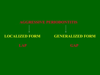
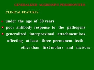
Presented by
Dr. P. Srujan Kumar
MDS Reader
AGGRESSIVE PERIODONTITIS
LOCALIZED FORM GENERALIZED FORM
LAP GAP
LOCALIZED AGGRESSIVE PERIODONTITIS
The term “JUVENILE PERIODONTITIS” was introduced
by CHAPUT in 1967 and by BUTLER in 1969
HISTORICAL BACKGROUND
In 1923 GOTTLIEB reported a fatal case of
influenza and called it as
“ diffuse atrophy of the alveolar bone ”
characterized by :
-- loss of collagen fibers
-- extensive bone loss
-- widened pdl space
In 1928 GOTTLIEB attributed this condition to
inhibition of continuous cementum formation,
which he termed the disease as
“deep cementopathia ”
In 1938 WANNENMACHER described incisor-first- molar
involvement and called the disease
“ parodontitis marginalis progressiva ”
He considered it as inflammatory process
In 1940 THOMA AND GOLDMAN
referred this disease as
“ paradontosis ”
The initial abnormality was located in the
al.bone rather then in the cementum
IN 1942 ORBAN AND WEINMANN
introduced the term
“ periodontosis ”
THREE STAGES
STAGE I
STAGE II
STAGE III
STAGE I :
degeneration of principal fibers of pdl
cessation of cementum formation
resorption of the alveolar bone
STAGE II :
rapid proliferation of the JE
earliest signs of inflammation
STAGE III :
progressive inflammation
deep, infrabony periodontal pockets
DEFINITION
BAER - 1971
“a disease of periodontium occurring in an
otherwise healthy adolescents which is characterized
by a rapid loss of alveolar bone about more than
one tooth of permanent dentition”
PREVELENCE
BAER
U.S POPULATION -- 0.1%
SAXBY
ENGLISH POPULATION -- 0.1%
MIGLANI
ASIAN POPULATION -- 0.1%
SAXEN
FRENCH POPULATION -- 0.1%
AGE AND SEX DISTRIBUTION
PUBERTY -- 21 Yrs OF AGE
MORE PREDILICTION FOR FEMALES
FEMALE : MALE = 3.5 : 1
DISTRIBUTION OF LESIONS
Classic distribution -- molar and incisors
Least distribution -- cuspid and premolars
THREE TYPES :
1. First molars and incisors
2. First molars and incisors and some
additional teeth [total of < 14 teeth ]
3. Generalized involvement
POSSIBLE REASONS FOR THE LIMITATIONS OF
PERIODONTAL DESTRUCTION TO 1st MOLARS AND
INCISORS ARE:
1] After initial colonisation of 1st per.molar and
incisors Aa bacilli are phagocytosed by
immune defenses [NEUTROPHILES, MONOCYTES] and
neutralize destructive factors. [ENDOTOXIN,
LEUCOTOXIN, COLLAGENASE]
2]Antagonist to Aa may develop there by
decreasing the destruction of the lesions
3] Aa may loose its leukotoxin producing
ability for unknown reasons. when this
happens the progress of the disease may
become
arrested
CLINICAL FINDINGS
1] The most striking feature of early Lap
is lack of clinical inflammation, despite the
presence of deep periodontal pockets
2] There may be a small amount of plaque,
which forms a thin film on tooth and rarely
mineralizes to become calculus
3] Disto-labial migration of max. incisors,
with diastema formation
4] Root surfaces become sensitive to thermal
and tactile stimuli
5] Deep, dull radiating pain may occur with
mastication
RADIOGRAPHIC FINDINGS
1] Vertical loss of alveolar bone around the
1st molars and incisors in healthy teenagers is
a diagnostic sign of classic lap
2] Arc- shaped bone loss extending from the
distal surface of second pm -- mesial surface of
2nd molar
3] Resorption of roots of 1st molar and incisors
4] Bilaterally symmetrical type of bone loss
resembling that of “ mirror images ”
CLINICAL COURSE
Lap progresses rapidly and rate of bone loss
is about 3- 4 times faster than in periodontitis
Bone resorption progresses until the
teeth are treated, exfoliated or extracted which
is termed as “ burn out” phenomenon
Rate of bone loss
= CP Age
= AP
GENERALIZED AGGRESSIVE PERIODONTITIS
CLINICAL FEATURES
under the age of 30 years
poor antibody response to the pathogens
generalized interproximal attachment loss
affecting at least three permanent teeth
other than first molars and incisors
occur episodically with periods of advanced
destruction followed by periods of quiescence
small amounts of bacterial plaque associated
with affected
teeth
TWO GINGIVAL TISSUE RESPONSES
ONE IS
severe, acutely inflamed, fiery red,
proliferating, ulcerated tissue
bleeding occur spontaneously
suppuration
SECOND IS
gingival tissues appear pink,
free of inflammation
despite mild clinical appearance,
deep pockets can be probed
RADIOLOGICAL FEATURES
generalized severe bone loss
osseous destruction of 25% --60% during a
9–week period
PREVALENCE / AGE AND SEX DISTRIBUTUON
SRILANKA -- 8%
U.S ADOLESCENTS AGED 14 -17 yrs -- 0.13%
BLACKS -- HIGHER RISK > WHITES
MALES -- MORE LIKELY > FEMALES
ETIOLOGY: [RISK FACTORS]
1. HEREDITY
Baer described the disease in twins, siblings
and first cousins as well as in parents and offspring
Transmitted as x- linked dominant disease
[if the father and mother has AP sons and daughters
has AP ]
2. BACTERIOLOGY
GRAM -- VE ANEROBIC RODS
MINIMAL -- ATTACHED PLAQUE
LARGER -- UNATTACHED PLAQUE
PREDOMINANT BACTERIA :
aggregatibacter actinomycetem comitans
capnocytophaga
TWO CLASSIFICATIONS
1ST CLASSIFICATION
GRAM – VE
1 Aa
2 capnocytophaga sub- species sputegena
3 Motile anaerobic rods
a, spirochetes b,mycoplasma
c,ekenella d,wolionella
e, black pigmented bacteroids
GRAM + VE
1 streptococcus
2 pepto streptococcus
2nd CLASSIFICATION
3 GROUPS
GROUP 1: Aa
GROUP 2: Aa, capnocytophaga
GROUP 3: Eubacterium saberium
3. IMMUNOLOGY
Functional defects of PMNs and monocytes
These defects can impair :
Chemo tactic attraction of PMNs to the site
Phagocytosis of microorganisms
Aa bacillus produce
4. VIRULENCE FACTORS
Bring about destructive process
i LEUKOTOXIN:
Destroys PMNs and monocytes there by inhibit
host defense
mechanism
ii ENDOTOXIN: [LPS]
Stimulate osteoclast -- bone resorption
iii COLLAGENASE:
Destroys gingival CT [collagen]
iv EPITHELIOTOXIN:
Facilitates penetration of Aa into CT through JE
and destroy collagen
v FIBROBLAST INHIBITING FACTOR:
Impair collagen formation
vi OSTEOCLAST ACTIVATING FACTOR [ OAF ] :
Brings about resorption of Al. bone
vii POLYCONAL B-LYMPHOCYTE ACTIVATING FACTOR:
Mediate inflammatory reaction by stimulating OAF
and bring about bone resorption
5. ENVIRONMENTAL FACTORS
Patients with GAP who smoke have more affected teeth
and loss of clinical attachment than non smokers
Smoking may not have the same impact on patients with LAP

AGGRESSIVE PERIODONTITIS.ppt

  • 2.
    Presented by Dr. P.Srujan Kumar MDS Reader
  • 3.
  • 4.
    LOCALIZED AGGRESSIVE PERIODONTITIS Theterm “JUVENILE PERIODONTITIS” was introduced by CHAPUT in 1967 and by BUTLER in 1969
  • 5.
    HISTORICAL BACKGROUND In 1923GOTTLIEB reported a fatal case of influenza and called it as “ diffuse atrophy of the alveolar bone ” characterized by : -- loss of collagen fibers -- extensive bone loss -- widened pdl space
  • 6.
    In 1928 GOTTLIEBattributed this condition to inhibition of continuous cementum formation, which he termed the disease as “deep cementopathia ”
  • 7.
    In 1938 WANNENMACHERdescribed incisor-first- molar involvement and called the disease “ parodontitis marginalis progressiva ” He considered it as inflammatory process
  • 8.
    In 1940 THOMAAND GOLDMAN referred this disease as “ paradontosis ” The initial abnormality was located in the al.bone rather then in the cementum
  • 9.
    IN 1942 ORBANAND WEINMANN introduced the term “ periodontosis ” THREE STAGES STAGE I STAGE II STAGE III
  • 10.
    STAGE I : degenerationof principal fibers of pdl cessation of cementum formation resorption of the alveolar bone STAGE II : rapid proliferation of the JE earliest signs of inflammation STAGE III : progressive inflammation deep, infrabony periodontal pockets
  • 11.
    DEFINITION BAER - 1971 “adisease of periodontium occurring in an otherwise healthy adolescents which is characterized by a rapid loss of alveolar bone about more than one tooth of permanent dentition”
  • 12.
    PREVELENCE BAER U.S POPULATION --0.1% SAXBY ENGLISH POPULATION -- 0.1% MIGLANI ASIAN POPULATION -- 0.1% SAXEN FRENCH POPULATION -- 0.1%
  • 13.
    AGE AND SEXDISTRIBUTION PUBERTY -- 21 Yrs OF AGE MORE PREDILICTION FOR FEMALES FEMALE : MALE = 3.5 : 1
  • 14.
    DISTRIBUTION OF LESIONS Classicdistribution -- molar and incisors Least distribution -- cuspid and premolars THREE TYPES : 1. First molars and incisors 2. First molars and incisors and some additional teeth [total of < 14 teeth ] 3. Generalized involvement
  • 15.
    POSSIBLE REASONS FORTHE LIMITATIONS OF PERIODONTAL DESTRUCTION TO 1st MOLARS AND INCISORS ARE: 1] After initial colonisation of 1st per.molar and incisors Aa bacilli are phagocytosed by immune defenses [NEUTROPHILES, MONOCYTES] and neutralize destructive factors. [ENDOTOXIN, LEUCOTOXIN, COLLAGENASE]
  • 16.
    2]Antagonist to Aamay develop there by decreasing the destruction of the lesions 3] Aa may loose its leukotoxin producing ability for unknown reasons. when this happens the progress of the disease may become arrested
  • 17.
    CLINICAL FINDINGS 1] Themost striking feature of early Lap is lack of clinical inflammation, despite the presence of deep periodontal pockets 2] There may be a small amount of plaque, which forms a thin film on tooth and rarely mineralizes to become calculus
  • 18.
    3] Disto-labial migrationof max. incisors, with diastema formation 4] Root surfaces become sensitive to thermal and tactile stimuli 5] Deep, dull radiating pain may occur with mastication
  • 20.
    RADIOGRAPHIC FINDINGS 1] Verticalloss of alveolar bone around the 1st molars and incisors in healthy teenagers is a diagnostic sign of classic lap 2] Arc- shaped bone loss extending from the distal surface of second pm -- mesial surface of 2nd molar
  • 21.
    3] Resorption ofroots of 1st molar and incisors 4] Bilaterally symmetrical type of bone loss resembling that of “ mirror images ”
  • 22.
    CLINICAL COURSE Lap progressesrapidly and rate of bone loss is about 3- 4 times faster than in periodontitis Bone resorption progresses until the teeth are treated, exfoliated or extracted which is termed as “ burn out” phenomenon
  • 23.
    Rate of boneloss = CP Age = AP
  • 26.
    GENERALIZED AGGRESSIVE PERIODONTITIS CLINICALFEATURES under the age of 30 years poor antibody response to the pathogens generalized interproximal attachment loss affecting at least three permanent teeth other than first molars and incisors
  • 27.
    occur episodically withperiods of advanced destruction followed by periods of quiescence small amounts of bacterial plaque associated with affected teeth
  • 28.
    TWO GINGIVAL TISSUERESPONSES ONE IS severe, acutely inflamed, fiery red, proliferating, ulcerated tissue bleeding occur spontaneously suppuration
  • 29.
    SECOND IS gingival tissuesappear pink, free of inflammation despite mild clinical appearance, deep pockets can be probed
  • 30.
    RADIOLOGICAL FEATURES generalized severebone loss osseous destruction of 25% --60% during a 9–week period
  • 32.
    PREVALENCE / AGEAND SEX DISTRIBUTUON SRILANKA -- 8% U.S ADOLESCENTS AGED 14 -17 yrs -- 0.13% BLACKS -- HIGHER RISK > WHITES MALES -- MORE LIKELY > FEMALES
  • 33.
    ETIOLOGY: [RISK FACTORS] 1.HEREDITY Baer described the disease in twins, siblings and first cousins as well as in parents and offspring Transmitted as x- linked dominant disease [if the father and mother has AP sons and daughters has AP ]
  • 34.
    2. BACTERIOLOGY GRAM --VE ANEROBIC RODS MINIMAL -- ATTACHED PLAQUE LARGER -- UNATTACHED PLAQUE PREDOMINANT BACTERIA : aggregatibacter actinomycetem comitans capnocytophaga
  • 35.
    TWO CLASSIFICATIONS 1ST CLASSIFICATION GRAM– VE 1 Aa 2 capnocytophaga sub- species sputegena 3 Motile anaerobic rods a, spirochetes b,mycoplasma c,ekenella d,wolionella e, black pigmented bacteroids GRAM + VE 1 streptococcus 2 pepto streptococcus
  • 36.
    2nd CLASSIFICATION 3 GROUPS GROUP1: Aa GROUP 2: Aa, capnocytophaga GROUP 3: Eubacterium saberium
  • 37.
    3. IMMUNOLOGY Functional defectsof PMNs and monocytes These defects can impair : Chemo tactic attraction of PMNs to the site Phagocytosis of microorganisms
  • 38.
    Aa bacillus produce 4.VIRULENCE FACTORS Bring about destructive process
  • 39.
    i LEUKOTOXIN: Destroys PMNsand monocytes there by inhibit host defense mechanism ii ENDOTOXIN: [LPS] Stimulate osteoclast -- bone resorption iii COLLAGENASE: Destroys gingival CT [collagen] iv EPITHELIOTOXIN: Facilitates penetration of Aa into CT through JE and destroy collagen
  • 40.
    v FIBROBLAST INHIBITINGFACTOR: Impair collagen formation vi OSTEOCLAST ACTIVATING FACTOR [ OAF ] : Brings about resorption of Al. bone vii POLYCONAL B-LYMPHOCYTE ACTIVATING FACTOR: Mediate inflammatory reaction by stimulating OAF and bring about bone resorption
  • 41.
    5. ENVIRONMENTAL FACTORS Patientswith GAP who smoke have more affected teeth and loss of clinical attachment than non smokers Smoking may not have the same impact on patients with LAP